

Paper Menu >>
Journal Menu >>
 Engineering, 2013, 5, 404-408 http://dx.doi.org/10.4236/eng.2013.510B082 Published Online October 2013 (http://www.scirp.org/journal/eng) Copyright © 2013 SciRes. ENG Research of Segmentation Algorithm s for Overlapping Chromosomes* Wenzhong Yan, Lei Bai Department of Computer, North China Institute of Science and Technology, Beijing, China Email: [email protected] Received 2013 ABSTRACT Chromosome segmentation is the most important step in the automatic chromosome analysis system. Since in almost every metaphase image partial touching and overlapping of chromosomes are a common phenomenon, how to separate these chromosomes correctly is a difficult yet vital problem. A number of attempts have been made to deal with this problem. This paper is focused on these attempts. Some algorithms are investigated. The principle and the realization of these algorithms are analyzed. Results of these algorithms are compared and discussed. Keywords: Chromosome; Segmentation; Overlapping; M-FISH 1. Introduction Human chromosome analysis is an essential task in cy- togenetics, especially in prenatal screening and genetic syndrome diagnosis, cancer pathology research and en- vironmentally induced mutagen dosimetry [1]. A com- puter-assisted system usually includes four processing steps: 1) image enhancement, 2) chromosome segmenta- tion (detection) and alignment, 3) feature computation and selection and 4) chromosome classification [2]. In these steps, chromosome segmentation is the most im- portant one because this step affects the performance of these systems. Since in almost every metaphase image partial touch- ing and overlapping of chromosomes are a common phe- nomenon (Figure 1 shows an example of overlapping chromosomes), finding solutions for automated separa- tion of overlapping chromosomes is difficult yet vital. Early studies found that a number of automated classifi- cation systems were somewhat successful in karyotyping the chromosomes under favorable imaging conditions. The typical case error rate was approximately 20% [3]. If the chromosomes were touching, overlapping or deformed, the classification error rate was substantially increased [4]. In order to solve the problems above, a number of at- tempts have been made to deal with segmentation of overlapping chromosomes. This paper is focused on these attempts. Some segmentation algorithms are investigated. The principle and the realization of these algorithms are analyzed. Results of these algorithms are compared and discussed. 2. Segmentation Algorithms A novel recursive searching algorithm was developed and implemented in an automated karyotyping system (AKS). The goal of this system is to label the chromo- somes from a metaphase image with minimal human intervention. It is unique in that it is designed to auto- matically process cells containing overlapping chromo- some. It also has four novel algorithmic features: the use of cross section sequence graphs, an over-segmentation- based segmentation strategy, the use of the “pale path” concept for banded chromosomes and the application of mathematical programming for cell-level chromosome classification in cells with overlapping chromosomes. The algorithm was tested using 29 cells (including 397 chromosomes) and achieved 82% accuracy. Three types of skeleton errors were found, which were missing frag- ments in the skeleton, extr a -fragments in the skeleton and hole information [5]. Figure 1. Example of overlapping chromosomes. * This research is sup ported by the Funda mental Research Fund s for the Central Universities (2011A010). 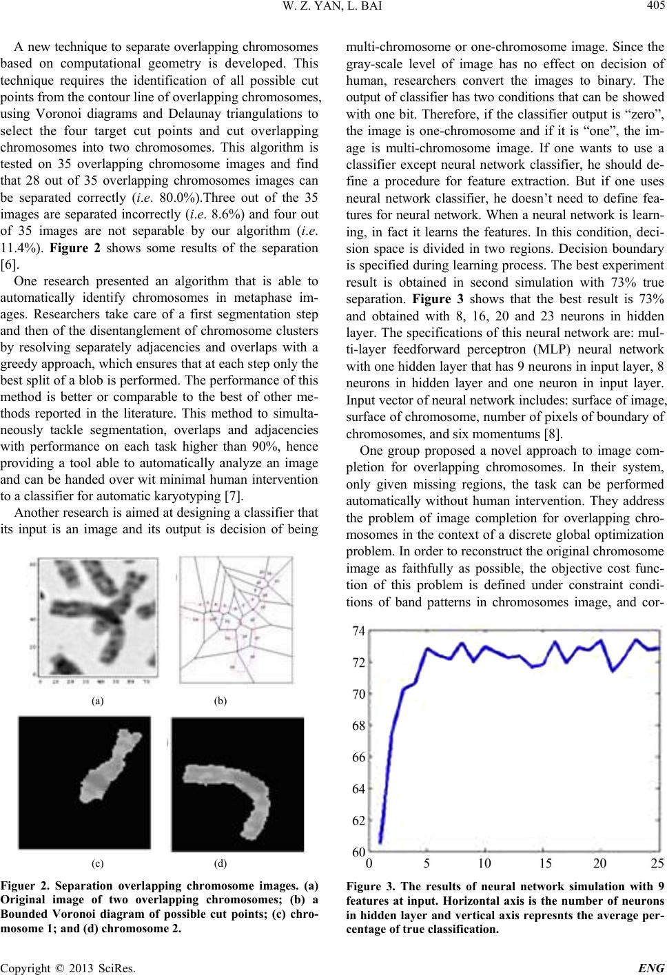 W. Z. YAN, L. BAI Copyright © 2013 SciRes. ENG 405 A new technique to separate overlapping chromosomes based on computational geometry is developed. This technique requires the identification of all possible cut points from the contour line of overlapping chromosomes, using Voronoi diagrams and Delaunay triangulations to select the four target cut points and cut overlapping chromosomes into two chromosomes. This algorithm is tested on 35 overlapping chromosome images and find that 28 out of 35 overlapping chromosomes images can be separated correctly (i.e. 80.0%).Three out of the 35 images are separated incorrectly (i.e. 8.6%) and four out of 35 images are not separable by our algorithm (i.e. 11.4%). Figure 2 shows some results of the separation [6]. One research presented an algorithm that is able to automatically identify chromosomes in metaphase im- ages. Researchers take care of a first segmentation step and then of the disentanglement of chromosome clusters by resolving separately adjacencies and overlaps with a greedy approach, which ensures that at each step only the best split of a blob is performed. The performance of this method is better or comparable to the best of other me- thods reported in the literature. This method to simulta- neously tackle segmentation, overlaps and adjacencies with performance on each task higher than 90%, hence providing a tool able to automatically analyze an image and can be handed over wit minimal human intervention to a classifier for automatic karyotyping [7]. Another research is aimed at designing a classifier that its input is an image and its output is decision of being (a) (b) (c) (d) Figuer 2. Separation overlapping chromosome images. (a) Original image of two overlapping chromosomes; (b) a Bounded Voronoi diagram of possible cut points; (c) chro- mosome 1; and (d) chromosome 2. mul ti-chromosome or one-chromosome image. Since the gray-scale level of image has no effect on decision of human, researchers convert the images to binary. The output of classifier has two conditions that can be showed with one bit. Therefore, if the classifier output is “zero”, the image is one-chromosome and if it is “one”, the im- age is multi-chromosome image. If one wants to use a classifier except neural network classifier, he should de- fine a procedure for feature extraction. But if one uses neural network classifier, he doesn’t need to define fea- tures for neural network. When a neural network is learn- ing, in fact it learns the features. In this condition, deci- sion space is divided in two regions. Decision boundary is specified during learning process. The best experiment result is obtained in second simulation with 73% true separation. Figure 3 shows that the best result is 73% and obtained with 8, 16, 20 and 23 neurons in hidden layer. The specifications of this neural network are: mul- ti-layer feedforward perceptron (MLP) neural network with one hidden layer that has 9 neurons in input layer, 8 neurons in hidden layer and one neuron in input layer. Input vector of neural network includes: surface of image, surface of chromosome, number of pixels of boundary of chromosomes, and six momentums [8]. One group proposed a novel approach to image com- pletion for overlapping chromosomes. In their system, only given missing regions, the task can be performed automatically without human intervention. They address the problem of image completion for overlapping chro- mosomes in the context of a discrete global optimization problem. In order to reconstruct the original chromosome image as faithfully as possible, the objective cost func- tion of this problem is defined under constraint condi- tions of band patterns in chromosomes image, and cor- Figure 3. The results of neural network simulation with 9 features at input. Horizontal axis is the number of neurons in hidden layer and vertical axis represnts the average per- centage of true classification. 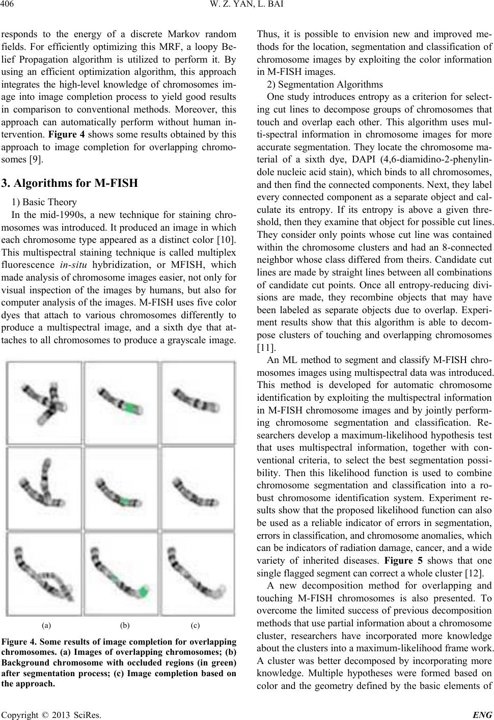 W. Z. YAN, L. BAI Copyright © 2013 SciRes. ENG 406 responds to the energy of a discrete Markov random fields. For efficiently optimizing this MRF, a loopy Be- lief Propagation algorithm is utilized to perform it. By using an efficient optimization algorithm, this approach integrates the high-level knowledge of chromosomes im- age into image completion process to yield good results in comparison to conventional methods. Moreover, this approach can automatically perform without human in- tervention. Figure 4 shows some results obtained by this approach to image completion for overlapping chromo- somes [9]. 3. Algorithms for M-FISH 1) Basic Theory In the mid-1990s, a new technique for staining chro- mosomes was introduced. It produced an image in which each chromosome type appeared as a distinct color [10]. This multispectral staining technique is called multiplex fluorescence in-situ hybridization, or MFISH, which made analysis of chromosome images easier, not only for visual inspection of the images by humans, but also for computer analysis of the images. M-FISH uses five color dyes that attach to various chromosomes differently to produce a multispectral image, and a sixth dye that at- taches to all chromosomes to produce a grayscale image. (a) (b) (c) Figure 4. Some results of image completion for overlapping chromosomes. (a) Images of overlapping chromosomes; (b) Background chromosome with occluded regions (in green) after segmentation process; (c) Image completion based on the approac h. Thus, it is possible to envision new and improved me- thods for the location, segmentation and classification of chromosome images by exploiting the color information in M-FISH images. 2) Segmentation Algorithms One study introduces entropy as a criterion for select- ing cut lines to decompose groups of chromosomes that touch and overlap each other. This algorithm uses mul- ti-spectral information in chromosome images for more accurate segmentation. They locate the chromosome ma- terial of a sixth dye, DAPI (4,6-diamidino-2-phenylin- dole nucleic acid stain), which binds to all chromosomes, and then find the connected components. Next, they label every connected component as a separate object and cal- culate its entropy. If its entropy is above a given thre- shold, then they examine that object for possible cut lines. They consider only points whose cut line was contained within the chromosome clusters and had an 8-connected neighbor whose class differed from theirs. Candidate cut lines are made by straight line s between all combination s of candidate cut points. Once all entropy-reducing divi- sions are made, they recombine objects that may have been labeled as separate objects due to overlap. Experi- ment results show that this algorithm is able to decom- pose clusters of touching and overlapping chromosomes [11]. An ML method to segment and classify M-FISH chro- moso me s i ma ge s using multispectr al data was introduced. This method is developed for automatic chromosome identification by exploiting the multispectral information in M-FISH chromosome images and by jointly perform- ing chromosome segmentation and classification. Re- searchers develop a maximum-likelihood hypothesis test that uses multispectral information, together with con- ventional criteria, to select the best segmentation possi- bility. Then this likelihood function is used to combine chromosome segmentation and classification into a ro- bust chromosome identification system. Experiment re- sults show that the proposed likelihood function can also be used as a reliable indicator of errors in segmentation, errors in classification, and chromosome anomalies, which can be indicators of radiation damage, cancer, and a wide variety of inherited diseases. Figure 5 shows that one single flagged segment can correct a whole cluster [12]. A new decomposition method for overlapping and touching M-FISH chromosomes is also presented. To overcome the limited success of previous decomposition methods that use partial information about a chromosome cluster, researchers have incorporated more knowledge about the clusters into a maximum-likelihood frame work. A cluster was better decomposed by incorporating more knowledge. Multiple hypotheses were formed based on color and the geometry defined by the basic elements of 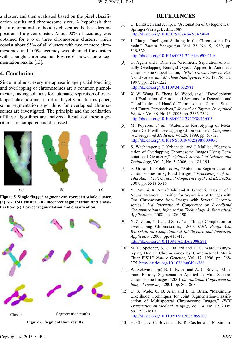 W. Z. YAN, L. BAI Copyright © 2013 SciRes. ENG 407 a cluster, and then evaluated based on the pixel classifi- cation results and chromosome sizes. A hypothesis that has a maximum-likelihood is chosen as the best decom- position of a given cluster. About 90% of accuracy was obtained for two or three chromosome clusters, which consist about 95% of all clusters with two or more chro- mosomes, and 100% accuracy was obtained for clusters with a single chromosome. Figure 6 shows some seg- mentation results [13]. 4. Conclusion Since in almost every metaphase image partial touching and overlapping of chromosomes are a common phenol- menon, finding solutions for automated separation of over- lapped chromosomes is difficult yet vital. In this paper, some segmentation algorithms for overlapped chromo- somes are investigated. The principle and the realization of these algorithms are analyzed. Results of these algo- rithms are compare d and discus s ed. (a) (b) (c) Figure 5. Singl e flagged segment can correct a whole cluster. (a) M-FISH cluster; (b) Incorrect segmentation and classi- fication; (c) Correct segmentation and classification. Figure 6. Segmentation results. REFERENCES [1] C. Lundsteen and J. Piper, “Automation of Cytogenetics,” Springer-Verlag, Berlin, 1989. http://dx.doi.org/10.1007/978-3-642-74738-0 [2] J. Liang, “Intelligent Splitting in the Chromosome Do- main,” Pattern Recognition, Vol. 22, No. 5, 1989, pp. 519-532. http://dx.doi.org/10.1016/0031-3203(89)90021-6 [3] G. Agam and I. Dinstein, “Geometric Separation of Par- tially Overlapping Nonrigid Objects Applied to Automatic Chromosome Classification,” IEEE Transactions on Pat- tern Analysis and Machine Intelligence, Vol. 19, No. 11, 1997, pp. 1212-1222. http://dx.doi.org/10.1109/34.632981 [4] X. W. Wang, B. Zheng, M. Wood, et al., “Development and Evaluation of Automated Systems for Detection and Classification of Handed Chromosomes: Current Status and Future Perspectives,” Journal of Physics D: Applied Physics, Vol.38, No.15, 2005, pp. 2536-2542. http://dx.doi.org/10.1088/0022-3727/38/15/003 [5] M. Popescu, et al., “Automatic Karyotyping of Meta- phase Cells with Overlapping Chromosomes,” Computers in Biology and Medicine, Vol.29, 1999, pp. 61-82. http://dx.doi.org/10.1016/S0010-4825(98)00040-7 [6] S. Wacharapong, J. Krisanadej and J. Mullica, “Segmen- tation of Overlapping Chromosome Images Using Com- putational Geometry,” Walailak Journal of Science and Technology, Vol. 2, No. 3, 2006, pp. 181-194. [7] E. Grisan, E. Poletti, et al., “Automatic Segmentation of Chromosomes in Q-Band Images,” Proceedings of the 29th Annual International Conference of the IEEE EMBS, 2007, pp. 5513-5516. [8] Y. Rahimi, R. Amirfattahi and R. Ghaderi, “Design of a Neural Network Classifier for Separation of Images with One Chromosome from Images with Several Chromo- somes,” 3rd International Conference on Broadband Communicaitons, Information Technology & Biomedical Applications, 2008, pp. 186-190. [9] X. Z. Zhou, Y. Lu and Z. Y. Yan, “Image Completion for Overlapping Chromosomes,” 2008 IEEE Pacific-Asia Workshop on Computational Intelligence and Industrial Application, 2008, pp. 413-417. http://dx.doi.org/10.1109/PACIIA.2008.271 [10] M. R. Speicher, S. G. Ballard and D. C. Ward, “Karyo- typing Human Chromosomes by Combinatorial Multi- Fluor FISH,” Nature Genetics, Vol. 12, 1996, pp. 368- 375. http://dx.doi.org/10.1038/ng0496-368 [11] W. Schwartzkopf, B. L. Evans and A. C. Bovik, “Mini- mum Entropy Segmentation Applied to Multi-Spectral Chromosome Images,” 2001 International Conference on Image Processing, 2001, pp. 865-868. [12] C. S. Wade, C. B. Alan and L. E. Brian, “Maximum- Likelihood Techniques for Joint Segmentation-Classifi- cation of Multispectral Chromosome Images,” IEEE Transaction on Medical Imaging, Vol. 24, No. 12, 2005, pp. 1593-1610. http://dx.doi.org/10.1109/TMI.2005.859207 [13] H. Choi, A. C. Bovik and K. R. Castleman, “Maximum-  W. Z. YAN, L. BAI Copyright © 2013 SciRes. ENG 408 Likelihood Decomposition of Overlapping and Touching M-FISH Chromosomes Using Geometry, Size and Color Information,” Proceedings of the 28th IEEE EMBS An- nual International Conference, 2006, pp. 3130-3133. |