

Paper Menu >>
Journal Menu >>
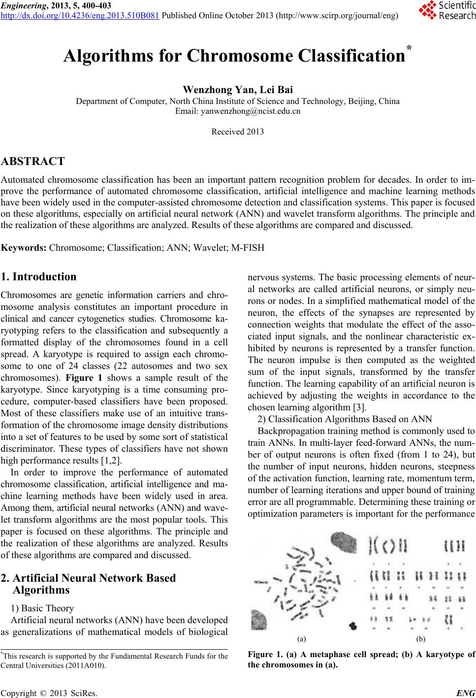 Engineering, 2013, 5, 400-403 http://dx.doi.org/10.4236/eng.2013.510B081 Published Online October 2013 (http://www.scirp.org/journal/eng) Copyright © 2013 SciRes. ENG Algorithms for Ch romosome Classification* Wenzhong Yan, Lei Bai Department of Computer, North China Institute of Science and Technology, Beijing, China Email: [email protected] Received 2013 ABSTRACT Automated chromosome classification has been an important pattern recognition problem for decades. In order to im- prove the performance of automated chromosome classification, artificial intelligence and machine learning methods have been widely used in the computer-assisted chromosome detection and classification systems. This paper is focused on these algorithms, especially on artificial neur al network (ANN) and wavelet transform algorithms. The princ iple and the realization of these algorithms are analyzed. Results of these algorithms are compared and discussed. Keywords: Chromosome; Classification; ANN; Wavelet; M-FISH 1. Introduction Chromosomes are genetic information carriers and chro- mosome analysis constitutes an important procedure in clinical and cancer cytogenetics studies. Chromosome ka- ryotyping refers to the classification and subsequently a formatted display of the chromosomes found in a cell spread. A karyotype is required to assign each chromo- some to one of 24 classes (22 autosomes and two sex chromosomes). Figure 1 shows a sample result of the karyotype. Since karyotyping is a time consuming pro- cedure, computer-based classifiers have been proposed. Most of these classifiers make use of an intuitive trans- formation of the chromosome image density distributions into a set of features to be used by some sort of statistical discriminator. These types of classifiers have not shown high perf o rmance re sults [1,2]. In order to improve the performance of automated chromosome classification, artificial intelligence and ma- chine learning methods have been widely used in area. Among them, artificial neural networks (ANN) and wave- let transform algorithms are the most popular tools. This paper is focused on these algorithms. The principle and the realization of these algorithms are analyzed. Results of these algorithms are compared and discussed. 2. Artificial Neural Network Based Algorithms 1) Basic Th eor y Artificial neural networks (ANN) have been developed as generalizations of mathematical models of biological nervous systems. The basic processing elements of neur- al networks are called artificial neurons, or simply neu- rons or nodes. In a simplified mathematical model of the neuron, the effects of the synapses are represented by connection weights that modulate the effect of the asso- ciated input signals, and the nonlinear characteristic ex- hibited by neurons is represented by a transfer function. The neuron impulse is then computed as the weighted sum of the input signals, transformed by the transfer function. The learning capability of an artificial neuron is achieved by adjusting the weights in accordance to the chosen learning algorithm [3]. 2) Classification Algorithms Based on ANN Backpropagation training method is commonly used to train ANNs. In multi-layer feed-forward ANNs, the num- ber of output neurons is often fixed (from 1 to 24), but the number of input neurons, hidden neurons, steepness of the activation function, learning rate, momentu m term, number of learning iter ation s and upp er boun d of train ing error are all programmable. Determining these training or optimization parameters is important for the performance (a) (b) Figure 1. (a) A metaphase cell spread; (b) A karyotype of the chromosomes in (a). *This research is supported by the Fundamental Research Funds for the Central Universities (2011A010). 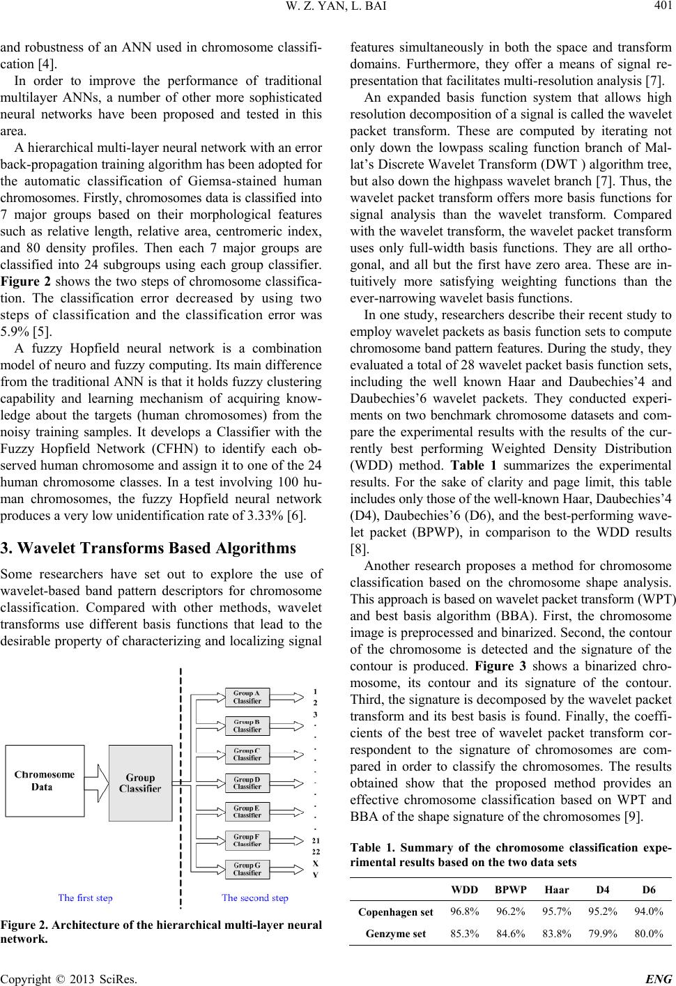 W. Z. YAN, L. BAI Copyright © 2013 SciRes. ENG 401 and robustness of an ANN used in chromosome classifi- cation [4]. In order to improve the performance of traditional multilayer ANNs, a number of other more sophisticated neural networks have been proposed and tested in this area. A hierarchical multi-layer neural network with an error back-propagation training algorithm has been adopted for the automatic classification of Giemsa-stained human chromosomes. Firstly, chro mosomes data is classified into 7 major groups based on their morphological features such as relative length, relative area, centromeric index, and 80 density profiles. Then each 7 major groups are classified into 24 subgroups using each group classifier. Figure 2 shows the two steps of chromosome classifica- tion. The classification error decreased by using two steps of classification and the classification error was 5.9% [5]. A fuzzy Hopfield neural network is a combination model of neuro and fuzzy computing. Its main difference from the traditiona l ANN is that it holds fuzzy clustering capability and learning mechanism of acquiring know- ledge about the targets (human chromosomes) from the noisy training samples. It develops a Classifier with the Fuzzy Hopfield Network (CFHN) to identify each ob- served human chromosome and assign it to one of the 24 human chromosome classes. In a test involving 100 hu- man chromosomes, the fuzzy Hopfield neural network produces a very low u nidenti f ication rate of 3. 33 % [6]. 3. Wavelet Transforms Based Algorithms Some researchers have set out to explore the use of wavelet-based band pattern descriptors for chromosome classification. Compared with other methods, wavelet transforms use different basis functions that lead to the desirable property of characterizing and localizing signal Figure 2. Architecture of the hierarchical multi-layer neural network. features simultaneously in both the space and transform domains. Furthermore, they offer a means of signal re- presentation that facilitates multi-resolution analysis [7]. An expanded basis function system that allows high resolution decomposition of a signal is called the wavelet packet transform. These are computed by iterating not only down the lowpass scaling function branch of Mal- lat’s Discrete Wavelet Transform (DWT ) algorithm tree, but also down the highpass wavelet branch [7]. Thus, the wavelet packet transform offers more basis functions for signal analysis than the wavelet transform. Compared with the wavelet transform, the wavelet packet transform uses only full-width basis functions. They are all ortho- gonal, and all but the first have zero area. These are in- tuitively more satisfying weighting functions than the ever-narrowing wavele t basis f unc t ions. In one study, researchers describe their recent study to employ wavelet packets as basis function sets to compute chromosome band pattern features. During the study, they evaluated a total of 28 wavelet packet basis function sets, including the well known Haar and Daubechies’4 and Daubechies’6 wavelet packets. They conducted experi- ments on two benchmark chromosome datasets and com- pare the experimental results with the results of the cur- rently best performing Weighted Density Distribution (WDD) method. Table 1 summarizes the experimental results. For the sake of clarity and page limit, this table includes only those of the well-known Haar, Daubechies’4 (D4), Daubechies’6 (D6), and the best-performing wave- let packet (BPWP), in comparison to the WDD results [8]. Another research proposes a method for chromosome classification based on the chromosome shape analysis. This approach is based on wavelet packet transform (WPT) and best basis algorithm (BBA). First, the chromosome image is preprocessed and binarized. Second, the contour of the chromosome is detected and the signature of the contour is produced. Figure 3 shows a binarized chro- mosome, its contour and its signature of the contour. Third, the signature is decomposed by the wavelet packet transform and its best basis is found. Finally, the coeffi- cients of the best tree of wavelet packet transform cor- respondent to the signature of chromosomes are com- pared in order to classify the chromosomes. The results obtained show that the proposed method provides an effective chromosome classification based on WPT and BBA of the shape signature of the chromosomes [9]. Table 1. Summary of the chromosome classification expe- rimental results based on the two data sets WDD BPWP Haar D4 D6 Copenhagen set 96.8% 96.2% 95.7% 95.2% 94.0% Genzyme set 85.3% 84.6% 83.8% 79.9% 80.0% 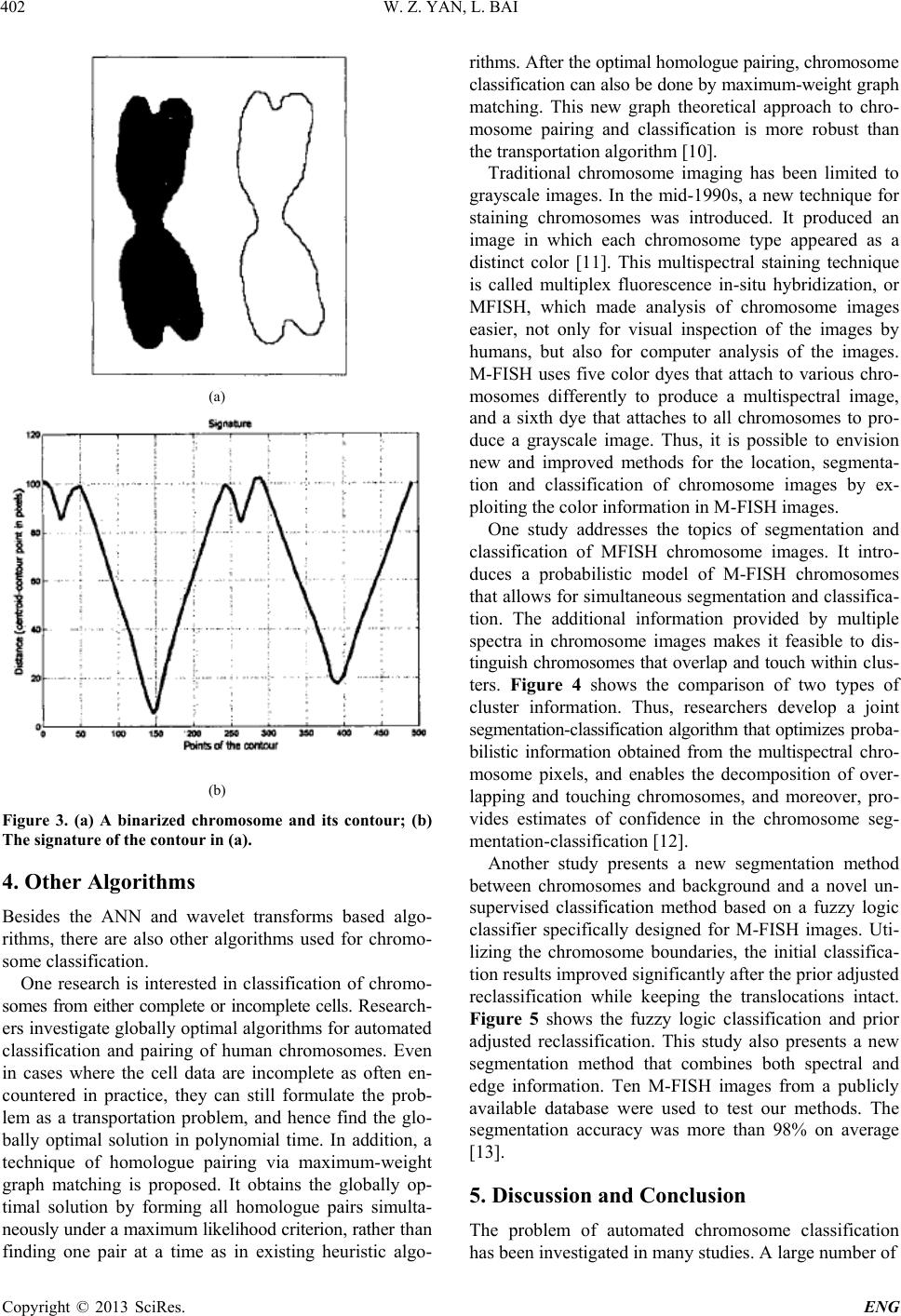 W. Z. YAN, L. BAI Copyright © 2013 SciRes. ENG 402 (a) (b) Figure 3. (a) A binarized chromosome and its contour; (b) The signature of the contour in (a). 4. Other Algorithms Besides the ANN and wavelet transforms based algo- rithms, there are also other algorithms used for chromo- some classification. One research is interested in classification of chromo- somes from either complete or incomplete cells. Research- ers investigate globally op timal algorithms for automated classification and pairing of human chromosomes. Even in cases where the cell data are incomplete as often en- countered in practice, they can still formulate the prob- lem as a transportation problem, and hence find the glo- bally optimal solution in polynomial time. In addition, a technique of homologue pairing via maximum-weight graph matching is proposed. It obtains the globally op- timal solution by forming all homologue pairs simulta- neously under a maximum likelihood criterion, rather than finding one pair at a time as in existing heuristic algo- rithms. After the optimal homologue pairing, chromosome classification can also be done by maximu m-weight graph matching. This new graph theoretical approach to chro- mosome pairing and classification is more robust than the transportation algorithm [10]. Traditional chromosome imaging has been limited to grayscale images. In the mid-1990s, a new technique for staining chromosomes was introduced. It produced an image in which each chromosome type appeared as a distinct color [11]. This multispectral staining technique is called multiplex fluorescence in-situ hybridization, or MFISH, which made analysis of chromosome images easier, not only for visual inspection of the images by humans, but also for computer analysis of the images. M-FISH uses five color dyes that attach to various chro- mosomes differently to produce a multispectral image, and a sixth dye that attaches to all chromosomes to pro- duce a grayscale image. Thus, it is possible to envision new and improved methods for the location, segmenta- tion and classification of chromosome images by ex- ploiting the color information in M-FISH images. One study addresses the topics of segmentation and classification of MFISH chromosome images. It intro- duces a probabilistic model of M-FISH chromosomes that allows for simultaneous segmentation and clas sifica- tion. The additional information provided by multiple spectra in chromosome images makes it feasible to dis- tinguish chromosomes that overlap and touch within clus- ters. Fig ure 4 shows the comparison of two types of cluster information. Thus, researchers develop a joint segmentation-classification algorith m that optimizes proba- bilistic information obtained from the multispectral chro- mosome pixels, and enables the decomposition of over- lapping and touching chromosomes, and moreover, pro- vides estimates of confidence in the chromosome seg- mentation-classification [12]. Another study presents a new segmentation method between chromosomes and background and a novel un- supervised classification method based on a fuzzy logic classifier specifically designed for M-FISH images. Uti- lizing the chromosome boundaries, the initial classifica- tion results improved significantly after the prior adjusted reclassification while keeping the translocations intact. Figure 5 shows the fuzzy logic classification and prior adjusted reclassification. This study also presents a new segmentation method that combines both spectral and edge information. Ten M-FISH images from a publicly available database were used to test our methods. The segmentation accuracy was more than 98% on average [13]. 5. Discussion and Conclusion The problem of automated chromosome classification has been investigated in many studies. A large number of 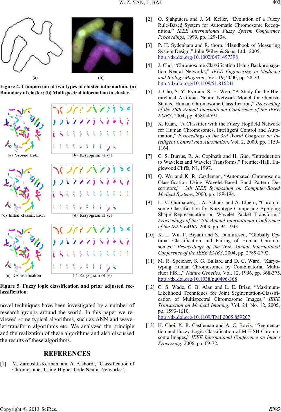 W. Z. YAN, L. BAI Copyright © 2013 SciRes. ENG 403 (a) (b) Figure 4. Comparison of two types of cluster information. (a) Boundary of cluster; (b) Multispectral information in cluster. Figure 5. Fuzzy logic classification and prior adjusted rec- lassification. novel techniques have been investigated by a number of research groups around the world. In this paper we re- viewed some typical algorithms, such as ANN and wave- let transform algorithms etc. We analyzed the principle and the realization of these algorithms and also discussed the results of these algorithms. REFERENCES [1] M. Zardoshti-Kermani and A. Afshordi, “Classification of Chromosomes Using Higher-Orde Neural Networks”. [2] O. Sjahputera and J. M. Keller, “Evolution of a Fuzzy Rule-Based System for Automatic Chromosome Recog- nition,” IEEE International Fuzzy System Conference Proceedings, 1999, pp. 129-134. [3] P. H. Sydenham and R. thorn, “Handbook of Measuring System Design,” John Wiley & Sons, Ltd., 2005. http://dx.doi.org/10.1002/0471497398 [4] J. Cho, “Chromosome Classification Using Backpropaga- tion Neural Networks,” IEEE Engineering in Medicine and Biology Magazine, Vol. 19, 2000, pp. 28-33. http://dx.doi.org/10.1109/51.816241 [5] J. Cho, S. Y. Ryu and S. H. Woo, “A Study for the Hie- rarchical Artificial Neural Network Model for Giemsa- Stained Human Chromosome Classification,” Proceeding of the 26th Annual International Conference of the IEEE EMBS, 2004, pp. 4588-4591. [6] X. Ruan, “A Classifier with the Fuzzy Hopfield Network for Human Chromosomes, Intelligent Control and Auto- mation,” Proceedings of the 3rd World Congress on In- telligent Control and Automation, Vol. 2, 2000, pp. 1159- 1164. [7] C. S. Burrus, R. A. Gopinath and H. Guo, “Introduction to Wavelets and Wavelet Transforms,” Prentice-Hall, En- glewood Cliffs, NJ, 1997. [8] Q. Wu and K. R. Castleman, “Automated Chromosome Classification Using Wavelet-Based Band Pattern De- scriptors,” 13th IEEE Symposium on Computer-Based Medical Systems, 2000, pp. 189-194. [9] L. V. Guimaraes, J. A. Schuck and A. Elbern, “Chromo- some Classification for Karyotype Composing Applying Shape Representation on Wavelet Packet Transform,” Proceedings of the 25th Annual International Conference of the IEEE EMBS, 2003, pp. 941-943. [10] X. L. Wu, P. Biy a ni and S. Dumitrescu, “Globally Op- timal Classification and Pairing of Human Chromo- somes,” Proceedings of the 26th Annual International Conference of the IEEE EMBS, 2004, pp. 2789-2792. [11] M. R. Speicher, S. G. Ballard and D. C. Ward, “Karyo- typing Human Chromosomes by Combinatorial Multi- fluor FISH,” Nature Genetics, Vol. 12, 1996, pp. 368-375. http://dx.doi.org/10.1038/ng0496-368 [12] C. S. Wade, C. B. Alan and L. E. Brian, “Maximum- Likelihood Techniques for Joint Segmentation-Classifi- cation of Multispectral Chromosome Images,” IEEE Transaction on Medical Imaging, Vol. 24, No. 12, 2005, pp. 1593-1610. http://dx.doi.org/10.1109/TMI.2005.859207 [13] H. Choi, K. R. Castleman and A. C. Bovik, “Segmenta- tion and Fuzzy-Logic Classification of M-FISH Chromo- some Images,” IEEE International Conference on Image Processing, 2006, pp . 69-72. |