

Paper Menu >>
Journal Menu >>
 Journal of Cancer Therapy, 2013, 4, 44-52 Published Online December 2013 (http://www.scirp.org/journal/jct) http://dx.doi.org/10.4236/jct.2013.411A006 Open Access JCT The PARP-1 Inhibitor Olaparib Causes Retention of γ-H2AX Foci in BRCA1 Heterozygote Cells Following Exposure to Gamma Radiation Emma C. Bourton1, Piers N. Plowman2, Amanda J. Harvey1, Sheba Adam Zahir1, Christopher N. Parris1 1Brunel Institute of Cancer Genetics and Pharmacogenomics, Division of Biosciences, School of Health Sciences and Social Care, Brunel University, Uxbridge, UK; 2Department of Radiotherapy, St Bartholomew’s Hospital, West Smithfield, London, UK. Email: [email protected] Received October 20th, 2013; revised November 16th, 2013; accepted November 24th, 2013 Copyright © 2013 Emma C. Bourton et al. This is an open access article distributed under the Creative Commons Attribution Li- cense, which permits unrestricted use, distribution, and reproduction in any medium, provided the original work is properly cited. ABSTRACT A novel treatment for cancer patients with homozygous deletions of BRCA1 and BRCA2 is to use drugs that inhibit the enzyme poly(ADP-ribose) polymerase (PARP). Specific inhibition of PARP-1 can induce synthetic lethality in irradi- ated cancer cells while theoretically leaving normal tissue unaffected. We recently demonstrated in a cell survival assay that lymphoblastoid cells with mono-allelic mutations of BRCA1 were hypersensitive to gamma radiation in the pres- ence of the PARP-1 inhibitor Olaparib compared to normal cells and mono-allelic BRCA2 cells. To determine if the enhanced radiation sensitivity was due to a persistence of DNA strand breaks, we performed γ-H2AX foci analysis in cells derived from two normal individuals, three heterozygous BRCA1 and three heterozygous BRCA2 cell lines. Cells were exposed to 2 Gy gamma radiation in the presence or absence of 5 µM Olaparib. Using immunofluorescence and imaging flow cytometry, foci were measured in untreated cells and at 0.5, 3, 5 and 24 hours post-irradiation. In all lymphoblastoid cells treated with 2 Gy gamma radiation, there was a predictable induction of DNA strand breaks, with a modest but significant retention of foci over 24 hours in irradiated cells treated with Olaparib (ANOVA P < 0.05). However, in mono-allelic BRCA1 cells, there was a failure to fully repair DNA double-strand breaks (DSB) in the pres- ence of Olaparib, evidenced by a significant retention of foci at 24 hours’ post irradiation (t-Test P < 0.05). These data show that the cellular hypersensitivity of mono-allelic BRCA1 lymphoblastoid cells to gamma radiation in the presence of the Olaparib is due to the retention of DNA DSB. These data may indicate that patients with inherited mutations in the BRCA1 gene treated with radiotherapy and PARP-1 inhibitors may experience elevated radiation-associated normal tissue toxicity. Keywords: BRCA1; BRCA2; Heterozygote; Radiosensitivity; PARP Inhibitor; Gamma-H2AX; Imaging Flow Cytometry 1. Introduction BRCA1 (BReast-CAncer susceptibility gene 1) and BRCA2 are tumour suppressor genes, the protein products of which contribute to DNA repair and transcriptional regu- lation. The BR CA proteins are activated in response to DNA damage such as double-strand breaks (DSB), form- ed by exposure to ionising radiation [1]. DSB repair is me- diated by two principal mechanisms: non-homologous end joining (NHEJ) and homologous recombination (HR). NHEJ occurs principally in non-cycling and G1 stage cells, where the DNA DSB are ligated together in an er- ror-prone manner. Here the BRCA1 protein is associated with the MRN complex, which senses DSB and is re- sponsible for DSB resolution. HR repairs DSB during the S and G2 phases of the cell cycle, when an intact sister chromatid can serve as a template for repair, a mecha- nism that is virtually error-free. Both BRCA1 and BRCA2 proteins are active in HR. BRCA1 is a signal mediator, whereas BRCA2 affects the initiation of repair by recruit- ing Rad51 to DSB. Hence BRCA1 is a versatile protein with multiple functional domains that interacts with many 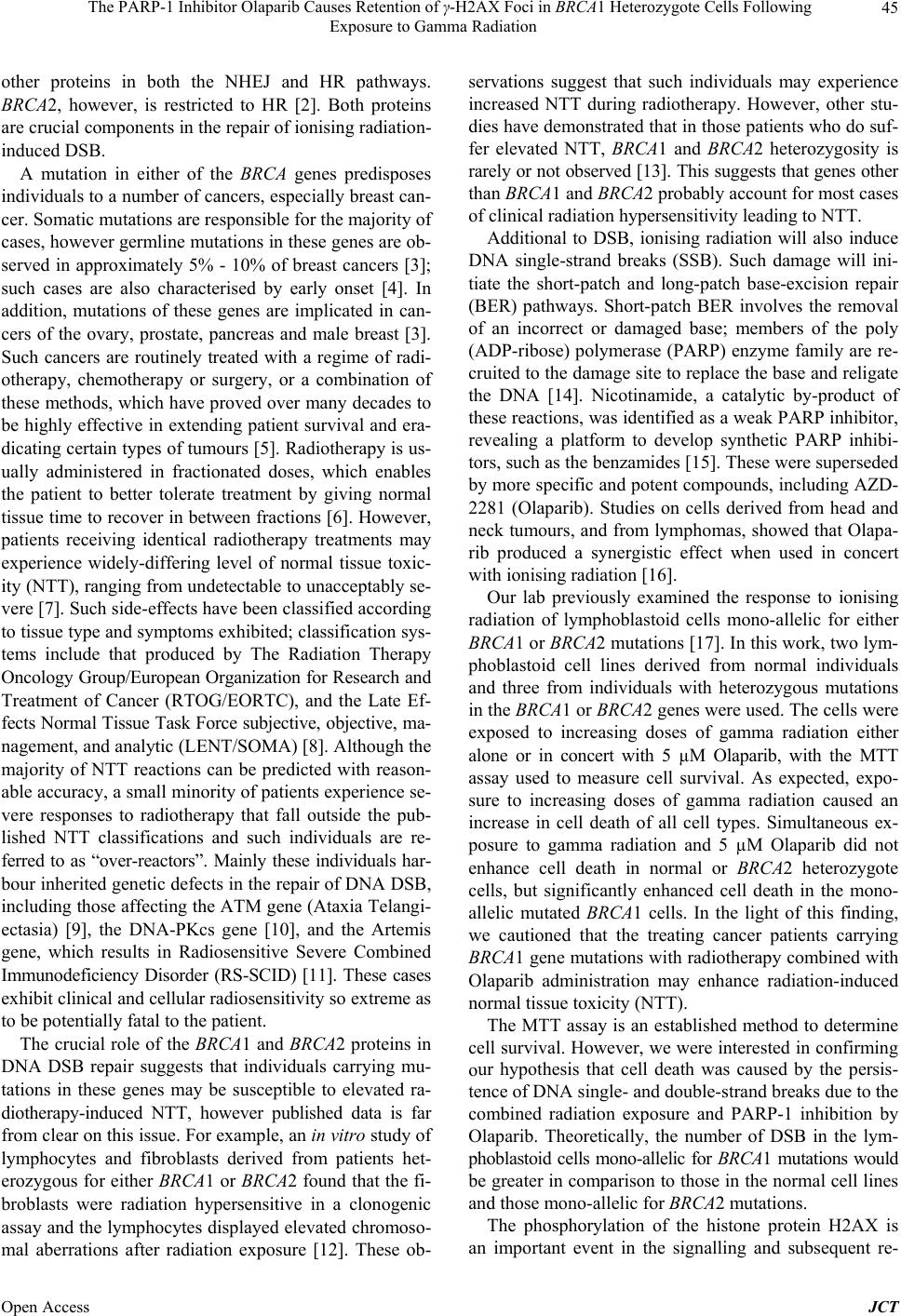 The PARP-1 Inhibitor Olaparib Causes Retention of γ-H2AX Foci in BRCA1 Heterozygote Cells Following Exposure to Gamma Radiation 45 other proteins in both the NHEJ and HR pathways. BRCA2, however, is restricted to HR [2]. Both proteins are crucial components in the repair of ionising radiation- induced DSB. A mutation in either of the BRCA genes predisposes individuals to a number of cancers, especially breast can- cer. Somatic mutations are responsible for the majority of cases, however germline mutations in these genes are ob- served in approximately 5% - 10% of breast cancers [3]; such cases are also characterised by early onset [4]. In addition, mutations of these genes are implicated in can- cers of the ovary, prostate, pancreas and male breast [3]. Such cancers are routinely treated with a regime of radi- otherapy, chemotherapy or surgery, or a combination of these methods, which have proved over many decades to be highly effective in extending patient survival and era- dicating certain types of tumours [5]. Radiotherapy is us- ually administered in fractionated doses, which enables the patient to better tolerate treatment by giving normal tissue time to recover in between fractions [6]. However, patients receiving identical radiotherapy treatments may experience widely-differing level of normal tissue toxic- ity (NTT), ranging from undetectable to unacceptably se- vere [7]. Such side-effects have been classified according to tissue type and symptoms exhibited; classification sys- tems include that produced by The Radiation Therapy Oncology Group/European Organization for Research and Treatment of Cancer (RTOG/EORTC), and the Late Ef- fects Normal Tissue Task Force subjective, objective, ma- nagement, and analytic (LENT/SOMA) [8]. Although the majority of NTT reactions can be predicted with reason- able accuracy, a small minority of patients experience se- vere responses to radiotherapy that fall outside the pub- lished NTT classifications and such individuals are re- ferred to as “over-reactors”. Mainly these individuals har- bour inherited genetic defects in the repair of DNA DSB, including those affecting the ATM gene (Ataxia Telangi- ectasia) [9], the DNA-PKcs gene [10], and the Artemis gene, which results in Radiosensitive Severe Combined Immunodeficiency Disorder (RS-SCID) [11]. These cases exhibit clinical and cellular radiosensitivity so extreme as to be potentially fatal to the patient. The crucial role of the BRCA1 and BRCA2 proteins in DNA DSB repair suggests that individuals carrying mu- tations in these genes may be susceptible to elevated ra- diotherapy-induced NTT, however published data is far from clear on this issue. For example, an in vitro study of lymphocytes and fibroblasts derived from patients het- erozygous for either BRCA1 or BRCA2 found that the fi- broblasts were radiation hypersensitive in a clonogenic assay and the lymphocytes displayed elevated chromoso- mal aberrations after radiation exposure [12]. These ob- servations suggest that such individuals may experience increased NTT during radiotherapy. However, other stu- dies have demonstrated that in those patients who do suf- fer elevated NTT, BRCA1 and BRCA2 heterozygosity is rarely or not observed [13]. This suggests that genes other than BRCA1 and BRCA2 probably account for most cases of clinical radiation hypersensitivity leading to NTT. Additional to DSB, ionising radiation will also induce DNA single-strand breaks (SSB). Such damage will ini- tiate the short-patch and long-patch base-excision repair (BER) pathways. Short-patch BER involves the removal of an incorrect or damaged base; members of the poly (ADP-ribose) polymerase (PARP) enzyme family are re- cruited to the damage site to replace the base and religate the DNA [14]. Nicotinamide, a catalytic by-product of these reactions, was identified as a weak PARP inhibitor, revealing a platform to develop synthetic PARP inhibi- tors, such as the benzamides [15]. These were superseded by more specific and potent compounds, including AZD- 2281 (Olaparib). Studies on cells derived from head and neck tumours, and from lymphomas, showed that Olapa- rib produced a synergistic effect when used in concert with ionising radiation [16]. Our lab previously examined the response to ionising radiation of lymphoblastoid cells mono-allelic for either BRCA1 or BRCA2 mutations [17]. In this work, two lym- phoblastoid cell lines derived from normal individuals and three from individuals with heterozygous mutations in the BRCA1 or BRCA2 genes were used. The cells were exposed to increasing doses of gamma radiation either alone or in concert with 5 µM Olaparib, with the MTT assay used to measure cell survival. As expected, expo- sure to increasing doses of gamma radiation caused an increase in cell death of all cell types. Simultaneous ex- posure to gamma radiation and 5 µM Olaparib did not enhance cell death in normal or BRCA2 heterozygote cells, but significantly enhanced cell death in the mono- allelic mutated BRCA1 cells. In the light of this finding, we cautioned that the treating cancer patients carrying BRCA1 gene mutations with radiotherapy combined with Olaparib administration may enhance radiation-induced normal tissue toxicity (NTT). The MTT assay is an established method to determine cell survival. However, we were interested in confirming our hypothesis that cell death was caused by the persis- tence of DNA single- and double-strand breaks due to the combined radiation exposure and PARP-1 inhibition by Olaparib. Theoretically, the number of DSB in the lym- phoblastoid cells mono-allelic for BRCA1 mutations would be greater in comparison to those in the normal cell lines and those mono-allelic for BRCA2 mutations. The phosphorylation of the histone protein H2AX is an important event in the signalling and subsequent re- Open Access JCT 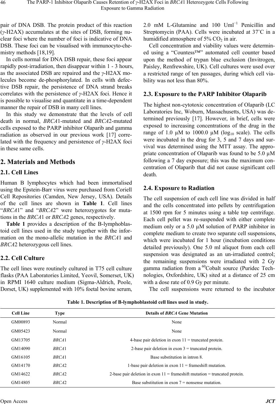 The PARP-1 Inhibitor Olaparib Causes Retention of γ-H2AX Foci in BRCA1 Heterozygote Cells Following Exposure to Gamma Radiation Open Access JCT 46 pair of DNA DSB. The protein product of this reaction (γ-H2AX) accumulates at the sites of DSB, forming nu- clear foci where the number of foci is indicative of DNA DSB. These foci can be visualised with immunocyto-che- mistry methods [18,19]. In cells normal for DNA DSB repair, these foci appear rapidly post-irradiation, then disappear within 1 - 3 hours, as the associated DSB are repaired and the γ-H2AX mo- lecules become de-phosphorylated. In cells with defec- tive DSB repair, the persistence of DNA strand breaks correlates with the persistence of γ-H2AX foci. Hence it is possible to visualise and quantitate in a time-dependent manner the repair of DSB in many cell lines. In this study we demonstrate that the levels of cell death in normal, BRCA1-mutated and BRCA2-mutated cells exposed to the PARP inhibitor Olaparib and gamma radiation as observed in our previous work [17] corre- lated with the frequency and persistence of γ-H2AX foci in these same cells. 2. Materials and Methods 2.1. Cell Lines Human B lymphocytes which had been immortalised using the Epstein-Barr virus were purchased from Coriell Cell Repositories (Camden, New Jersey, USA). Details of the cell lines are shown in Table 1. Cell lines “BRCA1” and “BRCA2” were heterozygotes for muta- tions in the BRCA1 or BRCA2 genes, respectively. Table 1 provides a description of the B-lymphoblas- toid cell lines used in the study together with the infor- mation on the mono-allelic mutation in the BRCA1 and BRCA2 heterozygous cell lines. 2.2. Cell Culture The cell lines were routinely cultured in T75 cell culture flasks (PAA Laboratories Limited, Yeovil, Somerset, UK) in RPMI 1640 culture medium (Sigma-Aldrich, Poole, Dorset, UK) supplemented with 10% foetal bovine serum, 2.0 mM L-Glutamine and 100 Uml−1 Penicillin and Streptomycin (PAA). Cells were incubated at 37˚C in a humidified atmosphere of 5% CO2 in air. Cell concentration and viability values were determin- ed using a “Countess™” automated cell counter based upon the method of trypan blue exclusion (Invitrogen, Paisley, Renfrewshire, UK). Cell cultures were used over a restricted range of ten passages, during which cell via- bility was not less than 80%. 2.3. Exposure to the PARP Inhibitor Olaparib The highest non-cytotoxic concentration of Olaparib (LC Laboratories Inc, Woburn, Massachusetts, USA) was de- termined previously [17]. However, in brief, cells were exposed to increasing concentrations of the drug in the range of 1.0 µM to 1000.0 µM (log10 scale). The cells were incubated in the drug for 3, 5 and 7 days and sur- vival was determined using the MTT assay. The appro- priate concentration of Olaparib was found to be 5.0 µM following a 7 day exposure; this was the maximum con- centration of Olaparib that did not cause significant cell death. 2.4. Exposure to Radiation The cell suspension of each cell line was divided in half and the cells concentrated into pellets by centrifugation at 1500 rpm for 5 minutes using a table top centrifuge. Each cell pellet was re-suspended with either complete medium only or a 5.0 µM solution of PARP inhibitor in complete medium to create two separate cell suspensions, which were incubated for 1 hour (incubation conditions detailed previously). One 5.0 ml aliquot from each cell suspension was designated as an un-irradiated control; the remaining suspensions were irradiated with 2 Gy gamma radiation from a 60Cobalt source (Puridec Tech- nologies, Oxfordshire, UK) sited at a distance of 25 cm with a dose rate of 0.9 Gy per minute. The cell suspensions were returned to the incubator Table 1. Description of B-lymphoblastoid c e l l lines used in study. Cell Line Type Details of BRCA Gene Mutation GM00893 Normal None GM05423 Normal None GM13705 BRCA1 4-base pair deletion in exon 11 = truncated protein. GM14090 BRCA1 2-base pair deletion in exon 3 = truncated protein. GM16105 BRCA1 Base substitution in intron 8. GM14170 BRCA2 1-base pair deletion in exon 11 = frameshift mutation. GM14622 BRCA2 2-base pair deletion in exon 11 = frameshift mutation = truncated protein. GM14805 BRCA2 Base substitution in exon 7 = nonsense mutation. 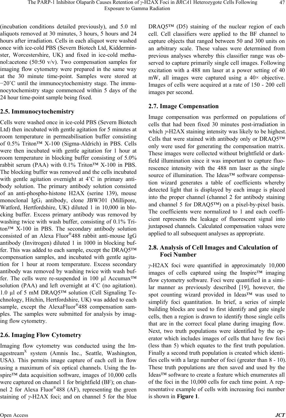 The PARP-1 Inhibitor Olaparib Causes Retention of γ-H2AX Foci in BRCA1 Heterozygote Cells Following Exposure to Gamma Radiation 47 (incubation conditions detailed previously), and 5.0 ml aliquots removed at 30 minutes, 3 hours, 5 hours and 24 hours after irradiation. Cells in each aliquot were washed once with ice-cold PBS (Severn Biotech Ltd, Kiddermin- ster, Worcestershire, UK) and fixed in ice-cold metha- nol:acetone (50:50 v/v). Two compensation samples for imaging flow cytometry were prepared in the same way at the 30 minute time-point. Samples were stored at −20˚C until the immunocytochemistry stage. The immu- nocytochemistry stage commenced within 5 days of the 24 hour time-point sample being fixed. 2.5. Immunocytochemistry Cells were washed once in ice-cold PBS (Severn Biotech Ltd) then incubated with gentle agitation for 5 minutes at room temperature in permeabilisation buffer consisting of 0.5% Triton™ X-100 (Sigma-Aldrich) in PBS. Cells were then incubated with gentle agitation for 1 hour at room temperature in blocking buffer consisting of 5.0% rabbit serum (PAA) with 0.1% Triton™ X-100 in PBS. The blocking buffer was removed and the cells incubated with gentle agitation overnight at 4˚C in primary anti- body solution. The primary antibody solution consisted of an anti-phospho-histone H2AX (serine 139), mouse monoclonal IgG1 antibody, clone JBW301 (Millipore, Watford, Hertfordshire, UK) diluted 1 in 10,000 in blo- cking buffer. Excess primary antibody was removed by washing twice with wash buffer, consisting of 0.1% Tri- ton™ X-100 in PBS. The secondary antibody solution consisted of an Alexa Fluor®488 rabbit anti-mouse IgG antibody (Invitrogen) diluted 1 in 1000 in blocking buf- fer. This was added to each sample, except the DRAQ5™ compensation samples, and incubated with gentle agita- tion for 1 hour at room temperature. Excess secondary antibody was removed by washing twice with wash buf- fer. The cells were re-suspended in 100 µl Accumax™ solution (PAA) and left overnight at 4˚C (no agitation). 1.0 µl of 5 mM DRAQ5™ solution (Cell Signaling Te- chnology, Hitchin, Hertfordshire, UK) was added to each sample, except the AlexaFluor®488 compensation sam- ples. The samples were submitted for analysis by imag- ing flow cytometry. 2.6. Imaging Flow Cytometry Imaging flow cytometry was conducted using the Im- agestreamX system (Amnis Inc., Seattle, Washington, USA). This permits image capture of each cell in flow using a maximum of six optical channels. Using the In- spire™ data acquisition software, images of 10,000 cells were captured on channel 1 for brightfield (BF); on chan- nel 2 for Alexa Fluor®488 (AF), representing the green staining of γ-H2AX foci; and on channel 5 for the blue DRAQ5™ (D5) staining of the nuclear region of each cell. Cell classifiers were applied to the BF channel to capture objects that ranged between 50 and 300 units on an arbitrary scale. These values were determined from previous analyses whereby this classifier range was ob- served to capture primarily single cell images. Following excitation with a 488 nm laser at a power setting of 40 mW, all images were captured using a 40× objective. Images of cells were acquired at a rate of 150 - 200 cell images per second. 2.7. Image Compensation Image compensation was performed on populations of cells that had been fixed 30 minutes post-irradiation in which -H2AX staining intensity was likely to be highest. Cells that were stained with antibody only or DRAQ5™ only were used for generating the compensation matrix. These images were collected without brightfield or dark- field illumination since it was important to capture fluo- rescence intensity with the 488 nm laser as the single source of illumination. The Ideas™ software compensa- tion wizard generates a table of coefficients whereby detected light that is displayed by each image is placed into the proper channel (channel 2 for antibody staining and channel 5 for DRAQ5™) on a pixel-by-pixel basis. The coefficients were normalized to 1 and each coeffi- cient represents the leakage of fluorescent signal into juxtaposed channels. Calculated compensation values were applied to all subsequent analyses as appropriate. 2.8. Analysis of Cell Images and Calculation of Foci Number γ-H2AX foci were quantified in approximately 10,000 images of cells captured using the Inspire™ imaging flow cytometry software. Foci were quantified in a simi- lar manner as previously described [19], however, the spot counting wizard provided in Ideas™ was used to simplify foci quantitation. In brief, a series of simple building blocks are used to first identify and gate single cells, then a region is drawn to identify those single cells that are in the correct focal plane during imaging flow. Next, two truth populations were identified by the op- erator which includes images of cells that have few foci (less than 5) which equates to the first truth population. Finally a second truth population is created which identi- fies cells with a large number of foci (greater than 8 - 10). These truth populations are then saved and used by the Ideas™ software to create a feature which enumerates all of the foci in the 10,000 cells for each time point. A rep- resentative example of cells with increasing foci number is shown in Figure 1. Open Access JCT  The PARP-1 Inhibitor Olaparib Causes Retention of γ-H2AX Foci in BRCA1 Heterozygote Cells Following Exposure to Gamma Radiation 48 Figure 1. Figure 1 shows representative images of increas- ing number of γ-H2AX foci in the lymphoblastoid cells cap- tured with imaging flow cytometry. The brightfield channel demonstrates the appearance of lymphoblastoid cells under light microscopy. The second channel represents the appea- rance of increasing numbers of γ-H2AX foci. The final co- lumn depicts the visualisation of the nuclear region of the cells by DRAQ5™ staining. Images of cells were captured at a magnification of 40×. 2.9. Statistical Analysis Using the data analysis programme in Microsoft Excel, two-way analysis of variance was used to compare the distribution of foci in radiation-exposed cells in the pre- sence or absence of Olaparib, across the complete time course of the experiment. A Student’s unpaired t-Test was utilised to determine if there were differences in foci retention at specific time points following radiation ex- posure in the presence or absence of Olaparib. Data were regarded as significantly different in the P value was less than 0.05. 3. Results 3.1. Gamma H2AX Foci Analysis in Normal Lymphoblastoid Cells Gamma H2AX foci analysis was performed in two nor- mal lymphoblastoid cell lines (GM00893 and GM05423). Cells were exposed to 2 Gy gamma radiation in the pre- sence or absence of the PARP-1 inhibitor Olaparib and gamma H2AX foci were measured in un-irradiated cells and at 30 minutes, 3, 5 and 24 hours post-irradiation. The profiles of foci induction combined for GM00893 and GM05423 are shown in Figure 2 . For normal cells with and without Olaparib exposure there is a predictable induction of foci at 30 minutes post-irradiation. In the cells not exposed to Olaparib there is a mean of 7.58 foci per cell, and in the Olaparib- Figure 2. Figure 2 shows the induction of γ-H2AX foci in the normal lymphoblastoid cells GM00893 and GM05423 plot- ted as an average graph. Data are derived from enumera- tion of foci in the images of approximately 10,000 cells cap- tured during imaging flow cytometry. Cells were treated with 2 Gy gamma radiation in the presence (black columns) or absence (white columns) of 5 m Olaparib. Foci were measured in untreated cells and those irradiated with 2 Gy gamma radiation at 0.5, 3, 5 and 24 hours post-irradiation. The data demo nstrate a slight but signi ficant retention of fo- ci in Olaparib-treated cells over a 24 hour period (ANOVA P > 0.05). However by comparing the 24 hours post-irra- diation time point alone, there was no significant difference in foci retention after irradiation with or without Olaparib (Student’s unpaired t-Test P < 0.05). treated cells this is slightly reduced at 6.83 foci per cell. Subsequently over a 24 hour period post-irradiation there is a decline in foci number in the normal cells, both Ola- parib-treated and untreated. At 24 hours this is reduced to near un-irradiated levels in the non-Olaparib-treated cells, at 2.38 foci per cell, and in the Olaparib-treated cells this figure is slightly higher at 3.24 foci per cell. Using analysis of variance to compare the time course distribution of post-2 Gy gamma radiation foci induction in the Olaparib-treated and untreated cells there is a sig- nificant difference in the distribution of foci (p = 0.012). However, using a Student’s t-Test to compare foci reten- tion at 24 hours in Olaparib-treated and untreated cells, there is no significant difference in foci numbers between treated and untreated cells (p = 0.315). This data indi- cates that Olaparib does not alter the repair of DNA DSB measured by gamma H2AX foci in lymphoblastoid cells derived from normal individuals. 3.2. Gamma H2AX Foci Analysis in BRCA1 Heterozygous Lymphoblastoid Cells Gamma H2AX foci analysis was performed following 2 Gy gamma radiation in three lymphoblastoid cell lines (GM13705, GM14090 and GM16105) (details of expe- riment and analysis as above). The profiles of foci induc- Open Access JCT  The PARP-1 Inhibitor Olaparib Causes Retention of γ-H2AX Foci in BRCA1 Heterozygote Cells Following Exposure to Gamma Radiation 49 tion combined for GM13705, GM14090 and GM16105 are shown in Figure 3. For BRCA1 heterozygous cells with and without Ola- parib exposure there is a predictable induction of foci at 30 minutes post-irradiation. In the cells not exposed to Olaparib there is a mean of 5.23 foci per cell, and in the Olaparib-treated cells this is approximately the same at 5.00 foci per cell. Subsequently over a 24 hour period post-irradiation there is a decline in foci number in the cells not treated with Olaparib; at 24 hours there is a mean of 2.00 foci per cell. However, in the Olaparib- treated cells there is retention of foci over the 24 hour period, so that the mean at 24 hours is 4.27 foci per cell, over twice the number of foci than in the untreated cells. Using analysis of variance to compare the time course distribution of post-2 Gy gamma radiation foci induction in the Olaparib-treated and untreated cells, there is a sig- nificant difference in the distribution of foci (p = 0.004). This significant difference is further verified with a Stu- dent’s t-Test. At 24 hours there is a significant retention of foci in the Olaparib-treated cells (p = 0.006). 3.3. Gamma H2AX Foci Analysis in BRCA2 Heterozygous Lymphoblastoid Cells Gamma H2AX foci analysis was performed following 2 Gy gamma radiation in three lymphoblastoid cell lines (GM14170, GM14622 and GM14805) (details of expe- riment and analysis as above). The profiles of foci induc- tion combined for GM14170, GM14622 and GM14805 are shown in Figure 4. For BRCA2 heterozygous cells with and without Ola- parib exposure there is a predictable induction of foci at 30 minutes post-irradiation. In the cells not exposed to Olaparib there is a mean of 5.16 foci per cell, and in the Olaparib-treated cells this is approximately the same at 5.90 foci per cell. Subsequently over a 24-hour period post-irradiation there is a decline in foci number in the BRCA2 heterozygous cells, both Olaparib-treated and untreated. At 24 hours this is reduced to slightly below un-irradiated levels in the non-Olaparib-treated cells, at 1.29 foci per cell, and in the Olaparib-treated cells this figure is slightly higher at 2.02 foci per cell. Using analysis of variance to compare the time course distribution of post-2 Gy gamma radiation foci induction in the Olaparib-treated and untreated cells there is a sig- nificant difference in the distribution of foci (p = 0.004). However, using a Student’s t-Test to compare foci reten- tion at 24 hours in Olaparib-treated and untreated cells, there is no significant difference in foci numbers (p = 0.181). This data indicates that Olaparib does not alter the repair of DNA DSB measured by gamma H2AX foci in lymphoblastoid cells derived from BRCA2 heterozy- gous individuals. Figure 3. Figure 3 shows the induction of γ-H2AX foci in three BRCA1 mono-allelic lymphoblastoid cell lines GM13705, GM14090 and GM16105 plotted as an average graph. Data are derived from enumerating the foci in the images of ap- proximately 10,000 cells captured during imaging flow cyto- metry. Cells were treated with 2 Gy gamma radiation in the presence (black columns) or absence (white columns) of 5 m Olaparib. Foci were measured in untreated cells and those irradiated with 2 Gy gamma radiation at 0.5, 3, 5 and 24 hours post-irradiation. The data demonstrate a slight but significant retention of foci in Olaparib treated cells over a 24 hour period (ANOVA P > 0.05). However by comparing foci retention at 24 hours post-irradiation alone there is a significant retention of foci in the presence of Olaparib (Student’s t-Test P > 0.05). 4. Discussion Radiotherapy is routinely employed in the clinical man- agement of cancer, frequently in conjunction with che- motherapy and/or surgery. The ionising radiation used in treatment creates single- and double-strand breaks in the DNA of both normal and tumour cells which if unrepair- ed can induce cell death. PARP inhibitors are chemothe- rapeutic agents prescribed to patients to accentuate the ionising radiation-induced DNA damage in tumour cells by inhibiting short-patch base-excision repair of DNA single-strand breaks. At replication forks where a SSB is encountered, the inhibition of PARP-1 by a drug such as Olaparib results in a DNA DSB [2], thus exacerbating ra- diation-induced DNA damage. These drugs are particular- ly effective in tumour cells homozygous for a mutation in the tumour suppressor genes BRCA1 and BRCA2 [16]. BRCA1 and BRCA2 proteins are involved in NHEJ and HR. BRCA1 is a versatile protein, participating in both the NHEJ and HR pathways, whereas BRCA2 is restrict- ed to HR. Hence cells where mutations in the BRCA genes have produced proteins with very little or no active function are likely to have reduced DSB repair capacity. NTT is the manifestation of damage to normal, non- cancerous tissue as a side-effect of anti-cancer therapy. There is no evidence to suggest that PARP inhibitors Open Access JCT  The PARP-1 Inhibitor Olaparib Causes Retention of γ-H2AX Foci in BRCA1 Heterozygote Cells Following Exposure to Gamma Radiation 50 Figure 4. Figure 4 shows the induction of γ-H2AX foci in three BRCA2 mono-allelic lymphoblastoid cell lines GM14170, GM14622 and GM14805 plotted as an average graph. Data are derived from enumerating the foci in the images of ap- proximately 10,000 cells captured during imaging flow cy- tometry. Cells were treated with 2 Gy gamma radiation in the presence (black columns) or absence (white columns) of 5 m Olaparib. Foci were measured in untreated cells and those irradiated with 2 Gy gamma radiation at 0.5, 3, 5 and 24 hours post-irradiation. The data demonstrate a slight but significant retention of foci in Olaparib treated cells over a 24 hour period (ANOVA P > 0.05). However by comparing 24 hours post-irradiation time point alone, there was no sig- nificant difference in foci retention after irradiation with or without Olaparib (Student’s unpaired t-Test P < 0.05). used as a single modality of treatment affect the survival of cells heterozygous for mutations in BRCA1 and BRCA2 i.e. non-cancer cells [20]. However, PARP inhibitors us- ed in conjunction with other forms of cytotoxic therapy (such as radiotherapy) may cause significant toxicity in these heterozygous/non-cancer tissue cells [17]. This has important implications for the treatment of those cancer patients carrying a BRCA1 or BRCA2 mutation; in a pre- vious paper we cautioned that such patients may be sus- ceptible to elevated levels of NTT [17]. The previous study examined the response to ionising radiation of lymphoblastoid cells mono-allelic for either BRCA1 or BRCA2 mutations, when the cells had been exposed to the PARP inhibitor Olaparib. Cell survival was measured using the MTT assay. The combined cyto- toxic treatment did not affect cell survival in normal or BRCA2 heterozygote cells, but significantly enhanced cell death in the BRCA1 heterozygote cells. We hypothesised that he enhanced cellular radiation sensitivity of BRCA1 heterozygous cells exposed to Ola- parib was due to DSB retention. The number of foci per cell in each of the cell lines was measured over a time course of 24 hours post-irra- diation, and the results combined depending on cell type (normal, BRCA1-mutated and BRCA2-mutated). The number of foci per cell in all cell types, both treated with Olaparib and untreated, increased considerably 30 min- utes post-irradiation, then declined over the time course as the DSB were repaired and the γ-H2AX proteins were de-phosphorylated. We did observe a slight retention of foci in all cell types at the 24 hour time point in those cells treated with Olaparib. The exception to this trend was observed in the BRCA1-mutated cells treated with Olaparib. They retained almost the same number of foci per cell over the 24 hours, whereas over the same time period the number of foci per cell in untreated BRCA1- mutated cells decreased almost to pre-irradiated levels. This confirms our hypothesis that the enhanced cell death detected by the MTT assay in heterozygous BRCA1 cells was a result of unrepaired strand breaks in the DNA of these cells. The reasons as to why this enhanced cell death is ob- served in heterozygous BRCA1 cells treated with Ola- parib, when compared with untreated BRCA1 cells, is likely due to the specific functions of the PARP-1 in- hibitor Olaparib and the BRCA1 protein. The DNA single strand breaks created by exposure to ionising radiation are repaired by the BER pathway which requires the ac- tion of the PARP-1 enzyme. Inhibition of this enzyme by a drug such as Olaparib means SSB remain unrepaired. Furthermore, SSB are converted to DSB at replication fork sites, thus triggering the DNA DSB repair pathway (which includes the phosphorylation of the histone pro- tein H2AX to γ-H2AX), and the formation of γ-H2AX foci [21]. The BRCA1 protein has a variety of roles within DNA DSB repair, both in NHEJ and HR, and also interacts with tumour suppressor genes (e.g. p53) and other cell cycle regulators. The ubiquitous nature of the BRCA1 protein in DNA repair means that a mutated/non-func- tioning protein may severely inhibit effective repair in both cycling and non-cycling cells. Another noticeable difference in the results arises from comparing the number of foci per cell detected in the normal cell lines with that reported in the BRCA-mutated cell lines. It can be seen from Figures 1-3 that normal cell lines report more foci per cell (and therefore more DNA DSB) at 30 minutes post-irradiation than either of the BRCA-mutated cell lines. There may be no single rea- son for this initially high level of reported DSB in normal cells, but a possible cause for this phenomenon includes the role of the BRCA1 protein in signal mediation imme- diately post-damage. Hence mutated BRCA1 proteins may inhibit the reporting of DNA damage in irradiated cells, although that does not explain the relatively low level of foci per cell in BRCA2-mutated cells 30 minutes post-ir- radiation. There may well be other, unidentified, reasons as to why this phenomenon occurs. Open Access JCT  The PARP-1 Inhibitor Olaparib Causes Retention of γ-H2AX Foci in BRCA1 Heterozygote Cells Following Exposure to Gamma Radiation 51 5. Conclusion In summary, this fundamental study of DSB repair kine- tics in a collection of lymphoblastoid cells mono-allelic for BRCA1 and BRCA2 indicates that the enhanced radia- tion sensitivity of BRCA1 heterozygous cells to radiation in the presence of Olaparib is caused by a persistence of DNA DSB. We reiterate that cancer patients with BRCA1 mutations may experience unexpectedly severe NTT when treated with radiotherapy and PARP inhibitors. 6. Acknowledgements Dr. Emma Bourton was supported by a grant from the Vidal Sassoon Foundation of America. We thank Mr. Hussein Al-Ali for assistance with fig- ure preparation. REFERENCES [1] K. Yoshida and Y. Miki, “Role of BRCA1 and BRCA2 as Regulators of DNA Repair, Transcription, and Cell Cycle in Response to DNA Damage,” Cancer Science, Vol. 95, No. 11, 2004, pp. 866-871. http://dx.doi.org/10.1111/j.1349-7006.2004.tb02195.x [2] R. Roy, J. Chun and S. N. Powell, “BRCA1 and BRCA2: Different Roles in a Common Pathway of Genome Pro- tection,” Nature Reviews. Cancer, Vol. 12, No. 1, 2011, pp. 68-78. http://dx.doi.org/10.1038/nrc3181 [3] A. R. Venkitaraman, “Functions of BRCA1 and BRCA2 in the Biological Response to DNA Damage,” Journal of Cell Science, Vol. 114, No. 20, 2001, pp. 3591-3598. [4] A. Bhattacharyya, U. S. Ear, B. H. Koller, R. R. Weichsel- baum and D. K. Bishop, “The Breast Cancer Susceptibil- ity Gene BRCA1 is Required for Subnuclear Assembly of Rad51 and Survival Following Treatment with the DNA Cross-Linking Agent Cisplatin,” The Journal of Biologi- cal Chemistry, Vol. 275, No. 31, 2000, pp. 23899-23903. http://dx.doi.org/10.1074/jbc.C000276200 [5] R. A. Weinberg, “The Rational Treatment of Cancer,” In: R. A. Weinberg, Ed., The Biology of Cancer, 2nd Edition, Garland Science, New York, 2013, pp. 797-876. [6] J. Bernier, E. J. Hall and A. Giaccia, “Radiation Oncolo- gy: A Century of Achievements,” Nature Reviews. Can- cer, Vol. 4, No. 9, 2004, pp. 737-747. http://dx.doi.org/10.1038/nrc1451 [7] S. L. Tucker, F. B. Geara, L. J. Peters and W. A. Brock, “How Much Could the Radiotherapy Dose Be Altered for Individual Patients Based on a Predictive Assay of Nor- mal-Tissue Radiosensitivity?” Radiotherapy and Oncol- ogy: Journal of the European Society for Therapeutic Ra- diology and Oncology, Vol. 38, No. 2, 1996, pp. 103-113. http://dx.doi.org/10.1016/0167-8140(95)01669-4 [8] U. Hoeller, S. Tribius, A. Kuhlmey, K. Grader, F. Fehlau- er and W. Alberti, “Increasing the Rate of Late Toxicity by Changing the Score? A Comparison of RTOG/EORTC and LENTA/SOMA Scores,” International Journal of Ra- diation Oncology, Biology, Physics, Vol. 55, No. 4, 2003, pp. 1013-1018. http://dx.doi.org/10.1016/S0360-3016(02)04202-5 [9] M. F. Lavin and Y. Shiloh, “The Genetic Defect in Ata- xia-Telangiectasia,” Annual Review of Immunology, Vol. 15, 1997, pp. 177-202. http://dx.doi.org/10.1146/annurev.immunol.15.1.177 [10] F. Abbaszadeh, P. H. Clingen, C. F. Arlett, P. N. Plowman, E. C. Bourton, M. Themis, E. M. Makarov, R. F. Newbold, M. H. L. Green and C. N. Parris, “A Novel Splice Variant of the DNA-PKcs Gene Is Associated with Clinical and Cellular Radiosensitivity in a Patient with Xeroderma Pig- mentosum,” Journal of Medical Genetics, Vol. 47, No. 3, 2010, pp. 176-181. http://dx.doi.org/10.1136/jmg.2009.068866 [11] D. Moshous, I. Callebaut, R. de Chasseval, B. Corneo, M. Cavazzana-Calvo, F. Le Deist, I. Tezcan, O. Sanal, Y. Bertrand, N. Philippe, A. Fischer and J.-P. de Villartay, “Artemis, A Novel DNA Double-Strand Break Repair/ V(D)J Recombination Protein, Is Mutated in Human Se- vere Combined Immune Deficiency,” Cell, Vol. 105, No. 2, 2001, pp. 177-186. http://dx.doi.org/10.1016/S0092-8674(01)00309-9 [12] T. A. Buchholz, X. Wu, A. Hussain, S. L. Tucker, G. B. Mills, B. Haffty, S. Bergh, M. Story, F. B. Geara and W. A. Brock, “Evidence of Haplotype Insufficiency in Hu- man Cells Containing a Germline Mutation in BRCA1 or BRCA2,” International Journal of Cancer, Vol. 97, No. 5, 2002, pp. 557-561. http://dx.doi.org/10.1002/ijc.10109 [13] T. Leong, J. Whitty, M. Keilar, S. Mifsud, J. Ramsey, G. Birrell, D. Venter, M. Southey and M. McKay, “Mutation Analysis of BRCA1 and BRCA2 Cancer Predisposition Genes in Radiation Hypersensitive Cancer Patients,” In- ternational Journal of Radiation Oncology, Biology, Phy- sics, Vol. 48, No. 4, 2000, pp. 959-965. [14] E. R. Plummer, “Inhibition of Poly(ADP-ribose) Polyme- rase in Cancer,” Current Opinion in Pharmacology, Vol. 6, No. 4, 2006, pp. 364-368. http://dx.doi.org/10.1016/j.coph.2006.02.004 [15] N. J. Curtin, “PARP Inhibitors for Cancer Therapy,” Ex- pert Reviews in Molecular Medicine, Vol. 7, No. 4, 2005, pp. 1-20. http://dx.doi.org/10.1017/S146239940500904X [16] D. Davar, J. H. Beumer, L. Hamieh and H. Tawbi, “Role of PARP Inhibitors in Cancer Biology and Therapy,” Current Medicinal Chemistry, Vol. 19, No. 23, 2012, pp. 3907-3921. http://dx.doi.org/10.2174/092986712802002464 [17] E. C. Bourton, H. A. Foster, P. N. Plowman, A. J. Harvey and C. N. Parris, “Hypersensitivity of BRCA1 Heterozy- gote Lymphoblastoid Cells to Gamma Radiation and PARP Inhibitors,” Journal of Genetic Syndrome & Gene Thera- py, Vol. 4, No. 5, 2013, pp. 146-151. [18] E. C. Bourton, P. N. Plowman, D. Smith, C. F. Arlett and C. N. Parris, “Prolonged Expression of the γ-H2AX DNA Repair Biomarker Correlates with Excess Acute and Chronic Toxicity from Radiotherapy Treatment,” Interna- tional Journal of Cancer, Vol. 129, No. 12, 2011, pp. 2928-2934. http://dx.doi.org/10.1002/ijc.25953 Open Access JCT  The PARP-1 Inhibitor Olaparib Causes Retention of γ-H2AX Foci in BRCA1 Heterozygote Cells Following Exposure to Gamma Radiation Open Access JCT 52 [19] E. C. Bourton, P. N. Plowman, S. Adam Zahir, G. Ulus Senguloglu, H. Serrai, G. Bottley and C. N. Parris, “Mul- tispectral Imaging Flow Cytometry Reveals Distinct Fre- quencies of γ-H2AX Foci Induction in DNA Double Strand Break Repair Defective Human Cell Lines,” Cytometry A, Vol. 81A, No. 2, 2012, pp. 130-137. http://dx.doi.org/10.1002/cyto.a.21171 [20] H. Farmer, N. McCabe, C. J. Lord, A. N. J. Tutt, D. A. Johnson, T. B. Richardson, M. Santarosa, K. J. Dillon, I. Hickson, C. Knights, N. M. B. Martin, S. P. Jackson, G. C. M. Smith and A. Ashworth, “Targeting the DNA Re- pair Defect in BRCA Mutant Cells as a Therapeutic Stra- tegy,” Nature, Vol. 434, No. 7035, 2005, pp. 917-921. http://dx.doi.org/10.1038/nature03445 [21] O. Fernandez-Capetillo, A. Lee, M. Nussenzweig and A. Nussenzweig, “H2AX: The Histone Guardian of the Ge- nome,” DNA Repair, Vol. 3, No. 8-9, 2004, pp. 959-967. http://dx.doi.org/10.1016/j.dnarep.2004.03.024 Abbreviations ATM: Ataxia Telangiectasia Mutated Gene BER: Base Excision Repair BRCA1: Breast Cancer Susceptibility Gene 1 BRCA2: Breast Cancer Susceptibility Gene 2 DNA: Deoxyribose Nucleic Acid DRAQ 5: 1,5-Bis{[2-(di-methylamino) ethyl]amino}-4, 8-dihydroxyanthracene-9, 10-dione DSB: Double Strand Break Gy: Gray HR: Homologous Recombination MRN: Mre11-Rad50-Nbs1 Complex NHEJ: Non Homologous End Joining NTT: Normal Tissue Toxicity PARP: Poly(ADP-ribose)polymerase PBS: Phosphate Buffered Saline |