

Paper Menu >>
Journal Menu >>
 Vol.2, No.9, 521-524 (2013) Case Reports in Clinical Medicine http://dx.doi.org/10.4236/crcm.2013.29136 Amiodarone-induced bronchiolitis obliterans organizing pneumonia in patient following percutaneous transluminal coronary angioplasty Massimo Bolognesi1*, Diletta Bolognesi2 1General Practice Medicine-Primary Care , C esena, Ita ly; *Corresponding Author: [email protected] 2Territorial Medicine-Primary Care, Cesena, Italy Received 23 September 2013; revised 20 October 2013; accepted 18 November 2013 Copyright © 2013 Massimo Bolognesi, Diletta Bolognesi. This is an open access article distributed under the Creative Commons Attribution License, which permits unrestricted use, distribution, and reproduction in any medium, provided the original work is properly cited. In accordance of the Creative Commons Attribution License all Copyrights © 2013 are reserved for SCIRP and the owner of the intellectual property Massimo Bolognesi, Diletta Bolognesi. All Copyright © 2013 are guarded by law and by SCIRP as a guardian. ABSTRACT Background: Many patients are affected by idio- pathicbronchiolitis obliterans organizing pneu- monia (BOOP). There are several know n causes of BOOP, and several systemic disorders have BOOP as an associated primary pulmonary le- sion. Numerous agents including cytotoxic and noncytotoxic drugs have the potential to cause pulmonary toxicity. Descriptions of amiodarone- related BOOP continue to be reported through- out the world. Case Report: We reported a pa- tient with original clinical presentation who de- veloped recurrent sustained ventricular tachy- cardia (SVT) despite the presence of implant able cardioverter-defibrillator (ICD), hypoxaemia and interstitial pneumonitis in both lung bases. After percutaneous transluminal coro nary angioplasty, he developed bronchiolitis obliterans organizing pneumonia (BOOP). Conclusions: To our know- ledge, such complications after percutaneous coronary procedure in patients with amiodarone therapy for arrhythmia prophylaxis, are not very frequent in literature. Keywords: Percutaneous Tr ansluminal Coronary Angioplasty; Amiodarone; BOOP 1. INTRODUCTION Bronchiolitis obliterans organizing pneumonia (BOOP) is a distinct entity with various clinical, rad iographic and histologic features [1]. The term Bronchiolitis obliterans organizing pneumonia was first described in the early 1980s as a clinical pathologic syndrome characterized symptomatically by subacute and chronic respiratory illness, histopathologically by granulation tissue in the bronchiolar lumen, alveolar ducts with some alveoli as- sociated with a variable degree of interstitial and air space infiltration by mononuclear cells with foamy macrophages [2]. In most cases, the aetiology remains unknown, although it has been associated with specific diseases and causes including bacterial or viral infections, diseases of the connective tissue, radiation therapy, mye- lodysplastic syndrome, cocaine abuse, human immuno- deficiency virus (HIV) infection, gastrointestinal disor- ders, coronary artery bypassing grafting, and more vari- ous pharmaceutical drugs [3]. Amiodarone is one of the principal drugs involved in pulmonary toxicity, espe- cially in patients undergoing cardiac surgery [4]. The manifestations of pulmonary toxicity from amiodarone, described in the literature include bronchiolitis obliter- ans with or without sign s of organizing pneumonia, with or without chronic interstitial fibrosis, pulmonary solitary or multiple masses or respiratory distress syndrome [5- 11]. A tissue biopsy specimen is needed for a precise diagnosis, but clinicoradiologic characteristics deter- mined through biopsy-based studies may provide suffi- cient diagnostic information. In fact, the ch est radiograph showed the typical bilateral patchy (alveolar) infiltrate and even more, the chest computed tomographic scan showed the same findings, with bilateral areas of con- solidation and ground glass opacities, usually with a pe- ripheral location [6]. High-resolution chest computed tomographic scans showed two types of linear opacities that usually occurred in the lower lobes, frequently asso- ciated with multifocal areas of consolidation, and usually completely resolved with treatment [7]. We report here a Copyright © 2013 SciRes. OPEN ACCESS 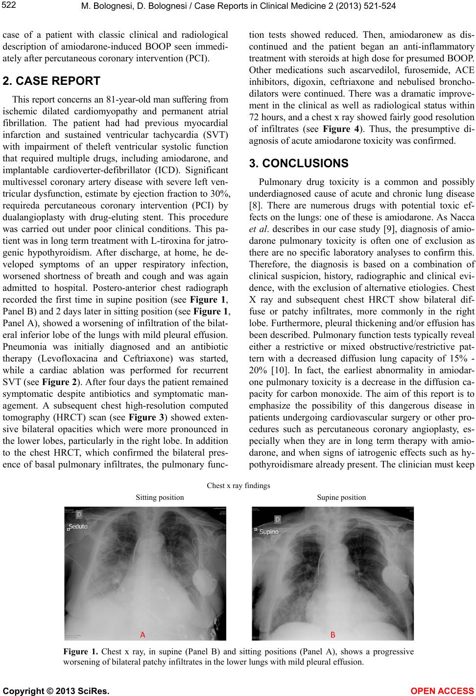 M. Bolognesi, D. Bolognesi / Case Reports in Clinical Medicine 2 (2013) 521-524 522 case of a patient with classic clinical and radiological description of amiodarone-induced BOOP seen immedi- ately after percutaneous coronary intervention (PCI). 2. CASE REPORT This report concerns an 81-year-old man suffering from ischemic dilated cardiomyopathy and permanent atrial fibrillation. The patient had had previous myocardial infarction and sustained ventricular tachycardia (SVT) with impairment of theleft ventricular systolic function that required multiple drugs, including amiodarone, and implantable cardioverter-defibrillator (ICD). Significant multivessel coronary artery disease with severe left ven- tricular dysfunction, estimate by ejection fraction to 30%, requireda percutaneous coronary intervention (PCI) by dualangioplasty with drug-eluting stent. This procedure was carried out under poor clinical conditions. This pa- tient was in long term treatment with L-tiroxin a for jatro- genic hypothyroidism. After discharge, at home, he de- veloped symptoms of an upper respiratory infection, worsened shortness of breath and cough and was again admitted to hospital. Postero-anterior chest radiograph recorded the first time in supine position (see Figure 1, Panel B) and 2 days later in sitting positio n (see Figure 1, Panel A), showed a worsening of infiltration of the bilat- eral inferior lobe of the lungs with mild pleu ral effu sion. Pneumonia was initially diagnosed and an antibiotic therapy (Levofloxacina and Ceftriaxone) was started, while a cardiac ablation was performed for recurrent SVT (see Figure 2). After four days the patient remained symptomatic despite antibiotics and symptomatic man- agement. A subsequent chest high-resolution computed tomography (HRCT) scan (see Figure 3) showed exten- sive bilateral opacities which were more pronounced in the lower lobes, particularly in the right lobe. In additio n to the chest HRCT, which confirmed the bilateral pres- ence of basal pulmonary infiltrates, the pulmonary func- tion tests showed reduced. Then, amiodaronew as dis- continued and the patient began an anti-inflammatory treatment with steroids at high dose for presumed BO OP. Other medications such ascarvedilol, furosemide, ACE inhibitors, digoxin, ceftriaxone and nebulised broncho- dilators were continued. There was a dramatic improve- ment in the clinical as well as radiological status within 72 hours, and a chest x ray sh owed f airly good resolu tion of infiltrates (see Figure 4). Thus, the presumptive di- agnosis of acute amiodarone toxicity was confirmed. 3. CONCLUSIONS Pulmonary drug toxicity is a common and possibly underdiagnosed cause of acute and chronic lung disease [8]. There are numerous drugs with potential toxic ef- fects on the lungs: one of these is amiodarone. As Nacca et al. describes in our case study [9], diagnosis of amio- darone pulmonary toxicity is often one of exclusion as there are no specific laboratory analyses to confirm this. Therefore, the diagnosis is based on a combination of clinical suspicion, history, radiographic and clinical evi- dence, with the exclusion of alternative etiologies. Chest X ray and subsequent chest HRCT show bilateral dif- fuse or patchy infiltrates, more commonly in the right lobe. Furthermore, pleural thickening and/or effusion has been described. Pulmonary function tests typically reveal either a restrictive or mixed obstructive/restrictive pat- tern with a decreased diffusion lung capacity of 15% - 20% [10]. In fact, the earliest abnormality in amiodar- one pulmonary toxicity is a decrease in the diffusion ca- pacity for carbon monoxide. The aim of this report is to emphasize the possibility of this dangerous disease in patients undergoing cardiovascular surgery or other pro- cedures such as percutaneous coronary angioplasty, es- pecially when they are in long term therapy with amio- darone, and when signs of iatrogenic effects such as hy- pothyroidismare already present. The clinician must keep Chest x ray findings Sitting position Supine position Figure 1. Chest x ray, in supine (Panel B) and sitting positions (Panel A), shows a progressive worsening of bilateral patchy infiltrates in the lower lungs with mild pleural effusion. Copyright © 2013 SciRes. OPEN ACCESS 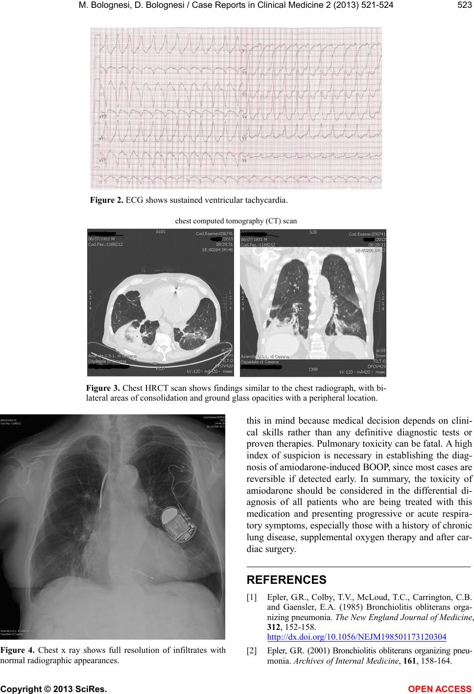 M. Bolognesi, D. Bolognesi / Case Reports in Clinical Medicine 2 (2013) 521-524 523 Figure 2. ECG shows sustained ventricular tachycardia. chest computed tomography (CT) scan Figure 3. Chest HRCT scan shows findings similar to the chest radiograph, with bi- lateral areas of consolidation and ground glass opacities with a peripheral location. Figure 4. Chest x ray shows full resolution of infiltrates with normal radiographic a pp ea ra nc es . this in mind because medical decision depends on clini- cal skills rather than any definitive diagnostic tests or proven therapies. Pulmonary toxicity can be fatal. A high index of suspicion is necessary in establishing the diag- nosis of amiodarone-induced BOOP, since most cases are reversible if detected early. In summary, the toxicity of amiodarone should be considered in the differential di- agnosis of all patients who are being treated with this medication and presenting progressive or acute respira- tory symptoms, especially those with a history of chronic lung disease, supplemental oxygen therapy and after car- diac surgery. REFERENCES [1] Epler, G.R., Colby, T.V., McLoud, T.C., Carrington, C.B. and Gaensler, E.A. (1985) Bronchiolitis obliterans orga- nizing pneumonia. The New England Journal of Medicine, 312, 152-158. http://dx.doi.org/10.1056/NEJM198501173120304 [2] Epler, G.R. (2001) Bronchiolitis obliterans organizing pneu- monia. Archives of Internal Medicine, 161, 158-164. Copyright © 2013 SciRes. OPEN ACCESS  M. Bolognesi, D. Bolognesi / Case Reports in Clinical Medicine 2 (2013) 521-524 524 http://dx.doi.org/10.1001/archinte.161.2.158 [3] Cordier, J.F. (1993) Cryptogenic organizing pneumonitis: bronchiolitis obliterans organizing pneumonia. Clinics in Chest Medicine, 14, 677-692. [4] Dimopoulou, I., Marathias, K., Daganou, M., et al. (1997) Low-dose amiodarone-related complications after cardiac operations. The Journal of Thoracic and Cardiovascular Surgery, 114, 31-37. http://dx.doi.org/10.1016/S0022-5223(97)70114-4 [5] Martin, W.J. and Rosenow, E.C. (1988) Amiodarone pul- monary toxicity: Recognition and pathogenesis. Chest, 93, 1067-1075. http://dx.doi.org/10.1378/chest.93.5.1067 [6] Costabel, U., Guzman, J. and Teschler, H. (1995) Bron- chiolitis obliterans with organizing pneumonia: Outcome. Thorax, 50, S59-S64. http://dx.doi.org/10.1136/thx.50.Suppl_1.S59 [7] Murphy, J.M., Schnyder, P., Verschakelen, J., Leuenber- ger, P. and Flower, C.D. (1999) Linear opacities on HRCT in bronchiolitis obliterans organizing pneumonia. European Radiology, 9, 1813-1817. http://dx.doi.org/10.1007/s003300050928 [8] Cooper Jr., J.A. (1997) Drug-induced lung disease. Ad- vanced Internal Medicine, 42, 231-268. [9] Nacca, N., Bhamidipati, C.M., Yuhico, L.S., Pinnamaneni, S. and Szombathy, R. (2012) Severe amiodarone induced pulmonarytoxicity. Journal of Thoracic Disease, 4, 667- 670. [10] Fabiani, I., Tacconi, D., Grotti, S., et al. (2011) Amiodar- one-induced pulmonary toxicity mimicking acute pulmo- nary edema. Journal of Cardiovascular Medicine, 12, 361-365. http://dx.doi.org/10.2459/JCM.0b013e328334a3f4 [11] Rossi, S.E., Erasmus, J.J., McAdams, H.P., Sporn, T.A. and Goodman, P.C. (2000) Pulmonary drug toxicity: Ra- diologic and pathologic manifestations. RadioGraphics, 20, 1245-1259. http://dx.doi.org/10.1148/radiographics.20.5.g00se08124 5 Copyright © 2013 SciRes. OPEN ACCESS |