

Paper Menu >>
Journal Menu >>
 Advances in Microbiology, 2013, 3, 1-6 Published Online December 2013 (http://www.scirp.org/journal/aim) http://dx.doi.org/10.4236/aim.2013.38A001 Open Access AiM Ultrastructural Analysis of in Vitr o Adherence and Production of Acid Proteases by Clinical Isolates of Candida parapsilosis Sensu Stricto Following Growth in the Presence of Keratinous Substrates from Human Source Ana Flávia L. Specian1, Luciana Furlaneto-Maia2, Célia G. T. J. Andrade3, Marcia Cristina Furlaneto1* 1Department of Microbiology, Paraná State University at Londrina, Londrina, Brazil 2Technological Federal University of Paraná, Londrina, Brazil 3Electronic Microscopy and Microanalysis Laboratory, Paraná State University at Londrina, Londrina, Brazil Email: *[email protected] Received September 25, 2013; revised October 25, 2013; accepted November 2, 2013 Copyright © 2013 Ana Flávia L. Specian et al. This is an open access article distributed under the Creative Commons Attribution License, which permits unrestricted use, distribution, and reproduction in any medium, provided the original work is properly cited. ABSTRACT Candida par a p silosis is an increasingly important human pathogen. However, little is known about its potential to cause disease. The aims of the present study were to analyse the production of acid proteinases by clinical isolates of C. parapsilosis in the presence of different keratinous substrates from human sources (stratum corneum, nail and hair) and to verify the capability of yeast cells to adhere and grow as biofilm on these substrates. By scanning electron micros- copy, it was observed that all C. parapsilosis sensu stricto isolates adhered to the keratinous substrates. For the isolate recovered from onychomycosis, the cell population attached to stratum corneum and hair keratin consisted mainly of blastoconidia. Differently, on nail keratin, pseudohyphae production was observed. Overall, there was a loose associa- tion between yeast cells and keratinous substrates. However, on stratum corneum, flocculent extracellular material was seen evolving cells from the onychomycosis isolate by forming a biofilm-like structure. The isolates recovered from onychomycosis and cutaneous lesion produced higher amount of acid proteinases in medium supplemented with nail keratin and stratum corneum keratin, respectively, than that in salt medium (absence of keratin). Furthermore, no dif- ferences were observed in the amount of acid proteinases produced by the isolate recovered from tracheal secretion in the media tested (absence and presence of keratin substrates). The information derived from this study will further our understanding of acid proteinase production by C. parapsilosis isolates an d provide an insight into pathogenic mecha- nisms in C. parapsilosis particularly from isolates recovered from superficial mycoses. Keywords: Adherence; Keratinic Substrates; SEM; Acid Proteinase 1. Introduction Candida parapsilosis is an opportunistic yeast pathogen that colonizes human skin and can spread nosocomially through hand carriage [1,2]. Over the past decade, the incidence of C. parapsilosis has dramatically increased. The yeast can cause candidiasis that can vary from rela- tively mild skin mycoses to life-threatening systemic or disseminated disease (reviewed in van Asbeck et al. [3]). Concerning superficial mycoses, C. parapsilosis has gained increasing recognition worldwide as the most common etiological agent causing Candida onychomy- cosis (reviewed in Trofa et al. [4]). In Brazil, C. parapsi- losis is the first or second most common cause of ony- chomycosis lesions [5-8]. For C. parapsilosis the colo- nized normal skin presumably serves as a reservoir of infection for the nails. Recently, we showed the capabil- ity of C. parapsilosis isolates exhibiting distinct pheno- types to grow as biofilm on human nail surfaces [9]. Several virulence factors of C. parapsilosis have been proposed, including adhesion, biofilm formation and secretion of hydrolases such as secreted aspartic pro- teinases (Saps) (reviewed in Trofa et al. [4]). We previ- *Corresponding author.  A. F. L. SPECIAN ET AL. 2 ously reported the production of proteinases and haemo- lytic factor by isolates of C. parapsilosis obtained from distinct clinical sources [10]. More recently, we have demonstrated that C. parapsilosis sensu stricto secretes a hemolytic factor into culture medium [11]. However, in contrast to the species Candida albicans, virulence traits of C. parapsilosis have not been exten- sively studied. Few studies have been undertaken to evaluate the adhesion ability of clinical strains of C. parapsilosis [9,12-14]. Furthermore, the relationship be- tween C. parapsilosis virulence and proteinase pheno- type is still unclear. In this study, we investigated for the first time the in vitro adherence pattern and production of acid proteases by clinical isolates of C. parapsilosis in the presence of keratinous substrates from human source, e.g., stratum corneum, nail and hair. 2. Materials and Methods 2.1. Candida Isolates and Identification Isolates of C. parapsilosis sensu stricto included in this study were recovered from fingernail onychomycosis (isolate 150.06), cutaneous candidiasis (isolate 220.07) and tracheal secretion (isolate 205.06) [15]. The identity of isolates was determined by the PCR technique as de- scribed by França et al. [10] using C. parapsilosis (for- merly C. parapsilosis group I) specific primers for URA3 gene (orotidine-5’-phosphate decarboxylase) [16] that allows to distinguish C. parapsilosis sensu stricto from the cryptospecies belonging to the C. parapsilosis com- plex. 2.2. Preparation of Keratin Substrates For the substrate stratum corneum, fragments of human sole from healthy volunteer were prepared as described previously [17] with modifications. The fragments were soaked in ethanol for 96 h following washes with steril- ized distillated water until a clean solution was obtained. The fragments were dried at 50˚C, grounded to a powder in liquid nitrogen and dried again at 50˚C. The substrates nail and hair were prepared as de- scribed previously [18] with modifications. Human’s hair and nail from healthy volunteer were cut to obtain small fragments ranging from 0.5 to 1 cm of size. The frag- ments were defatted by soaking for 4 d in chloroform- methanol 1:1 (v/v). The solvent was changed once a day. The fragments were then thoroughly washed with steril- ized distillated water and dried for 3 d at 50˚C. The nails fragments were grounded to a powder in presence of liq- uid nitrogen and dried again at 50˚C. The prepared sub- strates were autoclaved for 5 min at 115˚C and then added to previously sterilized basal minimal medium. 2.3. Growth Profiles Determination and Proteinase Production The isolates were pre-cultured in Sabouraud dextrose (4% dextrose, 1% peptone, 1% yeast extract) liquid medium and grown in submerged culture (180 rpm) at 37˚C for 18 h. For enzyme production, cell suspensions (104 cells/ml) were inoculated in basal minimal medium (MM) [19], with modifications, containing (g/l): K2HPO4—1.5 g; MgSO4·7H2O—0.025 g; CaCl2—0.025 g; FeSO4·7H2O— 0.015 g, ZnSO4·7H2O—0.005 g and 1% glucose, and MM amended with either 0.5% (w/v) stratum corneum, hair and nail (MM + SC, MM + H and MM + N, respec- tively) and grown in submerged culture (180 rpm) at 37˚C for up to 10 days. Media were buffered using a cit- rate buffer (0.1 M citric acid; 0.1 M sodium citrate), pH 5.0. Following growth, yeast cells were harvested by centrifugation at 8000 g for 20 min at 4˚C. The super- natants obtained were stored at −20 ˚C and assayed for protease activity. For growth rate assessment samples were removed at 12-h intervals. The growth rate was determined by haemocytometric counts. All experiments were repeated three times, and the results represent mean values SD. 2.4. Proteinase Assay Proteolytic activity was determined as previously de- scribed [20] with modifications, using haemoglobin (Sigma; St. Louis, MO) as substrate. Each assay included 50 l of culture supernatant and 0.2 ml of 20 mM citrate buffer, pH 4.0, containing haemoglobin (0.5 mg/ml). After incubation at 37˚C for 2 h, the reaction was stopped with trichloroacetic acid (TCA) 5% on ice, following incubation at 4˚C for 1 h. The mixture was centrifuged at 4000 g for 10 min at 4˚C. After this, three aliquots (150 μl each) of the reaction mixture were transferred to wells on a microtiter plate containing 100 μl of a Coomassie solution (0.025% Coomassie brilliant blue G-250, 11.75% ethanol, and 21.25% phosphoric acid). After 10 min to allow dye binding, the plate was read on an Asys HiTech UVM 340 microplate reader at an absorbance of 595 nm. Protease activity was calculated based on the absorbance difference between samples and controls. The control samples were added supernatants that were im- mediately treated with TCA. One unit (U) of proteolytic activity was defined as the amount of enzyme that caused an increase of 0.001 in absorbance unit, under standard assay conditions. The proteolytic activity is expressed as U/ml. 2.5. Scanning Electron Microscopy To verify the adhesion pattern of yeast cells with human keratin substrates, samples were fixed in 2.5% glutaral- Open Access AiM 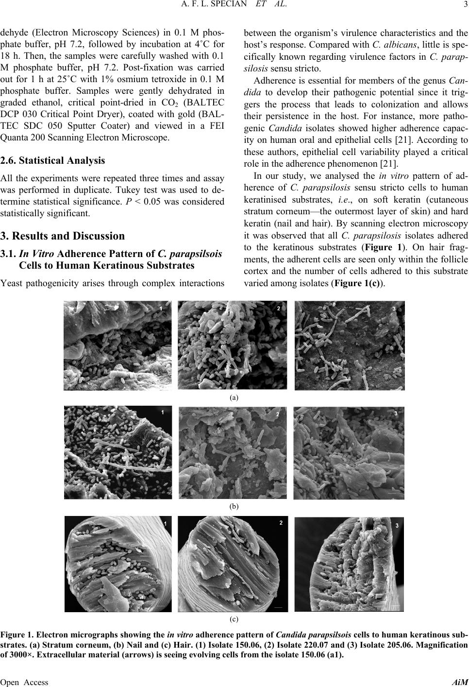 A. F. L. SPECIAN ET AL. Open Access AiM 3 dehyde (Electron Microscopy Sciences) in 0.1 M phos- phate buffer, pH 7.2, followed by incubation at 4˚C for 18 h. Then, the samples were carefully washed with 0.1 M phosphate buffer, pH 7.2. Post-fixation was carried out for 1 h at 25˚C with 1% osmium tetroxide in 0.1 M phosphate buffer. Samples were gently dehydrated in graded ethanol, critical point-dried in CO2 (BALTEC DCP 030 Critical Point Dryer), coated with gold (BAL- TEC SDC 050 Sputter Coater) and viewed in a FEI Quanta 200 Scanning Electron Microscope. between the organism’s virulence characteristics and the host’s response. Compared with C. albicans, little is spe- cifically known regarding virulence factors in C. parap- silosis sensu stricto. Adherence is essential for members of the genus Can- dida to develop their pathogenic potential since it trig- gers the process that leads to colonization and allows their persistence in the host. For instance, more patho- genic Candida isolates showed higher adherence capac- ity on human oral and epithelial cells [21]. According to these authors, epithelial cell variability played a critical role in the adherence phenomenon [21]. 2.6. Statistical Analysis In our study, we analysed the in vitro pattern of ad- herence of C. parapsilosis sensu stricto cells to human keratinised substrates, i.e., on soft keratin (cutaneous stratum corneum—the outermost layer of skin) and hard keratin (nail and hair). By scanning electron microscopy it was observed that all C. parapsilosis isolates adhered to the keratinous substrates (Figure 1). On hair frag- ments, the adherent cells are seen only within the follicle cortex and the number of cells adhered to this substrate varied among isolates (Figure 1(c)). All the experiments were repeated three times and assay was performed in duplicate. Tukey test was used to de- termine statistical significance. P < 0.05 was considered statistically significant. 3. Results and Discussion 3.1. In Vitro Adherence Pattern of C. parapsilsois Cells to Human Keratinous Substrates Yeast pathogenicity arises through complex interactions (a) (b) (c) Figure 1. Electron micrographs showing the in vitro adherence pattern of Candida parapsilsois cells to human keratinous sub- strates. (a) Stratum corneum, (b) Nail and (c) Hair. (1) Isolate 150.06, (2) Isolate 220.07 and (3) Isolate 205.06. Magnification of 3000×. Extracellular material (arrows) is seeing evolving cells from the isolate 150.06 (a1). 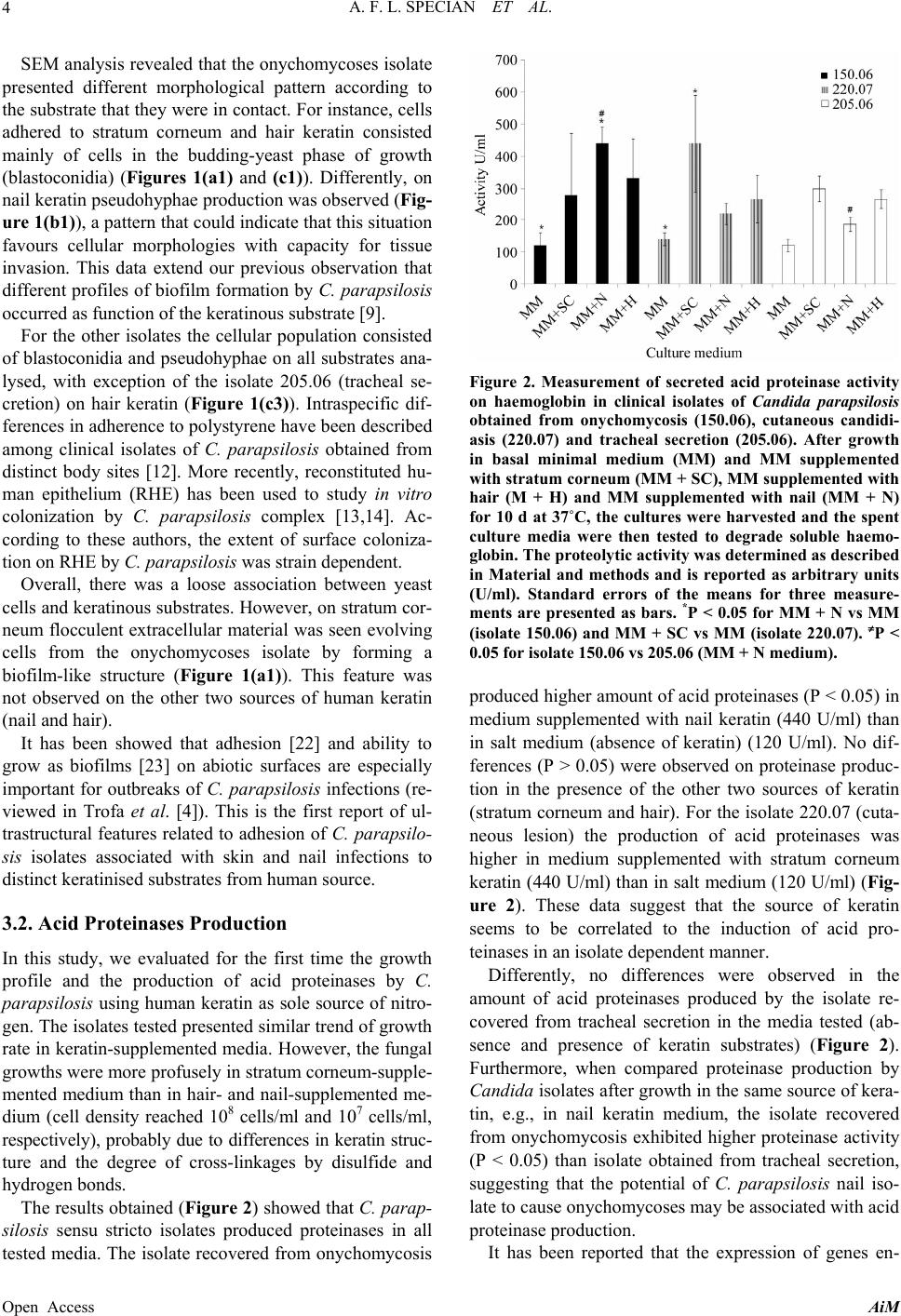 A. F. L. SPECIAN ET AL. 4 SEM analysis revealed that the onychomycoses isolate presented different morphological pattern according to the substrate that they were in contact. For instance, cells adhered to stratum corneum and hair keratin consisted mainly of cells in the budding-yeast phase of growth (blastoconidia) (Figures 1(a1) and (c1)). Differently, on nail keratin pseudohyphae production was observed (Fig- ure 1(b1)), a pattern that could indicate that this situation favours cellular morphologies with capacity for tissue invasion. This data extend our previous observation that different profiles of biofilm formation by C. parapsilosis occurred as function of the keratinous substrate [9]. For the other isolates the cellular population consisted of blastoconidia and pseudohyphae on all substrates ana- lysed, with exception of the isolate 205.06 (tracheal se- cretion) on hair keratin (Figure 1(c3)). Intraspecific dif- ferences in adherence to polystyrene have been described among clinical isolates of C. parapsilosis obtained from distinct body sites [12]. More recently, reconstituted hu- man epithelium (RHE) has been used to study in vitro colonization by C. parapsilosis complex [13,14]. Ac- cording to these authors, the extent of surface coloniza- tion on RHE by C. parapsilosis was strain dependent. Overall, there was a loose association between yeast cells and keratinous substrates. However, on stratum cor- neum flocculent extracellular material was seen evolving cells from the onychomycoses isolate by forming a biofilm-like structure (Figure 1(a1)). This feature was not observed on the other two sources of human keratin (nail and hair). It has been showed that adhesion [22] and ability to grow as biofilms [23] on abiotic surfaces are especially important for outbreaks of C. parapsilosis infections (re- viewed in Trofa et al. [4]). This is the first report of ul- trastructural features related to adhesion of C. parapsilo- sis isolates associated with skin and nail infections to distinct keratinised substrates from human source. 3.2. Acid Proteinases Production In this study, we evaluated for the first time the growth profile and the production of acid proteinases by C. parapsilosis using human keratin as sole source of nitro- gen. The isolates tested presented similar trend of growth rate in keratin-supplemented media. However, the fungal growths were more profusely in stratum corneum-supple- mented medium than in hair- and nail-supplemented me- dium (cell density reached 108 cells/ml and 107 cells/ml, respectively), probably due to differences in keratin struc- ture and the degree of cross-linkages by disulfide and hydrogen bonds. The results obtained (Figure 2 ) showed that C. parap- silosis sensu stricto isolates produced proteinases in all tested media. The isolate recovered from onychomycosis Figure 2. Measurement of secreted acid proteinase activity on haemoglobin in clinical isolates of Candida parapsilosis obtained from onychomycosis (150.06), cutaneous candidi- asis (220.07) and tracheal secretion (205.06). After growth in basal minimal medium (MM) and MM supplemented with stratum corneum (MM + SC), MM supplemented with hair (M + H) and MM supplemented with nail (MM + N) for 10 d at 37˚C, the cultures were harvested and the spent culture media were then tested to degrade soluble haemo- globin. The proteolytic activity was determined as described in Material and methods and is reported as arbitrary units (U/ml). Standard errors of the means for three measure- ments are presented as bars. *P < 0.05 for MM + N vs MM (isolate 150.06) and MM + SC vs MM (isolate 220.07). P < 0.05 for isolate 150.06 vs 205.06 (MM + N medium). produced higher amount of acid proteinases (P < 0.05) in medium supplemented with nail keratin (440 U/ml) than in salt medium (absence of keratin) (120 U/ml). No dif- ferences (P > 0.05) were observed on proteinase produc- tion in the presence of the other two sources of keratin (stratum corneum and hair). For the isolate 220.07 (cuta- neous lesion) the production of acid proteinases was higher in medium supplemented with stratum corneum keratin (440 U/ml) than in salt medium (120 U/ml) (Fig- ure 2). These data suggest that the source of keratin seems to be correlated to the induction of acid pro- teinases in an isolate dependent manner. Differently, no differences were observed in the amount of acid proteinases produced by the isolate re- covered from tracheal secretion in the media tested (ab- sence and presence of keratin substrates) (Figure 2). Furthermore, when compared proteinase production by Candida isolates after growth in the same source of kera- tin, e.g., in nail keratin medium, the isolate recovered from onychomycosis exhibited higher proteinase activity (P < 0.05) than isolate obtained from tracheal secretion, suggesting that the potential of C. parapsilosis nail iso- late to cause onychomycoses may be associated with acid proteinase production. It has been reported that the expression of genes en- Open Access AiM  A. F. L. SPECIAN ET AL. 5 coding aspartic proteinases (Saps) varied among different clinical isolates of C. parapsilosis complex when grown in contact with human oral epithelium [14]. According to these authors there is a trend relating Sap production and site of isolates recovering. In C. albicans, the most discussed hydrolytic enzymes are secreted aspartic proteinases (Saps), which are one of the well-known virulence factors of this species (re- viewed in Schaller et al. [24]). According to Monod and Borg-von [25] Saps play a role in fungal adherence and invasion of skin by C. albicans. The occurrence of Saps has been previously demonstrated in C. parapsilosis iso- lates obtained from distinct clinical samples [10,26-28] and it has being suggested that Saps are associated in superficial, but not with systemic invasion, caused by C. parapsilosis [26]. They observed the induction of Saps from C. parapsilosis cultivated in media containing bo- vine serum albumin (BSA) as a nitrogen source. Consid- ering the clinical point view, it is questionable whether these enzymes could have the required function of di- gesting keratinised tissue for parasitism. Recently we showed that the production of Saps, in BSA inducing medium, by C. parapsilosis isolates obtained from nail and skin was less expressive compared to isolates ob- tained from blood and tracheal secretion [10]. Although it has been established that C. parapsilosis is an oppor- tunistic pathogen related to the skin surface, and is also emerging as an important cause of onychomycosis, as far we know, in the present study we describe for the first time the production of acid proteases by isolates of C. parapsilosis recovered from superficial mycoses in the presence of keratinous substrates obtained from human sources. Nevertheless, the properties of the individual proteins that presumably account for the virulence of C. parapsilosis have not yet been elucidated to date. 4. Conclusion In conclusion, ultrastructural investigations of the in- terface of C. parapsilosis and the keratinised substrates from human source reveal important features, which may help to clarify the pathogenesis of superficial candidiasis. Our findings indicate the need for investigation of a pos- sible involvement of acid proteinase in the onychomyco- sis and cutaneous lesion due to this species. 5. Acknowledgements This work was supported by Conselho Nacional de De- senvolvimento Científico e Tecnológico (CNPq)—Brazil, Fundação Araucária—Paraná-Brazil and PROPPG/UEL- Brazil. A.F.L.S. was fellowship-holder of Coordenação de Aperfeiçoamento de Pessoal de Nível Superior (CAPES)—Brazil. REFERENCES [1] F. M. V. Lunel, J. F. G. M. Méis and A. Voss, “Nosoco- mial Fungal Infections: Candidemia,” Diagnostic Micro- biology and Infectious Disease, Vol. 34, No. 3, 1999, pp. 213-220. http://dx.doi.org/10.1016/S0732-8893(99)00035-8 [2] L. Saiman, E. Ludington and J. Dawson, “Risk Factors for Candida Species Colonization of Neonatal Intensive Care Unit Patients,” Pediatric Infectious Diseases Jour- nal, Vol. 20, No. 12, 2001, pp. 1119-1124. http://dx.doi.org/10.1097/00006454-200112000-00005 [3] E. C. van Asbeck, K. V. Clemins and D. A. Stevens, “Candida parapsilosis: A Review of Its Epidemiology, Pathogenesis, Clinical Aspects, Typing and Antimicrobial Susceptibility,” Critical Reviews in Microbiology, Vol. 35, No. 4, 2009, pp. 283-309. http://dx.doi.org/10.3109/10408410903213393 [4] D. Trofa, A. Gácser and J. D. Nosanchuk, “Candida parapsilosis: An Emerging Fungal Pathogen,” Clinical Microbiology Reviews, Vol. 21, No. 4, 2008, pp. 606-625. http://dx.doi.org/10.1128/CMR.00013-08 [5] R. S. N. Brilhante, R. A. Cordeiro, D. J. A. Medrano, M. F. G. Rocha, A. J. Monteiro, C. S. P. Cavalcante, T. E. F. Meireles and J. J. C. Sidrim, “Onychomycosis in Ceará (Northeast Brazil): Epidemiological and Laboratory As- pects,” Memórias do Instituto Oswaldo Cruz, Vol. 100, No. 2, 2005, pp. 131-135. http://dx.doi.org/10.1590/S0074-02762005000200005 [6] A. C. Oliveira, C. S. Shinobu, R. Longhini, S. L. Franco and T. I. E. Svidzinski, “Antifungal Activity of Propolis Extract against Yeasts Isolated from Onychomycosis Le- sions,” Memórias do Instituto Oswaldo Cruz, Vol. 101, No. 5, 2006, pp. 493-497. http://dx.doi.org/10.1590/S0074-02762006000500002 [7] V. T. Figueiredo, D. A. Santos, M. A. Resende and J. S. Hamdan, “Identification and in Vitro Antifungal Suscep- tibility Testing of 200 Clinical Isolates of Candida spp. Responsible for Fingernail Infections,” Mycopathologia, Vol. 164, No. 1, 2007, pp. 27-33. http://dx.doi.org/10.1007/s11046-007-9027-6 [8] E. A. Martins, L. V. Guerrer, K. C. Cunha, M. M. C. Soares and M. T. G. Almeida, “Onychomycosis: Clinical, Epidemiological and Mycological Study in the Munici- pality of São José do Rio Preto,” Revista da Sociedade Brasileira de Medicina Tropical, Vol. 40, No. 5, 2007, pp. 596-598. http://dx.doi.org/10.1590/S0037-86822007000500022 [9] M. T. Oliveira, A. F. L. Specian, C. G. T. J. Andrade, E. J. G. França, L. Furlaneto-Maia and M. C. Furlaneto, “In- teraction of Candida parapsilosis Isolates with Human Hair and Nail Surfaces Revealed by Scanning Electron Microscopy Analysis,” Micron, Vol. 41, No. 6, 2010, pp. 604-608. http://dx.doi.org/10.1016/j.micron.2010.03.011 [10] E. J. G. França, L. Furlaneto-Maia, R. M. B. Quesada, D. Favero, M. T. Oliveira and M. C. Furlaneto, “Haemolytic and Proteinase Activities in Clinical Isolates of Candida parapsilosis and Candida tropicalis with Reference to the Isolation Anatomic Site,” Mycoses, Vol. 54, No. 4, 2010, pp. e44-e51. Open Access AiM  A. F. L. SPECIAN ET AL. Open Access AiM 6 http://dx.doi.org/10.1111/j.1439-0507.2009.01825.x [11] D. Favero, L. Furlaneto-Maia, E. J. G. França, H. P. Góes and M. C. Furlaneto, “Hemolytic Factor Production by Clinical Isolates of Candida Species,” Current Microbi- ology, 2013. http://dx.doi.org/10.1007/s00284-013-0459-6 [12] A. Sanchez-Sousa, D. Tarrago, J. Velasco, M. E. Alvarez and F. Baquero, “Adherence to Polystyrene of Clinically Relevant Isolates of Candida Species,” Clinical Microbi- ology and Infection, Vol. 7, No. 7, 2001, pp. 379-382. http://dx.doi.org/10.1046/j.1198-743x.2001.00266.x [13] A. Gácser, W. Schafer, J. S. Nosanchuk, S. Salomon and J. D. Nosanchuk, “Virulence of Candida parapsilosis, Candida orthopsilosis, and Candida metapsilosis in Re- constituted Human Tissue Models,” Fungal Genetics and Biology, Vol. 44, No. 12, 2007, pp. 1336-1341. http://dx.doi.org/10.1016/j.fgb.2007.02.002 [14] S. Silva, M. Henriques, R. Oliveira, J. Azeredo, S. Malic, S. L. Hooper and D. W. Williams, “Characterization of Candida parapsilosis Infection of an in Vitro Reconsti- tuted Human Oral Epithelium,” European Journal of Oral Sciences, Vol. 117, No. 6, 2009, pp. 669-675. http://dx.doi.org/10.1111/j.1600-0722.2009.00677.x [15] M. C. Furlaneto, J. F. Rota, R. M. B. Quesada, L. Furlaneto-Maia, R. Rodrigues, S. Oda, M. T. Oliveira, R. Serpa and E. J. G. França, “Species Distribution and in Vitro Fluconazole Susceptibility of Clinical Candida Iso- lates in a Brazilian Tertiary-Care Hospital over a 3-Year Period,” Revista da Sociedade Brasileira de Medicina Tropical, Vol. 44, No. 5, 2011, pp. 595-599. http://dx.doi.org/10.1590/S0037-86822011000500013 [16] A. Tavanti, A. D. Davidson, N. A. R. Gow, M. C. J. Mai- den and F. C. Odds, “Candida orthopsilosis and Candida metapsilosis spp. nov. to Replace Candida parapsilosis Groups II and III,” Journal of Clinical Microbiology, Vol. 43, No. 1, 2005, pp. 284-292. http://dx.doi.org/10.1128/JCM.43.1.284-292.2005 [17] H. Gradisar, S. Kern and J. Friedrich, “Keratinase of Do- ratomyces microsporus,” Applied Microbiology and Bio- technology, Vol. 53, No. 2, 2000, pp. 196-200. http://dx.doi.org/10.1007/s002530050008 [18] J. Friedrich, H. Gradisar, D. Mandin and J. P. Chaumont, “Screening Fungi for Synthesis of Keratinolytic En- zymes,” Letters in Applied Microbiology, Vol. 28, No. 2, 1999, No. 2, pp. 127-130. [19] M. A. El-Naghy, M. S. El-Ktatny, E. M. Fadl-Allah and W. W. Nazeer, “Degradation of Chicken Feathers by Chrysosporium georgiae,” Mycopathologia, Vol. 143, No. 2, 1998, pp. 77-84. http://dx.doi.org/10.1023/A:1006953910743 [20] A. L. S. Santos, I. M. Carvalho, B. A. Silva, M. B. Portela, C. S. Alvino and R. M. A. Soares, “Secretion of Serine Peptidases by a Clinical Strain of Candida albicans: In- fluence of Growth Conditions and Cleavage of Human Serum Proteins and Extracellular Matrix Components,” FEMS Immunology and Medical Microbiology, Vol. 46, No. 2, 2006, pp. 209-220. http://dx.doi.org/10.1111/j.1574-695X.2005.00023.x [21] R. D. King, J. C. Lee and A. L. Morris, “Adherence of Candida albicans and Other Species to Mucosal Epithe- lial Cells,” Infection and Immunity, Vol. 27, No. 2, 1980, pp. 667-674. [22] D. M. Kuhn, J. Chandra, P. K. Mukherjee and M. A. Ghannoum, “Comparison of Biofilms Formed by Can- dida albicans and Candida parapsilosis on Bioprosthetic Surfaces,” Infection and Immunity, Vol. 70, No. 2, 2002, pp. 878-888. http://dx.doi.org/10.1128/IAI.70.2.878-888.2002 [23] D. M. Kuhn, P. K. Mukherjee and T. A. Clark, “Candida parapsilosis Characterization in an Outbreak Setting,” Emerging Infectious Diseases, Vol. 10, No. 6, 2004, pp. 1074-1081. http://dx.doi.org/10.3201/eid1006.030873 [24] M. Schaller, C. Borelli, H. C. Korting and B. Hube, “Hy- drolytic Enzymes as Virulence Factors of Candida albi- cans,” Mycoses, Vol. 48, No. 6, 2005, pp. 365-377. http://dx.doi.org/10.1111/j.1439-0507.2005.01165.x [25] M. Monod and Z. M. Borg-von, “Secreted Aspartic Pro- teases as Virulence Factors of Candida Species,” Bio- logical Chemistry, Vol. 383, No. 7-8, 2002, pp. 1087- 1093. http://dx.doi.org/10.1515/BC.2002.117 [26] F. De Bernardis, F. Mondello, R. San Millàn, J. Ponón and A. Cassone, “Biotyping and Virulence Properties of Skin Isolates of Candida parapsilosis,” Journal of Clini- cal Microbiology, Vol. 37, No. 11, 1999, pp. 3481-3486. [27] M. Dagdeviren, N. Cerikcioglu and M. Karavus, “Acid Proteinase, Phospholipase and Adherence Properties of Candida parapsilosis Strains Isolated from Clinical Spe- cimens of Hospitalized Patients,” Mycoses, Vol. 48, No. 5, 2005, pp. 321-326. http://dx.doi.org/10.1111/j.1439-0507.2005.01145.x [28] A. Tavanti, L. A. M. Hensgens, S. Mogavero, L. Majoros, S. Senesi and M. Campa, “Genotypic and Phenotypic Properties of Candida parapsilosis Sensu Strictu Strains Isolated from Different Geographic Regions and Body Sites,” BMC Microbiology, Vol. 10, 2010, pp. 2-11. http://dx.doi.org/10.1186/1471-2180-10-203 |