

Paper Menu >>
Journal Menu >>
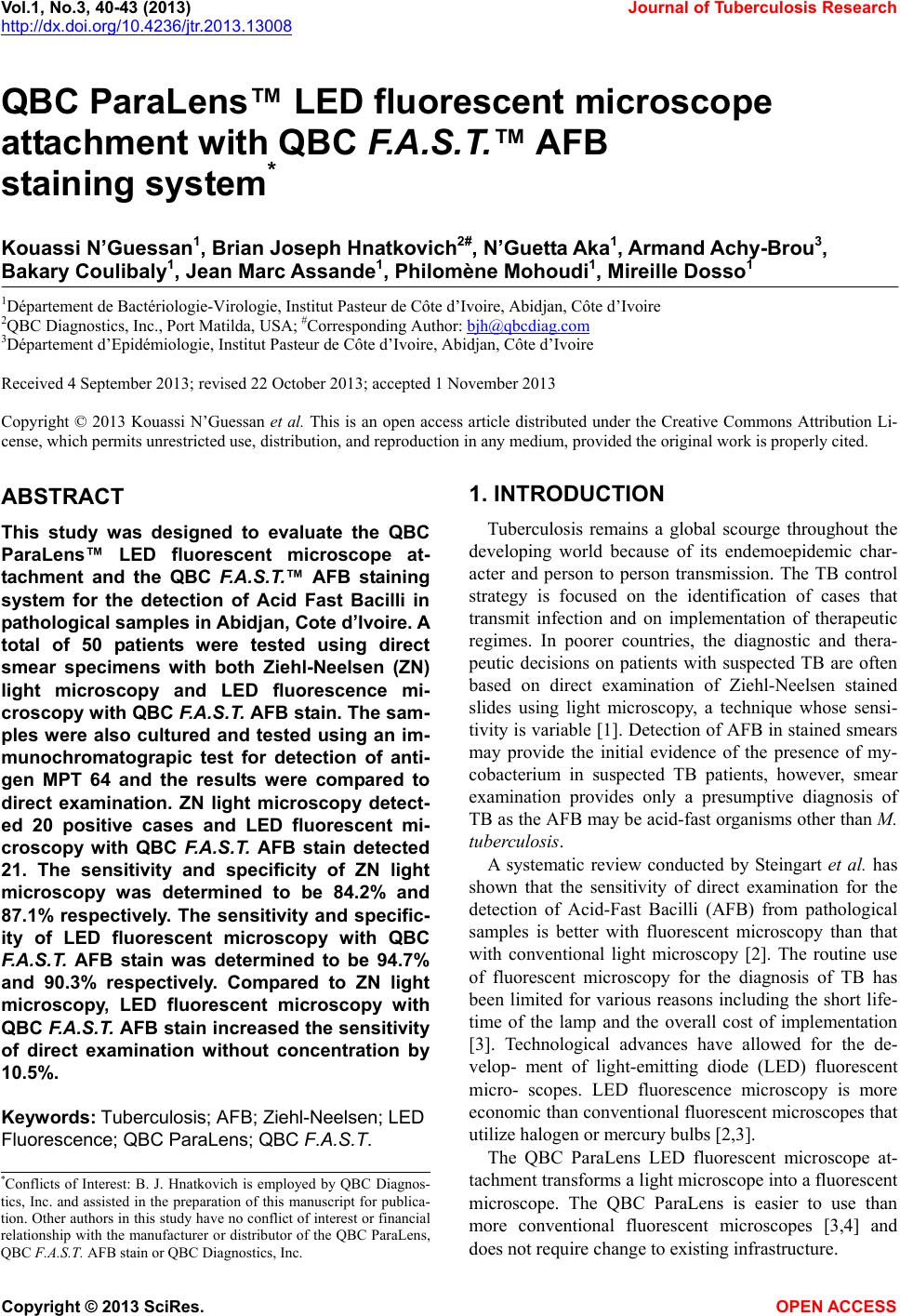 Vol.1, No.3, 40-43 (2013) Journal of Tubercul osis Resear ch http://dx.doi.org/10.4236/jtr.2013.13008 QBC ParaLens™ LED fluorescent microscope attachment with QBC F.A.S.T.™ AFB staining system* Kouassi N’Guessan1, Brian Joseph Hnatkovich2#, N’Guetta Aka1, Armand Achy-Brou3, Bakary Coulibaly1, Jean Marc Assande1, Philomène Mohoudi1, Mireille Dosso1 1Département de Bactériologie-Virologie, Institut Pasteur de Côte d’Ivoire, Abidjan, Côte d’Ivoire 2QBC Diagnostics, Inc., Port Matilda, USA; #Corresponding Author: [email protected] 3Département d’Epidémiologie, Institut Pasteur de Côte d’Ivoire, Abidjan, Côte d’Ivoire Received 4 September 2013; revised 22 October 2013; accepted 1 November 2013 Copyright © 2013 Kouassi N’Guessan et al. This is an open access article distributed under the Creative Commons Attribution Li- cense, which permits unrestricted use, distribution, and reproduction in any medium, provided the original work is properly cited. ABSTRACT This study was designed to evaluate the QBC ParaLens™ LED fluorescent microscope at- tachment and the QBC F.A.S.T.™ AFB staining system for the detection of Acid Fast Bacilli in pathological samples in Abidjan, Cote d’Ivoire. A total of 50 patients were tested using direct smear specimens with both Ziehl-Neelsen (ZN) light microscopy and LED fluorescence mi- croscopy with QBC F.A.S.T. AFB stain. The sam- ples were also cultured and tested using an im- munochromatograpic test for detection of anti- gen MPT 64 and the results were compared to direct examination. ZN light microscopy detect- ed 20 positive cases and LED fluorescent mi- croscopy with QBC F.A.S.T. AFB stain detected 21. The sensitivity and specificity of ZN light microscopy was determined to be 84.2% and 87.1% respectively. The sensitivity and specific- ity of LED fluorescent microscopy with QBC F.A.S.T. AFB stain was determined to be 94.7% and 90.3% respectively. Compared to ZN light microscopy, LED fluorescent microscopy with QBC F.A.S.T. AFB stain increased the sensitivity of direct examination without concentration by 10.5%. Keyw ords: Tuberculosis; AFB; Ziehl-Neelsen; LED Fluorescence; QBC ParaLens; QB C F.A.S.T. 1. INTRODUCTION Tuberculosis remains a global scourge throughout the developing world because of its endemoepidemic char- acter and person to person transmission. The TB control strategy is focused on the identification of cases that transmit infection and on implementation of therapeutic regimes. In poorer countries, the diagnostic and thera- peutic decisions on patients with suspected TB are often based on direct examination of Ziehl-Neelsen stained slides using light microscopy, a technique whose sensi- tivity is variable [1]. Detection of AFB in stained smears may provide the initial evidence of the presence of my- cobacterium in suspected TB patients, however, smear examination provides only a presumptive diagnosis of TB as the AFB may be acid-fast organisms other than M. tuberculosis. A systematic review conducted by Steingart et al. has shown that the sensitivity of direct examination for the detection of Acid-Fast Bacilli (AFB) from pathological samples is better with fluorescent microscopy than that with conventional light microscopy [2]. The routine use of fluorescent microscopy for the diagnosis of TB has been limited for various reasons including the short life- time of the lamp and the overall cost of implementation [3]. Technological advances have allowed for the de- velop- ment of light-emitting diode (LED) fluorescent micro- scopes. LED fluorescence microscopy is more economic than conventional fluorescent microscopes that utilize halogen or mercury bulbs [2 ,3]. The QBC ParaLens LED fluorescent microscope at- tachment transforms a light microscope into a fluorescent microscope. The QBC ParaLens is easier to use than more conventional fluorescent microscopes [3,4] and does not requir e change to existing infrastructure. *Conflicts of Interest: B. J. Hnatkovich is employed by QBC Diagnos- tics, Inc. and assisted in the preparation of this manuscript for publica- tion. Other authors in this study have no conflict of interest or financial relationship with the manufacturer or distributor of the QBC ParaLens, QBC F.A.S.T. AFB stain or QBC Diagnostics, Inc. Copyright © 2013 SciRes. OPEN A CCESS 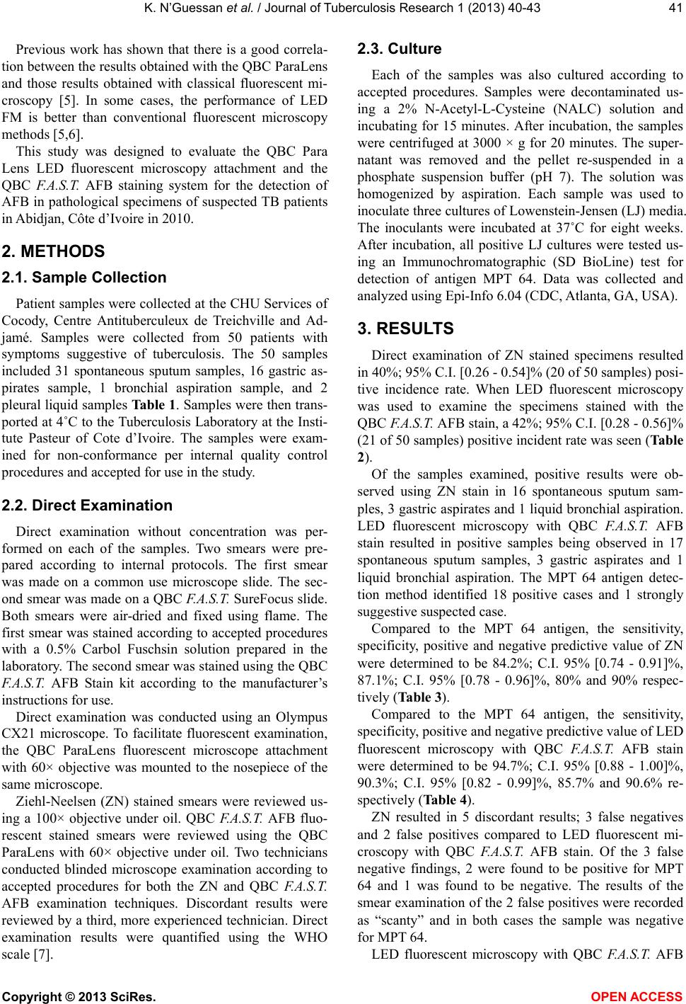 K. N’Guessan et al. / Journal of Tuberculosis Researc h 1 (2013) 40-43 41 Previous work has shown that there is a good correla- tion between the results obtained with the QBC ParaLens and those results obtained with classical fluorescent mi- croscopy [5]. In some cases, the performance of LED FM is better than conventional fluorescent microscopy methods [5,6]. This study was designed to evaluate the QBC Para Lens LED fluorescent microscopy attachment and the QBC F.A.S.T. AFB staining system for the detection of AFB in pathological specimens of suspected TB patients in Abidjan, Côte d’Ivo ire in 20 10. 2. METHODS 2.1. Sample Collection Patient samples were collected at the CHU Services of Cocody, Centre Antituberculeux de Treichville and Ad- jamé. Samples were collected from 50 patients with symptoms suggestive of tuberculosis. The 50 samples included 31 spontaneous sputum samples, 16 gastric as- pirates sample, 1 bronchial aspiration sample, and 2 pleural liquid samples Table 1. Samples were then tran s- ported at 4˚C to the Tubercu losis Laboratory at the Insti- tute Pasteur of Cote d’Ivoire. The samples were exam- ined for non-conformance per internal quality control procedures and accepted for use in the study. 2.2. Direct Examination Direct examination without concentration was per- formed on each of the samples. Two smears were pre- pared according to internal protocols. The first smear was made on a common use microscope slide. The sec- ond smear was made on a QBC F.A.S.T. SureFo cus slide. Both smears were air-dried and fixed using flame. The first smear was stained according to accepted procedures with a 0.5% Carbol Fuschsin solution prepared in the laboratory. The second smear was stained using the QBC F.A.S.T. AFB Stain kit according to the manufacturer’s instructions for use. Direct examination was conducted using an Olympus CX21 microscope. To facilitate fluorescent examination, the QBC ParaLens fluorescent microscope attachment with 60× objective was mounted to the nosepiece of the same microscope. Ziehl-Neelsen (ZN) stained smears were reviewed us- ing a 100× objective under oil. QBC F.A.S.T. AFB fluo- rescent stained smears were reviewed using the QBC ParaLens with 60× objective under oil. Two technicians conducted blinded microscope examination according to accepted procedures for both the ZN and QBC F.A.S.T. AFB examination techniques. Discordant results were reviewed by a third, more experienced technician. Direct examination results were quantified using the WHO scale [7]. 2.3. Culture Each of the samples was also cultured according to accepted procedures. Samples were decontaminated us- ing a 2% N-Acetyl-L-Cysteine (NALC) solution and incubating for 15 minutes. After incubation, the samples were centrifuged at 3000 × g for 20 minutes. The super- natant was removed and the pellet re-suspended in a phosphate suspension buffer (pH 7). The solution was homogenized by aspiration. Each sample was used to inoculate three cultures of Lowenstein-Jensen (LJ) media. The inoculants were incubated at 37˚C for eight weeks. After incubation, all positive LJ cultures were tested us- ing an Immunochromatographic (SD BioLine) test for detection of antigen MPT 64. Data was collected and analyzed using Epi-Info 6.04 (CDC, Atlanta, GA, USA). 3. RESULTS Direct examination of ZN stained specimens resulted in 40%; 95% C.I. [0.26 - 0.54] % (20 of 5 0 samples) posi- tive incidence rate. When LED fluorescent microscopy was used to examine the specimens stained with the QBC F.A.S.T. AFB stain, a 42%; 95% C.I. [0.28 - 0.56]% (21 of 50 samples) positive incident rate was seen (Table 2). Of the samples examined, positive results were ob- served using ZN stain in 16 spontaneous sputum sam- ples, 3 gastric aspirates and 1 liquid bronchial aspiration. LED fluorescent microscopy with QBC F.A.S.T. AFB stain resulted in positive samples being observed in 17 spontaneous sputum samples, 3 gastric aspirates and 1 liquid bronchial aspiration. The MPT 64 antigen detec- tion method identified 18 positive cases and 1 strongly suggestive suspected case. Compared to the MPT 64 antigen, the sensitivity, specificity, positive and negative predictive value of ZN were determined to be 84.2%; C.I. 95% [0.74 - 0.91]%, 87.1%; C.I. 95% [0.78 - 0.96]%, 80% and 90% respec- tively (Table 3). Compared to the MPT 64 antigen, the sensitivity, specificity, positive and negative predictive value of LED fluorescent microscopy with QBC F.A.S.T. AFB stain were determined to be 94.7%; C.I. 95% [0.88 - 1.00]%, 90.3%; C.I. 95% [0.82 - 0.99]%, 85.7% and 90.6% re- spectively (Table 4). ZN resulted in 5 discordant results; 3 false negatives and 2 false positives compared to LED fluorescent mi- croscopy with QBC F.A.S.T. AFB stain. Of the 3 false negative findings, 2 were found to be positive for MPT 64 and 1 was found to be negative. The results of the smear examination of the 2 false positives were recorded as “scanty” and in both cases the sample was negative for MPT 64. LED fluorescent microscopy with QBC F.A.S.T. AFB Copyright © 2013 SciRes. OPEN A CCESS 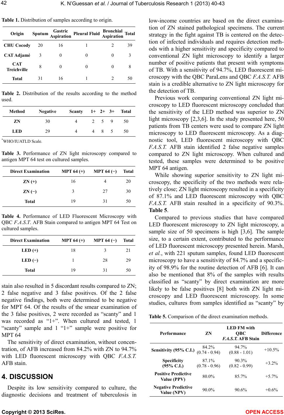 K. N’Guessan et al. / Journal of Tuberculosis Researc h 1 (2013) 40-43 42 Table 1. Distribution of samples according to origin. Origin Sputum Gastric Aspiration Pleural Fluid Bronchial Aspiration Total CHU Cocody 20 16 1 2 39 CAT Adjamé 3 0 0 0 3 CAT Treichville 8 0 0 0 8 Total 31 16 1 2 50 Table 2. Distribution of the results according to the method used. Method Negative Scanty 1+ 2+ 3+Total ZN 30 4 2 5 950 LED 29 4 4 8 550 *WHO/IUATLD Scale. Table 3. Performance of ZN light microscopy compared to antigen MPT 64 test on cultured samples. Direct Examination MPT 64 (+) MPT 64 (−) Total ZN (+) 16 4 20 ZN (−) 3 27 30 Total 19 31 50 Table 4. Performance of LED Fluorescent Microscopy with QBC F.A.S.T. AFB Stain compared to antigen MPT 64 Test on cultured samples. Direct Examination MPT 64 (+) MPT 64 (−) Total LED (+) 18 3 21 LED (−) 1 28 29 Total 19 31 50 stain also resulted in 5 disco rdan t results compared to ZN; 2 false negative and 3 false positives. Of the 2 false negative findings, both were determined to be negative for MPT 64. Of the results of the smear examination of the 3 false positives, 2 were recorded as “scanty” and 1 was recorded as “1+”. When cultured and tested, 1 “scanty” sample and 1 “1+” sample were positive for MPT 64 The sensitivity of direct examination, without concen- tration, of AFB increased from 84.2% with ZN to 94.7% with LED fluorescent microscopy with QBC F.A.S.T. AFB stain. 4. DISCUSSION Despite its low sensitivity compared to culture, the diagnostic decisions and treatment of tuberculosis in low-income countries are based on the direct examina- tion of ZN stained pathological specimens. The current strategy in the fight against TB is centered on the detec- tion of infected individuals and requires detection meth- ods with a higher sensitivity and specificity compared to conventional ZN light microscopy to identify a larger number of positive patients that present with symptoms of TB. With a sensitivity of 94.7%, LED fluorescent mi- croscopy with the QBC ParaLens and QBC F.A.S.T. AFB stain is a credible alternative to ZN light microscopy for the detection of TB. Previous work comparing conventional ZN light mi- croscopy to LED fluorescent microscopy concluded that the sensitivity of the LED method was superior to ZN light microscopy [2,3,6]. In the study presented here, 50 patients from TB centers were used to compare ZN light microscopy to LED fluorescent microscopy. As a diag- nostic tool, LED fluorescent microscopy with QBC F.A.S.T. AFB stain identified 2 false negative samples compared to ZN light microscopy. When cultured and tested, these samples were determined to be positive MPT 64 antigen. While showing superior sensitivity to ZN light mi- croscopy, the specificity of the two methods were rela- tively close; ZN light microscopy resulted in a specificity of 87.1% and LED fluorescent microscopy with QBC F.A.S.T. AFB stain resulted in a specificity of 90.3%. Table 5. Compared to previous studies that have compared LED fluorescent microscopy to ZN light microscopy, a sample size of 50 specimens is high [3,6]. The sample size, to a certain extent, contributed to the performance of LED fluorescent microscopy presented herein. Marsh, et al., with 221 sputum samples, found LED fluorescent microscopy to have a sens itivity of 84.7% and a sp ecific- ity of 98.9% for the routine detection of AFB [6]. It can also be mentioned that 8% of the samples with results classified as “scanty” by direct examination are more likely to be false positives [8] both with ZN light mi- croscopy and LED fluorescent microscopy. In some studies, cultures from samples identified as “scanty” by Table 5. Comparison of the direct examination methods. Performance ZN LED FM with QBC F.A.S.T. AFB Stain Difference Sensitivity (95% C.I.)84.2% (0.74 - 0.94) 94.7% (0.88 - 1.01) +10.5% Specificity (95% C.I.) 87.1% (0.78 - 0.96) 90.3% (0.82 - 0.99) +3.2% Positive Predictive Value (PPV) 80.0% 85.7% +5.7% Negative Predictive Value (NPV) 90.0% 90.6% +0.6% Copyright © 2013 SciRes. OPEN A CCESS  K. N’Guessan et al. / Journal of Tuberculosis Researc h 1 (2013) 40-43 Copyright © 2013 SciRes. 43 direct examination were found to be negative 95% of the time [9]. OPEN ACCESS In conclusion, we believe that LED fluorescent mi- croscopy with QBC F.A.S.T. AFB stain to be a credible alternative to ZN light microscopy for routine TB screening in countries endemic for pulmonary tuberculo- sis. 5. ACKNOWLEDGEMENTS We extend our sincere thanks to Mr. Alain Noudofinin for the finan- cial support he brought to this work and the equipment, reagents and consumables used in this study. REFERENCES [1] Salfinger, M. and Pfyffer, G.E. (1994) The new diagnostic mycobacteriology laboratory. European Journal of Cli- nical Microbiology & Infectious Diseases, 13, 961-979. http://dx.doi.org/10.1007/BF02111498 [2] Steingart, K.R., Henry, M., Ng, V., Hopewell, P.C., Ram- say, A., Cunningham, J., Urbanczik, R., Perkins, M., Aziz, M.A. and Pai, M. (2006) Fluorescence versus conven- tional sputum smear microscopy for tuberculosis: A sys- tematic review. The Lancet Infectious Diseases, 6, 570- 581. http://dx.doi.org/10.1016/S1473-3099(06)70578-3 [3] Affolabi, D., Torrea, G., Odoun, M., Senou, N., Ali, L.M., Anagonou, S. and Van Deun, A. (2010) Comparison of two LED fluorescence microscopy build-on modules for acid-fast-bacilli smear microscopy. The International Journal of Tuberculosis and Lung Disease, 14,160-164. [4] Kuhn, W., Armstrong, D., Atteberry, S., Dewbrey, E., Smith, D. and Hooper, N. (2010) Usefulness of the Pa- ralens fluorescent microscope adaptor for the identifica- tion of mycobacteria in both field and laboratory settings. The Open Microbiology Journal, 4, 30-33. http://dx.doi.org/10.2174/1874285801004010030 [5] Nguyen, V.H., Dinh, N.S., Anthony, R.M., Cobelens, F.G. and van Soolingen, D. (2007) Fluorescence microscopy for tuberculosis diagnosis. The Lancet Infectious Disea- ses, 7, 238-239. http://dx.doi.org/10.1016/S1473-3099(07)70059-2 [6] Marais, B.J., Brittle, W., Painczyk, K., Hesseling, A.C., Beyers, N., Wasserman, E., van Soolingen, D. and Warren, R.M. (2008) Use of light-emitting diode fluorescence mi- croscopy to detect acid fast bacilli in sputum. Clini cal In- fectious Diseases, 47, 203-207. http://dx.doi.org/10.1086/589248 [7] World Health Organization (1998) Laboratory services in tuberculosis control. Part II: Microscopy. WHO/TB/ 98.258. WHO, Geneva. [8] Van Deun, A., Salim, A.H., Cooreman, F., Daru, P., Das, A.P., Anug, K.J.M. and Reider, H.L. (2004) Scanty AFB smears: What’s in a name? The International Journal of Tuberculosis and Lung Disease, 8, 816-823. [9] Lawson, L., Yassin, M.A., Ramsay, A., Emenyonu, N.E., Squire, S.B. and Cuevas, L.E. (2005) Comparison of scanty AFB smears against culture in an area with high HIV prevalence. The International Journal of Tubercu- losis and Lung Disease, 9, 933-935. |