

Paper Menu >>
Journal Menu >>
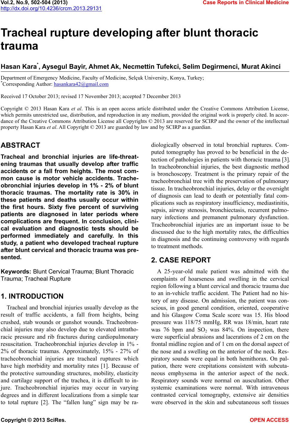 Vol.2, No.9, 502-504 (2013) Case Reports in Clinical Medicine http://dx.doi.org/10.4236/crcm.2013.29131 Tracheal rupture developing after blunt thoracic trauma Hasan Kara*, Aysegul Bayir, Ahmet Ak, Necmettin Tufekci, Selim Degirmenci, Murat Akinci Department of Emergency Medicine, Faculty of Medicine, Selçuk University, Konya, Turkey; *Corresponding Author: [email protected] Received 17 October 2013; revised 17 November 2013; accepted 7 December 2013 Copyright © 2013 Hasan Kara et al. This is an open access article distributed under the Creative Commons Attribution License, which permits unrestricted use, distribution, and reproduction in any medium, provided the original work is properly cited. In accor- dance of the Creative Commons Attribution License all Copyrights © 2013 are reserved for SCIRP and the owner of the intellectual property Hasan Kara et al. All Copyright © 2013 are guarded by law and by SCIRP as a guardian. ABSTRACT Tracheal and bronchial injuries are life-threat- ening traumas that usually develop after traffic accidents or a fall from heights. The most com- mon cause is motor vehicle accidents. Trache- obronchial injuries develop in 1% - 2% of blunt thoracic traumas. The mortality rate is 30% in these patients and deaths usually occur within the first hours. Sixty five percent of surviving patients are diagnosed in later periods where complications are frequent. In conclusion, clini- cal evaluation and diagnostic tests should be performed immediately and carefully. In this study, a patient who developed tracheal rupture after blunt cervical and thoracic trauma was pre- sented. Keyw ords: Blunt Cervical Traum a; Blunt Thoracic Trauma; Tracheal Rupture 1. INTRODUCTION Tracheal and bronchial injuries usually develop as the result of traffic accidents, a fall from heights, being crushed, stab wounds or gunshot wounds. Tracheobron- chial injuries may also develop due to elevated intratho- racic pressure and rib fractures during cardiopulmonary resuscitation. Tracheobronchial injuries develop in 1% - 2% of thoracic traumas. Approximately, 15% - 27% of tracheobronchial injuries are tracheal ruptures which have high morbidity and mortality rates [1]. Because of the protective surrounding structures, mobility, elasticity and cartilage support of the trachea, it is difficult to in- jure. Tracheobronchial injuries may occur in varying degrees and in different localizations from a simple tear to total rupture [2]. The “fallen lung” sign may be ra- diologically observed in total bronchial ruptures. Com- puted tomography has proved to be beneficial in the de- tection of pathologies in patients with thoracic trauma [3]. In tracheobronchial injuries, the best diagnostic method is bronchoscopy. Treatment is the primary repair of the tracheobronchial tree with the preservation of pulmonary tissue. In tracheobronchial injuries, delay or the oversight of diagnosis can lead to death or potentially fatal com- plications such as respiratory insufficiency, mediastinitis, sepsis, airway stenosis, bronchiectasis, recurrent pulmo- nary infections and premanent pulmonary dysfunction. Tracheobronchial injuries are an important issue to be discussed due to the high mortality rates, the difficulties in diagnosis and the continuing controversy with regards to treatment methods. 2. CASE REPORT A 25-year-old male patient was admitted with the complaints of hoarseness and swelling in the cervical region following a blunt cervical and thoracic trauma due to an in-vehicle traffic accident. The Patient had no his- tory of any disease. On admission, the patient was con- scious, in good general condition, oriented, cooperative and his Glasgow Coma Scale score was 15. His blood pressure was 118/75 mmHg, RR was 18/min, heart rate was 76 bpm and SO2 was 84%. On inspection, there were superficial abrasions and lacerations of 2 cm on the frontal midline region and of 1 cm on the dorsal aspect of the nose and a swelling on the anterior of the neck. Res- piratory sounds were equal in both hemithorax. On pal- pation, there were crepitations consistent with subcuta- neous emphysema in the anterior aspect of the neck. Respiratory sounds were normal on auscultation. Other systemic examinations were normal. With intravenous contrasted cervical tomography, extensive air densities were observed in the skin and subcutaneous soft tissues Copyright © 2013 SciRes. OPEN ACCESS 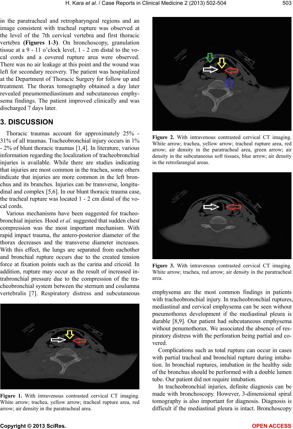 H. Kara et al. / Case Reports in Clinical Medicine 2 (2013) 502-504 503 in the paratracheal and retropharyngeal regions and an image consistent with tracheal rupture was observed at the level of the 7th cervical vertebra and first thoracic vertebra (Figures 1-3). On bronchoscopy, granulation tissue at a 9 - 11 o’clock level, 1 - 2 cm distal to the vo- cal cords and a covered rupture area were observed. There was no air leakage at this point and the wound was left for secondary recovery. The patient was hospitalized at the Department of Thoracic Surgery for follow up and treatment. The thorax tomography obtained a day later revealed pneumomediastinum and subcutaneous emphy- sema findings. The patient improved clinically and was discharged 7 days later. 3. DISCUSSION Thoracic traumas account for approximately 25% - 31% of all traumas. Tracheobronchial injury occurs in 1% - 2% of blunt thoracic traumas [1,4]. In literature, various information regarding the localization of tracheobronchial injuries is available. While there are studies indicating that injuries are most common in the trachea, some others indicate that injuries are more common in the left bron- chus and its branches. Injuries can be transverse, longitu- dinal and complex [5,6]. In our blunt thoracic trauma case, the tracheal rupture was located 1 - 2 cm distal of the vo- cal cords. Various mechanisms have been suggested for tracheo- bronchial injuries. Hood et al. suggested that sudden chest compression was the most important mechanism. With rapid impact trauma, the antero-posterior diameter of the thorax decreases and the transverse diameter increases. With this effect, the lungs are separated from eachother and bronchial rupture occurs due to the created tension force at fixation points such as the carina and cricoid. In addition, rupture may occur as the result of increased in- trabronchial pressure due to the compression of the tra- cheobronchial system between the sternum and coulumna vertebralis [7]. Respiratory distress and subcutaneous Figure 1. With intravenous contrasted cervical CT imaging. White arrow; trachea, yellow arrow; tracheal rupture area, red arrow; air density in the paratracheal area. Figure 2. With intravenous contrasted cervical CT imaging. White arrow; trachea, yellow arrow; tracheal rupture area, red arrow; air density in the paratracheal area, green arrow; air density in the subcutaneous soft tissues, blue arrow; air density in the retrofarangial areas. Figure 3. With intravenous contrasted cervical CT imaging. White arrow; trachea, red arrow; air density in the paratracheal area. emphysema are the most common findings in patients with tracheobronchial injury. In tracheobronchial ruptures, mediastinal and cervical emphysema can be seen without pneumothorax development if the mediastinal pleura is durable [8,9]. Our patient had subcutaneous emphysema without penumothorax. We associated the absence of res- piratory distress with the perforation being partial and co- vered. Complications such as total rupture can occur in cases with partial tracheal and bronchial rupture during intuba- tion. In bronchial ruptures, intubation in the healthy side of the bronchus should be performed with a double lumen tube. Our patient did not require intubation. In tracheobronchial injuries, definite diagnosis can be made with bronchoscopy. However, 3-dimensional spiral tomography is also important for diagnosis. Diagnosis is difficult if the mediastinal pleura is intact. Bronchoscopy Copyright © 2013 SciRes. OPEN ACCESS 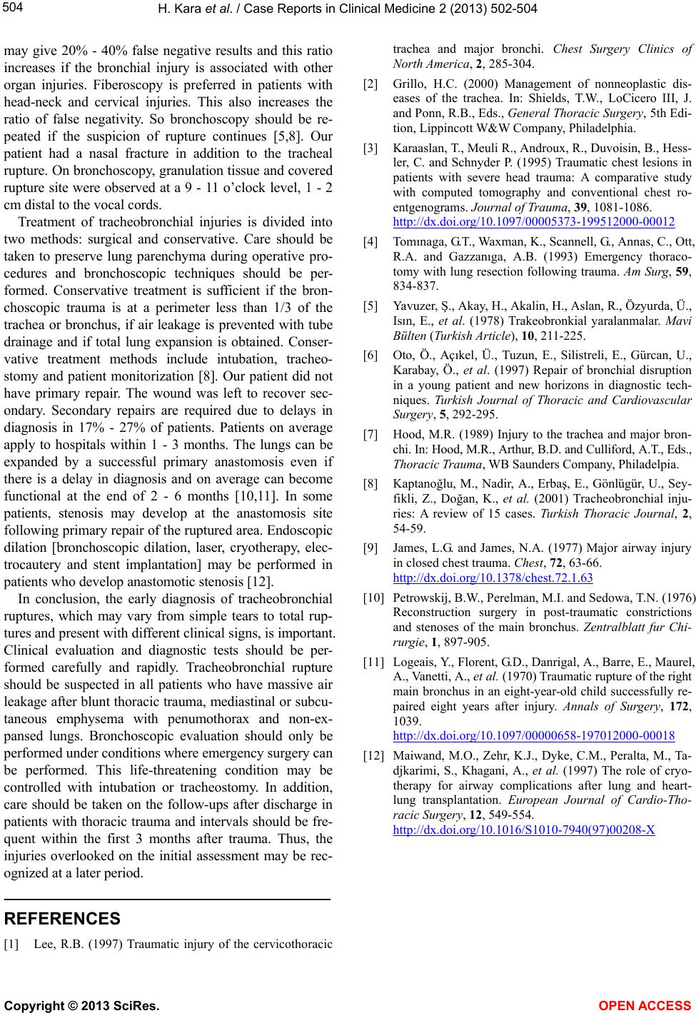 H. Kara et al. / Case Reports in Clinical Medicine 2 (2013) 502-504 Copyright © 2013 SciRes. OPEN ACCESS 504 may give 20% - 40% false negative results and this ratio increases if the bronchial injury is associated with other organ injuries. Fiberoscopy is preferred in patients with head-neck and cervical injuries. This also increases the ratio of false negativity. So bronchoscopy should be re- peated if the suspicion of rupture continues [5,8]. Our patient had a nasal fracture in addition to the tracheal rupture. On bronchoscopy, granulation tissue and covered rupture site were observed at a 9 - 11 o’clock level, 1 - 2 cm distal to the vocal cords. Treatment of tracheobronchial injuries is divided into two methods: surgical and conservative. Care should be taken to preserve lung parenchyma during operative pro- cedures and bronchoscopic techniques should be per- formed. Conservative treatment is sufficient if the bron- choscopic trauma is at a perimeter less than 1/3 of the trachea or bronchus, if air leakage is prevented with tube drainage and if total lung expansion is obtained. Conser- vative treatment methods include intubation, tracheo- stomy and patient monitorization [8]. Our patient did not have primary repair. The wound was left to recover sec- ondary. Secondary repairs are required due to delays in diagnosis in 17% - 27% of patients. Patients on average apply to hospitals within 1 - 3 months. The lungs can be expanded by a successful primary anastomosis even if there is a delay in diagnosis and on average can become functional at the end of 2 - 6 months [10,11]. In some patients, stenosis may develop at the anastomosis site following primary repair of the ruptured area. Endoscopic dilation [bronchoscopic dilation, laser, cryotherapy, elec- trocautery and stent implantation] may be performed in patients who develop anastomotic stenosis [12]. In conclusion, the early diagnosis of tracheobronchial ruptures, which may vary from simple tears to total rup- tures and present with different clinical signs, is important. Clinical evaluation and diagnostic tests should be per- formed carefully and rapidly. Tracheobronchial rupture should be suspected in all patients who have massive air leakage after blunt thoracic trauma, mediastinal or subcu- taneous emphysema with penumothorax and non-ex- pansed lungs. Bronchoscopic evaluation should only be performed under conditions where emergency surgery can be performed. This life-threatening condition may be controlled with intubation or tracheostomy. In addition, care should be taken on the follow-ups after discharge in patients with thoracic trauma and intervals should be fre- quent within the first 3 months after trauma. Thus, the injuries overlooked on the initial assessment may be rec- ognized at a later period. REFERENCES [1] Lee, R.B. (1997) Traumatic injury of the cervicothoracic trachea and major bronchi. Chest Surgery Clinics of North America, 2, 285-304. [2] Grillo, H.C. (2000) Management of nonneoplastic dis- eases of the trachea. In: Shields, T.W., LoCicero III, J. and Ponn, R.B., Eds., General Thoracic Surgery, 5th Edi- tion, Lippincott W&W Company, Philadelphia. [3] Karaaslan, T., Meuli R., Androux, R., Duvoisin, B., Hess- ler, C. and Schnyder P. (1995) Traumatic chest lesions in patients with severe head trauma: A comparative study with computed tomography and conventional chest ro- entgenograms. Journal of Trauma, 39, 1081-1086. http://dx.doi.org/10.1097/00005373-199512000-00012 [4] Tom ınaga, G.T., Waxman, K., Scannell, G., Annas, C., Ott, R.A. and Gazzanıga, A.B. (1993) Emergency thoraco- tomy with lung resection following trauma. Am Surg, 59, 834-837. [5] Yavuzer, Ş., Akay, H., Akalin, H., Aslan, R., Özyurda, Ü., Isın, E., et al. (1978) Trakeobronkial yaralanmalar. Mavi Bülten (Turkish Art icle), 10, 211-225. [6] Oto, Ö., Açıkel, Ü., Tuzun, E., Silistreli, E., Gürcan, U., Karabay, Ö., et al. (1997) Repair of bronchial disruption in a young patient and new horizons in diagnostic tech- niques. Turkish Journal of Thoracic and Cardiovascular Surgery, 5, 292-295. [7] Hood, M.R. (1989) Injury to the trachea and major bron- chi. In: Hood, M.R., Arthur, B.D. and Culliford, A.T., Eds., Thoracic Trauma, WB Saunders Company, Philadelpia. [8] Kaptanoğlu, M., Nadir, A., Erbaş, E., Gönlügür, U., Sey- fikli, Z., Doğan, K., et al. (2001) Tracheobronchial inju- ries: A review of 15 cases. Turkish Thoracic Journal, 2, 54-59. [9] James, L.G. and James, N.A. (1977) Major airway injury in closed chest trauma. Chest, 72, 63-66. http://dx.doi.org/10.1378/chest.72.1.63 [10] Petrowskij, B.W., Perelman, M.I. and Sedowa, T.N. (1976) Reconstruction surgery in post-traumatic constrictions and stenoses of the main bronchus. Zentralblatt fur Chi- rurgie, 1, 897-905. [11] Logeais, Y., Florent, G.D., Danrigal, A., Barre, E., Maurel, A., Vanetti, A., et al. (1970) Traumatic rupture of the right main bronchus in an eight-year-old child successfully re- paired eight years after injury. Annals of Surgery, 172, 1039. http://dx.doi.org/10.1097/00000658-197012000-00018 [12] Maiwand, M.O., Zehr, K.J., Dyke, C.M., Peralta, M., Ta- djkarimi, S., Khagani, A., et al. (1997) The role of cryo- therapy for airway complications after lung and heart- lung transplantation. European Journal of Cardio-Tho- racic Surgery, 12, 549-554. http://dx.doi.org/10.1016/S1010-7940(97)00208-X |