

Paper Menu >>
Journal Menu >>
 Open Journal of Veterinary Medicine, 2013, 3, 315-318 Published Online December 2013 (http://www.scirp.org/journal/ojvm) http://dx.doi.org/10.4236/ojvm.2013.38051 Open Access OJVM Effect of a Wildlife-Livestock Interface on the Prevalence of Intra-Erythrocytic Hemoparasites in Cattle Richard M. Kabuusu1,2*, Ruth Ale xa n de r1, Annet M. Kabuusu2, Sylvia N. Muwanga3, Patrick Atimnedi4, Calum Macpherson2,5 1Pathobiology Academic Program St. George’s Grenada, School of Veterinary Medicine, St. George’s University, St. George’s, Grenada 2Graduate Studies Program, St. George’s University, St. George’s, Grenada 3Department of Wildlife and Animal Resources Management, Faculty of Veterinary Medicine, Makerere University, Kampala, Uganda 4Uganda Wildlife Authority (UWA), Kampala, Uganda 5School of Medicine, St. George’s University, St. George’s, Grenada Email: *[email protected] Received October 22, 2013; revised November 22, 2013; accepted November 29, 2013 Copyright © 2013 Richard M. Kabuusu et al. This is an open access article distributed under the Creative Commons Attribution Li- cense, which permits unrestricted use, distribution, and reproduction in any medium, provided the original work is properly cited. In accordance of the Creative Commons Attribution License all Copyrights © 2013 are reserved for SCIRP and the owner of the intel- lectual property Richard M. Kabuusu et al. All Copyright © 2013 are guarded by law and by SCIRP as a guardian. ABSTRACT We conducted a cross-sectional study to establish the effect of proximity of livestock to a wildlife-livestock interface on the relative abundance of intra-erythrocytic hemoparasites in cattle. Blood samples were obtained from 131 randomly- selected cattle raised around Queen Elizabeth National Park. Cattle-farm location was determined by using Global Posi- tioning System device from an arbitrarily reference point. Giemsa-stained blood smears were examined microscopically for intra-erythrocytic hemoparasites. Correlational analysis was used to examine the relationship between farm location and prevalence, wh ereas risk ratios were u sed to determine the strength of mixed hemoparasitic infections among cattle, using a significant level of α = 0.05. The location of a cattle farm significantly predicted the prevalence of Anaplasma (rs = 0.33, p < 0.05) and Theileria ( rs = 0.57, p < 0.01) but, far m’s proximity to QEN P did not explain the var iation in the prevalen ce of Bab esia (rs = 0.14, p < 0.2). Althou gh mix ed infections occurred in 15% of sampled cattle, concurren t infection of cattle with A. marginale and B. bigemina [RR = 36; 95% CI (7.191); p < 0.001] was the only statistically significant mixed infection which was record ed. This study demonstrated that unlike the pr evalence of B. bigemina, the prevalence of T. parva and A. marginale in livestock sign ificantly increased with close proximity to a wildlife-liv estock interface. Keywords: Wildlife- L i v estock I n t erface; Geographical Information System; Proximity; Ankole Long-Horned Cattle; Intra-Erythrocytic Hemoparasites 1. Introduction Wildlife-livestock interfaces are characterized by conflict between livestock keepers and wildlife conservation au- thorities especially as it relates to the transmission and prevention of diseases common to both wildlife and do- mesticated animals [1]. Livestock keepers living within the wildlife-livestock interface mostly practice pastoral farming as a sustain- able management system [2]. This management system is characterized by bidirectional movement of domesticated cattle and wild herbivores in search of water and pasture with little regard to defined boundaries, limited access to veterinary services, use of local plant species for pro- phylaxis and chemotherapy, and if inadequate at all any record keeping [2,3]. Such characteristics of the wildlife- livestock interface are fundamentally responsible for patterns of distribution of ticks and tick borne diseases (TTBDs) between livestock and wildlife [2,4]. Cattle keepers raising animals around wildlife national parks have identified Theileriosis (East coast fever) caused by Theileria parva and vectored by Rhepicephalus appen- diculatus; Anaplasmosis caused by Anaplasma margi- *Corresponding author. 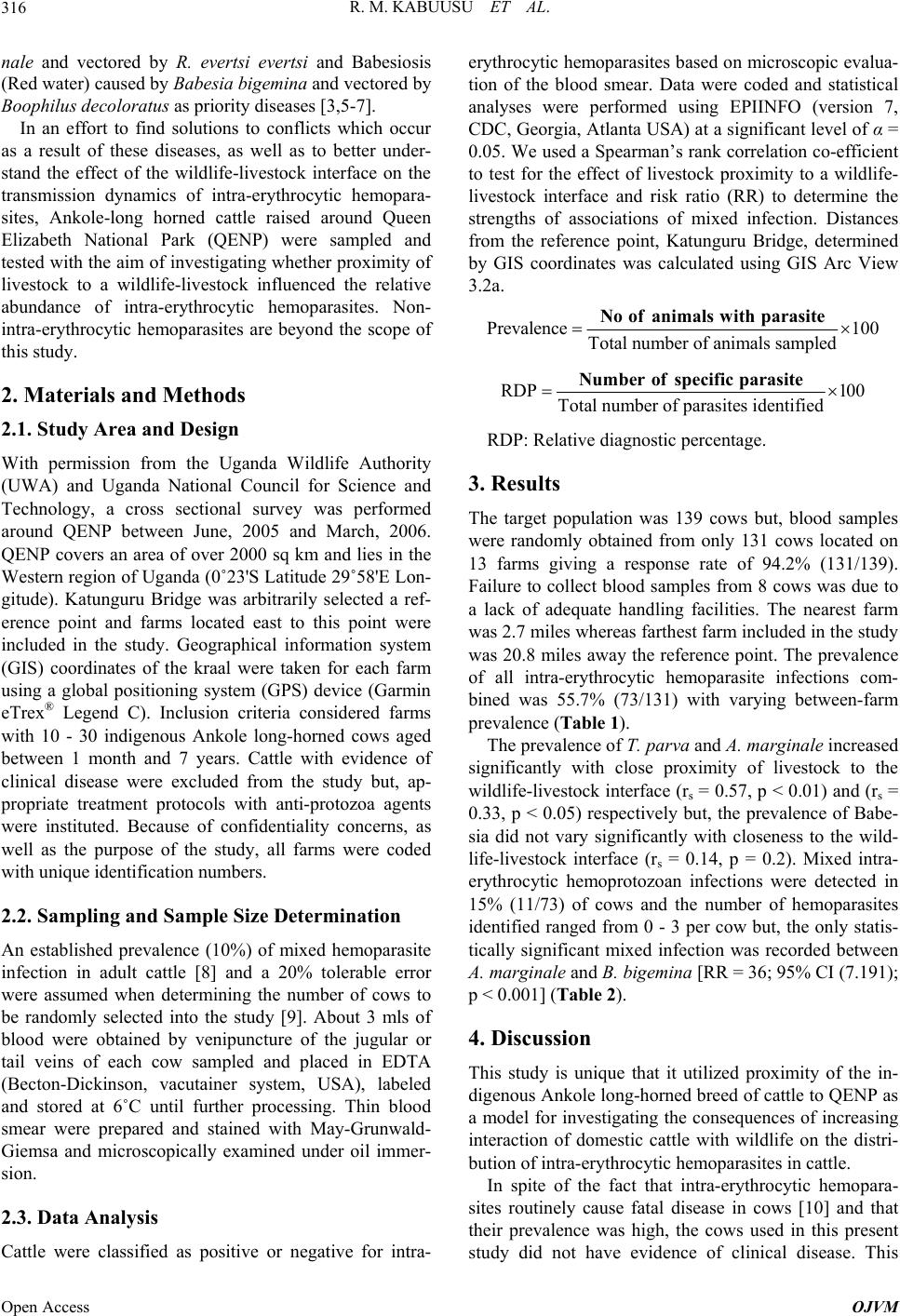 R. M. KABUUSU ET AL. 316 nale and vectored by R. evertsi evertsi and Babesiosis (Red water) caused by Babesia bigemina and vectored by Boophilus decoloratus as priority diseases [3,5-7]. In an effort to find solutions to conflicts which occur as a result of these diseases, as well as to better under- stand the effect of the wildlife-livestock interface on the transmission dynamics of intra-erythrocytic hemopara- sites, Ankole-long horned cattle raised around Queen Elizabeth National Park (QENP) were sampled and tested with the aim of investigating whether pro ximity of livestock to a wildlife-livestock influenced the relative abundance of intra-erythrocytic hemoparasites. Non- intra-erythrocytic hemoparasites are beyond the scope of this study. 2. Materials and Methods 2.1. Study Area and Design With permission from the Uganda Wildlife Authority (UWA) and Uganda National Council for Science and Technology, a cross sectional survey was performed around QENP between June, 2005 and March, 2006. QENP covers an area of over 2000 sq km and lies in the Western region of Uganda (0˚23'S Latitude 29˚58'E Lon- gitude). Katunguru Bridge was arbitrarily selected a ref- erence point and farms located east to this point were included in the study. Geographical information system (GIS) coordinates of the kraal were taken for each farm using a global positioning system (GPS) device (Garmin eTrex® Legend C). Inclusion criteria considered farms with 10 - 30 indigenous Ankole long-horned cows aged between 1 month and 7 years. Cattle with evidence of clinical disease were excluded from the study but, ap- propriate treatment protocols with anti-protozoa agents were instituted. Because of confidentiality concerns, as well as the purpose of the study, all farms were coded with unique identification numbers. 2.2. Sampling and Sample Size Determination An established prevalence (10%) of mixed hemoparasite infection in adult cattle [8] and a 20% tolerable error were assumed when determining the number of cows to be randomly selected into the study [9]. About 3 mls of blood were obtained by venipuncture of the jugular or tail veins of each cow sampled and placed in EDTA (Becton-Dickinson, vacutainer system, USA), labeled and stored at 6˚C until further processing. Thin blood smear were prepared and stained with May-Grunwald- Giemsa and microscopically examined under oil immer- sion. 2.3. Data Analysis Cattle were classified as positive or negative for intra- erythrocytic hemoparasites based on microscopic evalua- tion of the blood smear. Data were coded and statistical analyses were performed using EPIINFO (version 7, CDC, Georgia, Atlanta USA) at a significant level of α = 0.05. We used a Spearman’s rank correlation co-efficient to test for the effect of livestock proximity to a wildlife- livestock interface and risk ratio (RR) to determine the strengths of associations of mixed infection. Distances from the reference point, Katunguru Bridge, determined by GIS coordinates was calculated using GIS Arc View 3.2a. Prevalence 100 Total number of animals sampled No ofanimals withparasite RDP 100 Total number of parasite s id entifie d Number ofspecific parasite RDP: Relative diagnostic percentage. 3. Results The target population was 139 cows but, blood samples were randomly obtained from only 131 cows located on 13 farms giving a response rate of 94.2% (131/139). Failure to collect blood samples from 8 cows was due to a lack of adequate handling facilities. The nearest farm was 2.7 miles whereas farthest farm included in the study was 20.8 miles away the reference point. The prevalence of all intra-erythrocytic hemoparasite infections com- bined was 55.7% (73/131) with varying between-farm prevalence (Table 1). The prevalence of T. parva and A. marginale increased significantly with close proximity of livestock to the wildlife-livestock interface (rs = 0.57, p < 0.01) and (rs = 0.33, p < 0.05) respectively but, the prevalence of Babe- sia did not vary significantly with closeness to the wild- life-livestock interface (rs = 0.14, p = 0.2). Mixed intra- erythrocytic hemoprotozoan infections were detected in 15% (11/73) of cows and the number of hemoparasites identified ranged from 0 - 3 per cow but, the only statis- tically significant mixed infection was recorded between A. marginale and B. bigemina [RR = 36 ; 95% CI (7.191); p < 0.001] (Table 2). 4. Discussion This study is unique that it utilized proximity of the in- digenous Ankole lon g-horned breed of cattle to Q ENP as a model for investigating the consequences of increasing interaction of domestic cattle with wildlife on the distri- bution of intra-erythrocytic hemoparasites in cattle. In spite of the fact that intra-erythrocytic hemopara- sites routinely cause fatal disease in cows [10] and that their prevalence was high, the cows used in this present study did not have evidence of clinical disease. This Open Access OJVM 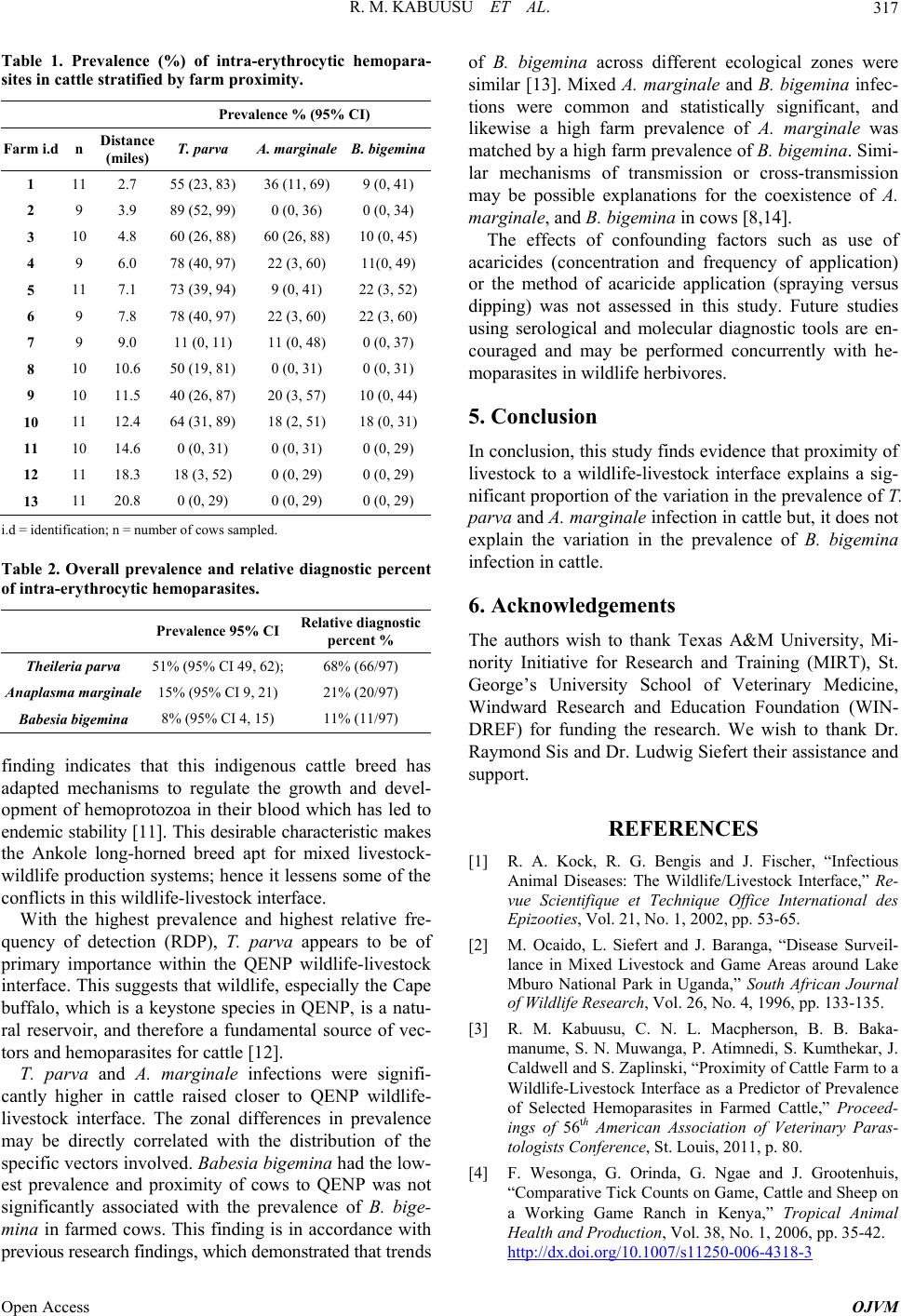 R. M. KABUUSU ET AL. 317 Table 1. Prevalence (%) of intra-erythrocytic hemopara- sites in cattle stratified by farm proximity. Prevalence % (95% CI) Farm i.d n Distance (miles) T. parva A. marginale B. bigemina 1 11 2.7 55 (23, 83)36 (11, 69) 9 (0, 41) 2 9 3.9 89 (52, 99)0 (0, 36) 0 (0, 34) 3 10 4.8 60 (26, 88)60 (26, 88) 10 (0, 45) 4 9 6.0 78 (40, 97)22 (3, 60) 11(0, 49) 5 11 7.1 73 (39, 94)9 (0, 41) 22 (3, 52) 6 9 7.8 78 (40, 97)22 (3, 60) 22 (3, 60) 7 9 9.0 11 (0, 11) 11 (0, 48) 0 (0, 37) 8 10 10.6 50 (19, 81)0 (0, 31) 0 (0, 31) 9 10 11.5 40 (26, 87)20 (3, 57) 10 (0, 44) 10 11 12.4 64 (31, 89)18 (2, 51) 18 (0, 31) 11 10 14.6 0 (0, 31) 0 (0, 31) 0 (0, 29) 12 11 18.3 18 (3, 52 ) 0 (0, 29) 0 (0, 29) 13 11 20.8 0 (0, 29) 0 (0, 29) 0 (0, 29) i.d = identification; n = number of cows sam pled. Table 2. Overall prevalence and relative diagnostic percent of intra-erythrocytic hemoparasites. Prevalence 95% CI Relative diagnostic percent % Theileria parva 51% (95% CI 49, 62); 68% (66/97) Anaplasma marginale 15% (95% CI 9, 21) 21% (20/97) Babesia bigemina 8% (95% CI 4, 15) 11% (11/97) finding indicates that this indigenous cattle breed has adapted mechanisms to regulate the growth and devel- opment of hemoprotozoa in their blood which has led to endemic stability [11]. Th is desirable characteristic ma kes the Ankole long-horned breed apt for mixed livestock- wildlife production systems; henc e it lessens some of the conflicts in this wildlife-livestock interface. With the highest prevalence and highest relative fre- quency of detection (RDP), T. parva appears to be of primary importance within the QENP wildlife-livestock interface. This suggests that wildlife, especially the Cap e buffalo, which is a keystone species in QENP, is a natu- ral reservoir, and therefore a fundamental source of vec- tors and hemoparasites for cattle [12]. T. parva and A. marginale infections were signifi- cantly higher in cattle raised closer to QENP wildlife- livestock interface. The zonal differences in prevalence may be directly correlated with the distribution of the specific vectors involved. Babesia bigemina had the low- est prevalence and proximity of cows to QENP was not significantly associated with the prevalence of B. bige- mina in farmed cows. This finding is in accordance with previous research findings, whi ch demonstrated that trends of B. bigemina across different ecological zones were similar [13]. Mixed A. marginale and B. bigemina infec- tions were common and statistically significant, and likewise a high farm prevalence of A. marginale was matched by a high farm prevalence of B. bigemina. Simi- lar mechanisms of transmission or cross-transmission may be possible explanations for the coexistence of A. marginale, and B. bigemina in cows [8,14]. The effects of confounding factors such as use of acaricides (concentration and frequency of application) or the method of acaricide application (spraying versus dipping) was not assessed in this study. Future studies using serological and molecular diagnostic tools are en- couraged and may be performed concurrently with he- moparasites in wildlife herbivores. 5. Conclusion In conclusion, this study finds evidence that proximity of livestock to a wildlife-livestock interface explains a sig- nificant proportion of the variation in the prevalence of T. parva and A. marginale infection in cattle but, it does not explain the variation in the prevalence of B. bigemina infection in cattle. 6. Acknowledgements The authors wish to thank Texas A&M University, Mi- nority Initiative for Research and Training (MIRT), St. George’s University School of Veterinary Medicine, Windward Research and Education Foundation (WIN- DREF) for funding the research. We wish to thank Dr. Raymond Sis and Dr. Ludwig Siefert their assistance and support. REFERENCES [1] R. A. Kock, R. G. Bengis and J. Fischer, “Infectious Animal Diseases: The Wildlife/Livestock Interface,” Re- vue Scientifique et Technique Office International des Epizooties, Vol. 21, No. 1, 2002, pp. 53-65. [2] M. Ocaido, L. Siefert and J. Baranga, “Disease Surveil- lance in Mixed Livestock and Game Areas around Lake Mburo National Park in Uganda,” South African Journal of Wildlife Research, Vol. 26, No. 4, 1996, pp. 133-135. [3] R. M. Kabuusu, C. N. L. Macpherson, B. B. Baka- manume, S. N. Muwanga, P. Atimnedi, S. Kumthekar, J. Caldwell and S. Zaplinski, “Proximity of Cattle Farm to a Wildlife-Livestock Interface as a Predictor of Prevalence of Selected Hemoparasites in Farmed Cattle,” Proceed- ings of 56th American Association of Veterinary Paras- tologists Conference, St. Louis, 2011, p. 80. [4] F. Wesonga, G. Orinda, G. Ngae and J. Grootenhuis, “Comparative Tick Counts on Game, Cattle and Sheep on a Working Game Ranch in Kenya,” Tropical Animal Health and Production, Vol. 38, No. 1, 2006, pp. 35-42. http://dx.doi.org/10.1007/s11250-006-4318-3 Open Access OJVM  R. M. KABUUSU ET AL. Open Access OJVM 318 [5] J. Okello-Onen, E. M. Tukahirwa, B. D. Perry, G. J. Rowlands, S. M. Nagda, G. Musisi, E. Bode, R. Heinonen, W. Mwayi and J. Opuda-Asibo, “Population Dynamics of Ticks on Indigenouscattle in a Pastoral Dry to Semi-Arid Range and Zone of Uganda,” Experimental & Applied Acarology, Vol. 23, No. 1, 1999, pp. 79-88. http://dx.doi.org/10.1023/A:1006058317111 [6] Z. I. Rajput, S.-H. Hu, A. G. Arijo, M. Habib and M. Khalid, “Comparative Study of Anaplasma Parasites in Tick Carrying Buffaloes and Cattle,” Journal of Zhejiang University Science B, Vol. 6, No. 11, 2005, pp. 1057- 1062. [7] F. Kabi, J. W. Mugon a, G. W . Nasinyama an d J . Walubengo, “Seroprevalence of Tick-Borne Infections among the Nkedi Zebu and Ankole Cattle in Soroti District, U ga nd a,” Journal of Protozoology Research, Vol. 18, No. 2, 2008, pp. 61-70. [8] J. W. Magona and J. S. P. Mayende, “Occurence of Con- current Trypanosomosis, Theileriosis, Anaplasmosis and Helminthosis in Friesian, Zebu and Sahiwal Cattle in Uganda,” Onderstepoort Journal of Veterinary Research, Vol. 69, No. 2, 2002, pp. 133-140. [9] S. W. Martin, A. H. Meek and P. Willeberg, “Veterinary Epidemiology. Principles and Methods,” Iowa State Uni- versity Press, Ames, 1987. [10] D. Shannon, “Field Cases of East Coast Fever in Grade Cattle in Uganda,” Tropical Animal Health and Produc- tion, Vol. 9, No. 1, 1977, pp. 29-35. http://dx.doi.org/10.1007/BF02297388 [11] R. W. Paling, C. Mpagala, B. Luttinhuizen and G. Sibo- mana, “Exposure of Ankole Cattle and Crossbred Cattle to Theileriosis in Rwanda,” Tropical Animal Health and Production, Vol. 23, No. 4, 1991, pp. 203-214. http://dx.doi.org/10.1007/BF02357101 [12] M. Ocaido, R. T. Muwazi and J. Opuda-Asibo, “Disease Incidence in Ranch and Pastoral Livestock Herds around Lake Mburo National Park, in South Western Uganda,” Tropical Animal Health and Production, Vol. 41, No. 7, 2009, pp. 1299-1308. http://dx.doi.org/10.1007/s11250-009-9315-x [13] C. Rubaire-Akiiki, J. Okello-Onen, G. W. Nasinyama, M. Vaarst, E. K. Kabagambe, W. Mwayi, D. Musunga and W. Wandukwa, “The Prevale nce of Seru m Anti bodie s to T ick- Borne Infections in Mbale Dist rict, Uganda: The Effect of Agroecologicalzone, Grazing Management and Age of Cattle,” Journal of Insect Science, Vol. 4, No. 8, 2004, p. 8. [14] C. P. Otim, “Advances in Disease Control: Ticks and T i c k- Borne Diseases,” Uganda Journal of Agricultural Sci- ences, Vol. 5, No. 1, 2000, pp. 79-85. |