

Paper Menu >>
Journal Menu >>
 Neuroscience & Medicine, 2013, 4, 277-279 Published Online December 2013 (http://www.scirp.org/journal/nm) http://dx.doi.org/10.4236/nm.2013.44040 Open Access NM 277 Anterior Cerebral Infarction by Fronto-Basal Meningioma Paul Macaire Ossou-Nguiet1*, Dieu Donné Gnonlonfoun2, Karen Obondzo-Aloba1, Hugues Brieux Ekouélé-Mbaki1, Gilbert Fabrice Otiobanda3 1Department of Neurology, University Hospital of Brazzaville, Brazzaville, Congo; 2Department of Neurology, University Hospital of Cotonou, Cotonou, Benin; 3Department of Intensive Care, University Hospital of Brazzaville, Brazzaville, Congo Email: *[email protected] Received August 31st, 2013; revised September 25th, 2013; accepted October 23rd, 2013 Copyright © 2013 Paul Macaire Ossou-Nguiet et al. This is an open access article distributed under the Creative Commons Attribution License, which permits unrestricted use, distribution, and reproduction in any medium, provided the original work is properly cited. ABSTRACT The association between stroke and meningioma is rarely reported in the data. In most etiology classification, there is no compressive cause. The association between meningioma and stroke is increasingly reported. We report a case of 52- year-old woman, previously healthy, presented with sudden right hemiplegia with some transient mood disorders. The CT-scan revealed acute left anterior cerebral artery infarction associated with fronto-basale meningioma infiltrating both anterior cerebral arteries. She was treated by corticosteroid and rehabilitation, with partial recovery. No stroke pre- vention was used before surgical treatment. Keywords: Infarction; Anterior Cerebral Artery; Meningioma 1. Introduction Mechanisms and causes of cerebral infarction are well studied and documented in the literature, and usually two mechanisms are identified: hemodynamics and throm- botic or thromboembolic causes [1]. Compressive causes by tumor are not reported in the main etiological classi- fications as Trial of Org1017 2 in Acute Stroke Treatment (TOAST) classification and ASCO (atherosclerosis, small vessel disease, cardiac source, other causes) [2]. Men- ingioma is the second most common primary brain tumor with incidence rates of 3 to 13 cases per 100,000. That incidence increases with female gender and age [3 ], an d i t is the most common benign tumor in adult, which is usu- ally manifested by slow progressive signs. The occur- rence of stroke associated with meningioma is increas- ingly reported, but there is no cohort study. We report a case of sudden neurological deficit due to calloso-mar- ginal branch of the anterior cerebral artery infarction as- sociated with infiltrating fronto-basal meningioma. 2. Case It was a patient of 52 years, with no particular history, admitted to the neurology department for motor deficit of the right side of the body. The examination revealed some transient episodes of mood disorders without headache or vomiting. Physical examination on admis- sion noted a right hemiplegia predominantly on lower limb and the presence of grasping reflexe, without other elements of the frontal lobe involvement. The blood pressure was 135/80 mmHg, regular heart without added noise. The CT scan without injection showed a tumor lesion isodensefrontobasale parenchyma associated with infarction in calloso-marginal territory of the anterior cerebral artery (Figures 1(A) and (B)). A secondary in- jection revealed homogeneous contrast enhancement of the lesion characteristic of meningioma. That tumor le- sion infiltrates both anterior cerebral arteries (Figures 1(C) and (D)). Lipid and inflammatory balance, echocar- diography, the 24 hours ECG-recording, and echo-Dop- pler of the supra-aortic trunks founded no abnormalities. It was not possible to make vascular exploration with MRI or angiography. The possible tumor origin of the infarct was selected. The treatment consisted to corticos- teroid based on prednisolone 1 mg/kg associated with and motor rehabilitation for 1 month, with partial recov- ery, then the patient was transferred to neurosurgery de- partment for surgical management, no stroke prevention was conducted before surgery. *Corresponding a uthor. 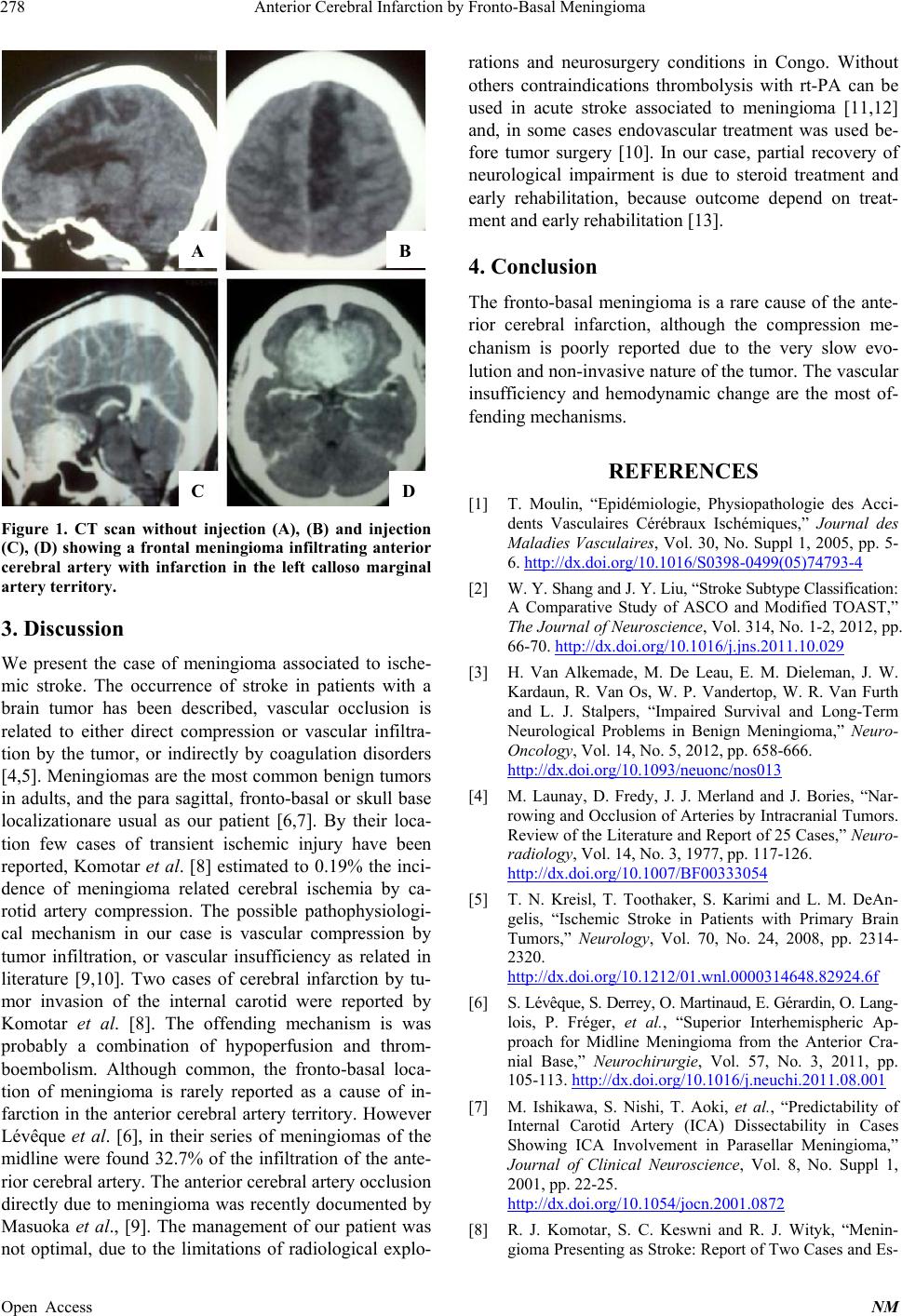 Anterior Cerebral Infarction by Fronto-Basal Meningioma 278 A B C D Figure 1. CT scan without injection (A), (B) and injection (C), (D) showing a frontal meningioma infiltrating anterior cerebral artery with infarction in the left calloso marginal artery territory. 3. Discussion We present the case of meningioma associated to ische- mic stroke. The occurrence of stroke in patients with a brain tumor has been described, vascular occlusion is related to either direct compression or vascular infiltra- tion by the tumor, or indirectly by coagulation disorders [4,5]. Meningiomas are the most common benign tumors in adults, and the para sagittal, fronto-basal or skull base localizationare usual as our patient [6,7]. By their loca- tion few cases of transient ischemic injury have been reported, Komotar et al. [8] estimated to 0.19% the inci- dence of meningioma related cerebral ischemia by ca- rotid artery compression. The possible pathophysiologi- cal mechanism in our case is vascular compression by tumor infiltration, or vascular insufficiency as related in literature [9,10]. Two cases of cerebral infarction by tu- mor invasion of the internal carotid were reported by Komotar et al. [8]. The offending mechanism is was probably a combination of hypoperfusion and throm- boembolism. Although common, the fronto-basal loca- tion of meningioma is rarely reported as a cause of in- farction in the anterior cerebral artery territory. However Lévêque et al. [6], in their series of meningiomas of the midline were found 32.7% of the infiltration of the ante- rior cerebral artery. The anterior cerebral artery occlusion directly due to meningioma was recently documented by Masuoka et al., [9]. The management of our patient was not optimal, due to the limitations of radiological explo- rations and neurosurgery conditions in Congo. Without others contraindications thrombolysis with rt-PA can be used in acute stroke associated to meningioma [11,12] and, in some cases endovascular treatment was used be- fore tumor surgery [10]. In our case, partial recovery of neurological impairment is due to steroid treatment and early rehabilitation, because outcome depend on treat- ment and early rehabilitation [13]. 4. Conclusion The fronto-basal meningioma is a rare cause of the ante- rior cerebral infarction, although the compression me- chanism is poorly reported due to the very slow evo- lution and non-inv asive nature of th e tumor. The vascu lar insufficiency and hemodynamic change are the most of- fending mechanisms. REFERENCES [1] T. Moulin, “Epidémiologie, Physiopathologie des Acci- dents Vasculaires Cérébraux Ischémiques,” Journal des Maladies Vasculaires, Vol. 30, No. Suppl 1, 2005, pp. 5- 6. http://dx.doi.org/10.1016/S0398-0499(05)74793-4 [2] W. Y. Shang and J. Y. Liu, “Stroke Subtype Classification: A Comparative Study of ASCO and Modified TOAST,” The Journal of Neuroscience, Vol. 314, No. 1-2, 2012, pp. 66-70. http://dx.doi.org/10.1016/j.jns.2011.10.029 [3] H. Van Alkemade, M. De Leau, E. M. Dieleman, J. W. Kardaun, R. Van Os, W. P. Vandertop, W. R. Van Furth and L. J. Stalpers, “Impaired Survival and Long-Term Neurological Problems in Benign Meningioma,” Neuro- Oncology, Vol. 14, No. 5, 2012, pp. 658-666. http://dx.doi.org/10.1093/neuonc/nos013 [4] M. Launay, D. Fredy, J. J. Merland and J. Bories, “Nar- rowing and Occlusion of Arteries by Intracranial Tumors. Review of the Literature and Report of 25 Cases,” Neuro- radiology, Vol. 14, No. 3, 1977, pp. 117-126. http://dx.doi.org/10.1007/BF00333054 [5] T. N. Kreisl, T. Toothaker, S. Karimi and L. M. DeAn- gelis, “Ischemic Stroke in Patients with Primary Brain Tumors,” Neurology, Vol. 70, No. 24, 2008, pp. 2314- 2320. http://dx.doi.org/10.1212/01.wnl.0000314648.82924.6f [6] S. Lévêque, S. Derrey , O. Mart inaud, E. Gérardin, O. Lang- lois, P. Fréger, et al., “Superior Interhemispheric Ap- proach for Midline Meningioma from the Anterior Cra- nial Base,” Neurochirurgie, Vol. 57, No. 3, 2011, pp. 105-113. http://dx.doi.org/10.1016/j.neuchi.2011.08.001 [7] M. Ishikawa, S. Nishi, T. Aoki, et al., “Predictability of Internal Carotid Artery (ICA) Dissectability in Cases Showing ICA Involvement in Parasellar Meningioma,” Journal of Clinical Neuroscience, Vol. 8, No. Suppl 1, 2001, pp. 22-25. http://dx.doi.org/10.1054/jocn.2001.0872 [8] R. J. Komotar, S. C. Keswni and R. J. Wityk, “Menin- gioma Presenting as Stroke: Report of Two Cases and Es- Open Access NM  Anterior Cerebral Infarction by Fronto-Basal Meningioma Open Access NM 279 timation of Incidence,” Journal of Neurology, Neurosur- gery & Psychiatry, Vol. 74, No. 1, 2003, p. 136. http://dx.doi.org/10.1136/jnnp.74.1.136 [9] J Masuoka, F Yoshioka, H Ohgushi, M Kawashima, T. Matsushima, “Meningioma Manifesting as Ce rebral Infarc- tion. Case Report,” Neurologia Medico-Chirurgica (To- kyo), Vol. 50, No. 7, 2010, pp. 585-587. http://dx.doi.org/10.2176/nmc.50.585 [10] F. Fernandez-Valverde, E. Jimenez-Gomez, E. Roldan- Romero and F. Bravo-Rodriguez, “Presurgical Endovas- cular Treatment of Symptomatic Carotid Stenosis Caused by a Meningioma of the Planumsphenoidale,” Revue Neu- rologique, Vol. 56, No. 3, 2013, pp. 157-160. [11] R. F. Baiense, A. Abrahão, I. F. Ricarte, T. G. Fukuda, R. A. Oliveira, G. S. Silva, M. Costa and M. J. Teixeira, “Successful Intra-Arterial Thrombolysis in a Patient with an Intracranial Meningioma,” Journal of Stroke and Cere- brovascular Diseases, 2013, in press. [12] T. Etgen, I. Steinich and L. Gsottschneider, “Thromboly- sis for Ischemic Stroke in Patients with Brain Tumors,” ournal of Stroke and Cerebrovascular Diseases, 2013, in press. [13] M. Bartolo, C. Zucchella, A. Pace, G. Lanzetta, C. Vec- chione, M. Bartolo, G. Grillea, M. Serrao, C. Tassorelli, G. Sandrini and F. Pierelli, “Early Rehabilitation after Surgery Improves Functional Outcome in Inpatients with Brain Tumours,” Journal of Neuro-Oncology, Vol. 107, No. 3, 2012, pp. 537-544. http://dx.doi.org/10.1007/s11060-011-0772-5 |