

Paper Menu >>
Journal Menu >>
 Neuroscience & Medicine, 2013, 4, 263-266 Published Online December 2013 (http://www.scirp.org/journal/nm) http://dx.doi.org/10.4236/nm.2013.44037 Open Access NM 263 Polymicrogyria of the Unilateral Temporal Lobe in a Transsexual Patient—Case Report Ana Starcevic1, Dusica Markovic Zigic2, Branislav Filipovic1 1Institute of Anatomy “Niko Miljanic”, Faculty of Medicine, University of Belgrade, Belgrade, Serbia; 2Psychiatry Hospital, Clinical Center Dedinje “Dr Dragisa Misovic”, Belgrade, Serbia. Email: [email protected] Received April 19th, 2013; revised May 18th, 2013; accepted June 14th, 2013 Copyright © 2013 Ana Starcevic et al. This is an open access article distributed under the Creative Commons Attribution License, which permits unrestricted use, distribution, and reproduction in any medium, provided the original work is properly cited. ABSTRACT Introduction: Polymicrogyria, the appearance of irregular small and over numbered gyri on the surface of the brain, has been reported as the most frequent finding, when exists, on the temporal lobe. Case Presentation: A patient, male, age of 35, came to the regular head and brain MRI scan due to psychiatric and hormonal treatment within transsexu al- ism. There were no data of epilepsy in a patient’s history. MRI scans were acquired with a 1.5 T Siemens Magnetom system with a standard head coil. Three-dimensional 3D RAGE, contiguous 1.0 mm slice, and 1 acquisition sequence underwent final 3D rendering and subsequent volumetry. On the definitive 3D image, we have rev ealed an unusual gy- risation on the left temporal lobe which had a picture of partial polymicrogyria. Conclusion: This is a unique finding, to our knowledge, of the unilateral temporal polymicrogyria in a person with transsexualism. Although polymicrogyria is mostly related to epileptifom attacks, its impact on the transsexualism appearance is opened to be examined. Keywords: Transsexualism; Polymicrogyria; MRI 1. Introduction Transsexuality is characterized by a strong conviction of having been born in the wrong body and is also consid- ered to be the extreme end of the spectrum of gender identity disorders characterized by a pursuit of sex reas- signment surgery [1]. Transsexuals experience them- selves as being of the opposite sex, despite having the biological characteristics of one sex. There can be seen a strong psychological rejection of the actual body. Fe- male-to-male transsexuals and male-to-female transsexu- als are characterized by persistent male or female identi- fication and constant feeling of being trapped in the wrong body, which is seems to be their permanent kind of a obsessive thought and final life goal as gender iden- tification is the basic human psychological characteris- tics. There might be many factors that can influence the development of this disorder and it has been implicated that genetic, hormonal and psychological factors are the most dominant. There is also a fact that there must be a morphological substrate like in every disorder that serves like a base or material for development of the disorder. Polymicrogyria is one of the most common malforma- tions of cortical development. It is often associated with specific syndromes, and has a wide variety of clinical presentation [2-4]. It is considered to be a cortical mal- formation development in which normal process of cor- tical development is somehow stopped by different fac- tors. Some of them are defined in the late stage of neu- ronal migration or in the early stage of neuronal organi- sation [4,5]. Polymicrogyria is a malformation with many faces, so it is of major significance to have that in mind when examing the MRI scans. It is mostly related to epi- leptic attacks, and its impact on the transsexualism ap- pearance is opened to be examined. 2. Case Presentation Our patient is male to female transsexual, thirty five years of age. He was reported to Counsiling Service for Sexual and Gender Identity Disorder. During the inter- view, he gave data on feeling different since childhood as a female trapped in a male body. He mentioned he liked to play more with h is girlfriends and felt like he is one of them. It was at the age of four. His mother thought it was 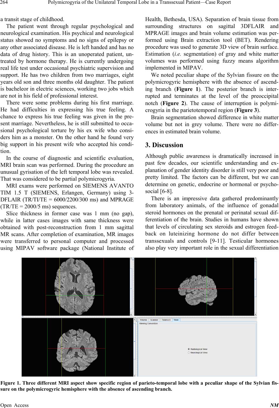 Polymicrogyria of the Unilateral Temporal Lobe in a Transsexual Pa ti en t—Case Report 264 a transit stage of childhood. The patient went through regular psychological and neurological examination. His psychical and neurological status showed no symptoms and no signs of epilepsy or any other associated disease. He is left handed and has no data of drug history. This is an unoperated patient, un- treated by hormone therapy. He is currently undergoing real life test under occasional psychiatric supervision and support. He has two children from two marriages, eight years old son and three months old daughter. The patient is bacheleor in electric sciences, working two jobs which are not in his field of professional interest. There were some problems during his first marriage. He had difficulties in expressing his true feeling. A chance to express his true feeling was given in the pre- sent marriage. Nevertheless, he is still submitted to occa- sional psychological torture by his ex wife who consi- ders him as a monster. On the other hand he found very big support in his present wife who accepted his condi- tion. In the course of diagnostic and scientific evaluation, MRI brain scan w as performed. During th e procedure an unusual gyrisation of the left temporal lobe was revealed. That was considered to be partial polymicrogyria. MRI exams were performed on SIEMENS AVANTO TIM 1.5 T (SIEMENS, Erlangen, Germany) using 3- DFLAIR (TR/TI/TE = 6000/2200/300 ms) and MPRAGE (TR/TE = 2000/5 ms) sequences. Slice thickness in former case was 1 mm (no gap), while in latter cases images with same thickness were obtained with post-reconstruction from 1 mm sagittal MR scans. After completion of examination, MR images were transferred to personal computer and processed using MIPAV software package (National Institute of Health, Bethesda, USA). Separation of brain tissue from surrounding structures on sagittal 3DFLAIR and MPRAGE images and brain volume estimation was per- formed using Brain extraction tool (BET). Rendering procedure was used to generate 3D view of brain surface. Estimation (i.e. segmentation) of gray and white matter volumes was performed using fuzzy means algorithm implemented in MIPAV. We noted peculiar shape of the Sylvian fissure on the polymicrogyric hemisphere with the absence of ascend- ing branch (Figure 1). The posterior branch is inter- rupted and terminates at the level of the preoccipital notch (Figure 2). The cause of interruption is polymi- crogyria in the parietotemporal region (Figure 3). Brain segmentation showed difference in white matter volume but not in grey volume. There were no differ- ences in estimated brain volume. 3. Discussion Although public awareness is dramatically increased in past few decades, our scientific understanding and ex- planation of gender iden tity disorder is still very poor and pretty limited. The factors can be different, but we can determine on genetic, endocrine or hormonal or psycho- social [6-8]. There is an impressive data gathered predominantly from laboratory animals, of the influence of gonadal steroid hormones on the prenatal or perinatal sexual dif- ferentiation of the brain. Studies in humans have shown that levels of circulating sex steroids and estrogen feed- back on luteinizing hormone do not differ between transsexuals and controls [9-11]. Testicular hormones also play very important ro le in the sexual differen tiation Figure 1. Three different MRI aspect show specific region of parieto-temporal lobe with a peculiar shape of the Sylvian fis- sure on the polymicrogyric hemisphere with the absence of ascending branch. Open Access NM 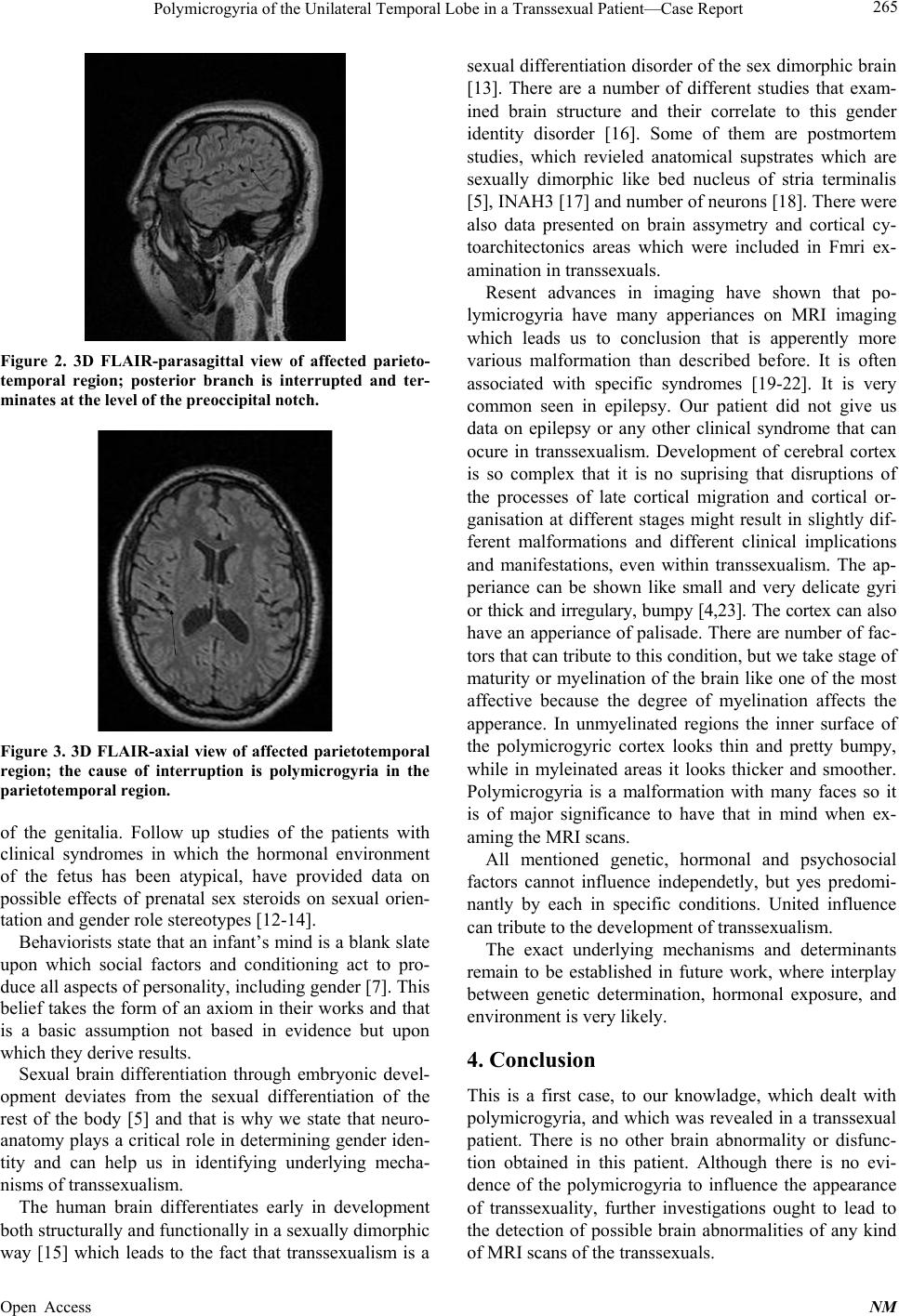 Polymicrogyria of the Unilateral Temporal Lobe in a Transsexual Pa ti en t— Case Report 265 Figure 2. 3D FLAIR-parasagittal view of affected parieto- temporal region; posterior branch is interrupted and ter- minates at the level of the preoccipital notc h. Figure 3. 3D FLAIR-axial view of affected parietotemporal region; the cause of interruption is polymicrogyria in the parietotemporal region. of the genitalia. Follow up studies of the patients with clinical syndromes in which the hormonal environment of the fetus has been atypical, have provided data on possible effects of prenatal sex steroids on sexual orien- tation and gender role stereotypes [12-14]. Behaviorists state that an infant’s mind is a blank slate upon which social factors and conditioning act to pro- duce all aspects of personality, including gender [7]. This belief takes the form of an axiom in their works and that is a basic assumption not based in evidence but upon which they derive results. Sexual brain differentiation through embryonic devel- opment deviates from the sexual differentiation of the rest of the body [5] and that is why we state that neuro- anatomy plays a critical role in determining gender iden- tity and can help us in identifying underlying mecha- nisms of transsexualism. The human brain differentiates early in development both structurally and functionally in a sexually dimorphic way [15] which leads to the fact that transsexualism is a sexual differentiation disorder of the sex dimorphic brain [13]. There are a number of different studies that exam- ined brain structure and their correlate to this gender identity disorder [16]. Some of them are postmortem studies, which revieled anatomical supstrates which are sexually dimorphic like bed nucleus of stria terminalis [5], INAH3 [17] and number of neurons [18]. There were also data presented on brain assymetry and cortical cy- toarchitectonics areas which were included in Fmri ex- amination in transsexuals. Resent advances in imaging have shown that po- lymicrogyria have many apperiances on MRI imaging which leads us to conclusion that is apperently more various malformation than described before. It is often associated with specific syndromes [19-22]. It is very common seen in epilepsy. Our patient did not give us data on epilepsy or any other clinical syndrome that can ocure in transsexualism. Development of cerebral cortex is so complex that it is no suprising that disruptions of the processes of late cortical migration and cortical or- ganisation at different stages might result in slightly dif- ferent malformations and different clinical implications and manifestations, even within transsexualism. The ap- periance can be shown like small and very delicate gyri or thick and irregulary, bumpy [4,23]. The cortex can also have an apperiance of palisade. There are number of fac- tors that can tribute to this cond ition, bu t we take stage of maturity or myelination of the brain like one of the most affective because the degree of myelination affects the apperance. In unmyelinated regions the inner surface of the polymicrogyric cortex looks thin and pretty bumpy, while in myleinated areas it looks thicker and smoother. Polymicrogyria is a malformation with many faces so it is of major significance to have that in mind when ex- aming the MRI scans. All mentioned genetic, hormonal and psychosocial factors cannot influence independetly, but yes predomi- nantly by each in specific conditions. United influence can tribute to the development of transsexualism. The exact underlying mechanisms and determinants remain to be established in future work, where interplay between genetic determination, hormonal exposure, and environment is very likely. 4. Conclusion This is a first case, to our knowladge, which dealt with polymicrogyria, and which was revealed in a transsexual patient. There is no other brain abnormality or disfunc- tion obtained in this patient. Although there is no evi- dence of the polymicrogyria to influence the appearance of transsexuality, further investigations ought to lead to the detection of possible brain abnormalities of any kind of MRI scans of the transsexuals. Open Access NM  Polymicrogyria of the Unilateral Temporal Lobe in a Transsexual Pa ti en t—Case Report 266 REFERENCES [1] P. T. Cohen-Kettenis and L. J. Gooren, “Transsexualism, a Review of Etiology, Diagnosis and Treatment,” Journal of Psychosomatic Research, Vol. 46, No. 4, 1999, pp. 315-333. http://dx.doi.org/10.1016/S0022-3999(98)00085-3 [2] A. J. Barkovich, “Current Concepts of Polymicrogyria,” Neuroradiology, Vol. 52, No. 6, 2010, pp. 479-487. http://dx.doi.org/10.1007/s00234-009-0644-2 [3] R. J. Leventer, A. Jansen, D. T. Pilz, N. Stoodley, C. Marini, F. Dubeau, et al., “Clinical and Imaging Hetero- geneity of Polymicrogyria: A Study of 328 Patients,” Brain, Vol. 133, No. 5, 2010, 1415-1427. http://dx.doi.org/10.1093/brain/awq078 [4] A. J. Barkovich, P. Gressens and P. Evrard, “Formation, Maturation and Disorders of Brain Neocortex,” AJNR American Journal of Neuroradiology, Vol. 13, 1992, pp. 423-446. [5] J. N. Zhou, M. A. Hofman, L. J. Gooren and D. F. Swaab, “A Sex Difference in the Human Brain and Its Relation to Transsexuality,” Nature, Vol. 378, No. 6552, 1995, pp. 68-70. http://dx.doi.org/10.1038/378068a0 [6] S. M. Corsello, V. Di Donna and P. Senes, et al., “Biolo- gical Aspects of Gender Disorders,” Minerva Endocri- nologica, Vol. 36, No. 4, 2011, pp. 325-339. [7] A. M. Bao and D. F. Swaab, “Sex Differences in the Brain, Behaviour, and Neuropsychiatric Disorders,” Neu- roscientist, Vol. 16, No. 5, 2010, pp. 550-565. http://dx.doi.org/10.1177/1073858410377005 [8] C. Kraus, “Am I My Brain or My Genitals? A Nature- Culture Controversy in the Hermaphrodite Debate from the Mid-1960s to the Late 1990s,” Gesnerus, Vol. 68, No. 1, 2011, pp. 80-106. [9] A. A. Lawrence, “A Critique of the Brain-Sex Theory of Transsexualism,” Transsexual Women’s Resources, 2007. [10] I. Savic, A. Garcia-Falgueras and D. F. Swaab, “Sexual Differentiation of the Human Brain in Relation to Gender IDENTITY and Sexual Orientation,” Progress in Brain Research, Vol. 186, 2010, pp. 41-62. http://dx.doi.org/10.1016/B978-0-444-53630-3.00004-X [11] D. F. Swaab, L. J. Gooren and M. A. Hofman, “Gender and Sexual Orientation in Relation to Hypothalamic Structures,” Hormone Research, Vol. 38, No. 2, 1992, pp. 51-61. http://dx.doi.org/10.1159/000182597 [12] D. F. Swaab, “Sexual Differentiation of the Human Brain: Relevance for Gender Identity, Transsexualism and Sex- ual Orientation,” Gynecological Endocrinology, Vol. 19, No. 6, 2004, pp. 301-312. http://dx.doi.org/10.1080/09513590400018231 [13] L. Gooren, “The Biology of Human Psychosexual Differ- entiation,” Hormones and Behavior, Vol. 50, No. 4, 2006, pp. 589-601 http://dx.doi.org/10.1016/j.yhbeh.2006.06.011 [14] A. M. Bao and D. F. Swaab, “Sexual Differentiation of the Human Brain: Relation to Gender Identity, Sexual Orientation and Neuropsychiatric Disorders,” Frontiers in Neuroendocrinology, Vol. 32, No. 2, 2011, pp. 214-226. http://dx.doi.org/10.1016/j.yfrne.2011.02.007 [15] D. F. Swaab, “Sexual Differentiation of the Brain and Behaviour,” Best Practice & Research Clinical Endocri- nology & Metabolism, Vol. 21, No. 3, 2007, pp. 431-444. http://dx.doi.org/10.1016/j.beem.2007.04.003 [16] W. C. Chung, G. J. De Vries and D. F. Swaab, “Sexual Differentiation of the Bed Nucleus of the Stria Terminalis in Hu ma ns May Extend int o Adulthood,” Neuroradiology, Vol. 22, No. 3, 2002, pp. 1027-1033. [17] A. Garcia-Falgueras and D. F. Swaab, “A Sex Difference in the Hypothalamic Uncinate Nucleus: Relationship to Gender Identity,” Brain, Vol. 131, No. 12, 2008, pp. 3132- 3146. http://dx.doi.org/10.1093/brain/awn276 [18] F. P. M. Kruijver, J. N. Zhou, C. W. Pool, M. A. Hofman, L. J. Gooren and D. F. Swaab, “Male-to-Female Trans- sexuals Have Female Neuron Numbers in a Limbic Nu- cleus,” The Journal of Clinical Endocrinology & Metabo- lism, Vol. 85, No. 5, 2000, p. 2034. http://dx.doi.org/10.1210/jc.85.5.2034 [19] M. Rijpkema, D. Everaerd, C. van der Pol, B. Franke, I. Tendolkar and G. Fernández, “Normal Sexual Dimor- phism in the Human Basal Ganglia,” Human Brain Map- ping, Vol. 33, No. 5, 2011, pp. 1346-1252. http://dx.doi.org/10.1002/hbm.21283 [20] J. Brabec, J. Krásený and P. Petrovický, “Volumetry of Striatum and Pallidum in Man—Anatomy, Cytoarchitec- ture, Connections, MRI and Aging,” Sbornik Lekaisky, Vol. 104, No. 1, 2003, pp. 13-65. [21] Y. Miyahira, J. Yu, K. Hiramatsu, Y. Shimazaki and Y. Takeda, “Brain Volumetric MRI Study in Healthy Elderly Persons Using Statistical Parametric Mapping,” Seishin Shinkeigaku Zasshi, Vol. 106, No. 2, 2004, pp. 138-151. [22] L. G. Almeida Montes, J. Ricardo-Garcell, L. B. Barajas De La Torre, et al., “Clinical Correlations of Grey Matter Reductions in the Caudate Nucleus of Adults with Atten- tion Deficit Hyperactivity Disorder,” Journal of Psychia- try and Neuroscience, Vol. 35, No. 4, 2010, pp. 238-246. http://dx.doi.org/10.1503/jpn.090099 [23] A. J. Barkovich, “MRI Analysis of Sulcation Morphology in Polymicrogyria,” Epilepsia, Vol. 51, No. 1, 2010, pp. 17-22. http://dx.doi.org/10.1111/j.1528-1167.2009.02436.x Open Access NM |