

Paper Menu >>
Journal Menu >>
 Advances in Infectious Diseases, 2013, 3, 263-268 Published Online December 2013 (http://www.scirp.org/journal/aid) http://dx.doi.org/10.4236/aid.2013.34040 Open Access AID 263 Invasive Fungal Sinusitis in Immunocompromised Patients: A Multicenter, University Hospital Experience in Shiraz Mohsen Moghadami1, Hossein Ruzbahani2, Parisa Badiee3*, Abolhassan Faramarzi4, Payam Peymani1, Kamran Bagheri Lankarani1 1Health Policy Research Center, Shiraz University of Medical Sciences, Shiraz, Iran; 2Department of Internal Medicine, Shiraz Uni- versity of Medical Sciences, Shiraz, Iran; 3Alborzi Clinical Microbiology Research Center, Shiraz University of Medical Sciences, Shiraz, Iran; 4Department of Otolaryngology, Shiraz University of Medical Sciences, Shiraz, Iran. Email: [email protected] Received September 28th, 2013; revised October 28th, 2013; accepted November 6th, 2013 Copyright © 2013 Mohsen Moghadami et al. This is an open access article distributed under the Creative Commons Attribution Li- cense, which permits unrestricted use, distribution, and reproduction in any medium, provided the original work is properly cited. ABSTRACT Objective: It is to determine the causes of invasive fungal sinusitis in p atients of Shiraz University hospitals, Iran. Me- thods: This cross-sectional study was conducted during 18 months (from 21 March 2009 till 22 September 2010) in three Shiraz University Hospitals. Thirty-six patients with sings of invasive fungal sinusitis were enrolled, and tissue samples were investigated for histop athology, culture and antifungal suscep tibility test. The laboratory results with host factor and sinus computed tomography scan were evaluated for classification of patients as proven, probable and possi- ble invasive fungal sinusitis. Results: Thirty-five patients have involved with at least one risk factor (immune compro- mised disease, diabetes mellitus, or use of immune suppressed drugs). Radiological findings of parasinus invasion or necrosis were present in 20 patients. By histopathology, 21 patients were considered as proven, from these, 17 samples had positive growth. The culture aetiology agents were 4 Candida, 8 Aspergillus, and 5 Mucor. All positive culture samples were matched with histopathology findings. Significant associations were considered for radiologic finding and histopathology and culture (p < 0.05). From 8 patients with mucormycosis histopathology, 6 suffered from diabetes mellitus. None of the antifungal agents were effective on these three types of infections. Conclusion: DM is the most common predisposing factor for IFS followed by ALL and AML. The most common aetiology of IFS was found to be Aspergillus fumigant followed by Mucormycosis and Candida. None of antifungal agents could successfully cover all the species. Keywords: Fungal Sinusitis; Mucor Mycosis; Aspergillus; Invasive Fungal 1. Introduction Opportunistic fungal infections are usually results of im- munosuppression and immunodeficiency. Currently sev- eral etiologies of immunosuppression lead to increase prevalence of invasive fungal infection (IFI) including leukemia, diabetes mellitus (DM), AIDS, solid organ transplantation, bone marrow transplan tation and chemo- therapy [1]. All these conditions result in neutropenia which should be treated with wide-spectrum antibiotics. IFI is considered as an important complication of neutro- penia which is suspected with persistent fever for 72 - 96 hours after treatment with broad-spectrum antibacterial antibiotic [1-3]. However, since the culture methods are insensitive and radiologic findings are nonspecific, the diagnosis of invasive fungal infection remains a chal- lenge [4]. An intern ational consensus on th e diagnosis of opportunistic invasive fungal infections in immunocom- promised patients with cancer and hematopoietic stem cell transplants was established by the Invasive Fungal Infections Cooperativ e Group of the Europ ean Organiza- tion for Research and Treatment of Cancer (EORTC) and the Mycoses Study Group of the National Institu te of Al- lergy and Infectious Diseases (MSG-NIAID) [5,6]. Delay in the treatment of invasive fungal infection during neu- tropenia causes high mortality in p a tients with transplants and hematological malig nancy [4,5,7 ]. The unc ertainty in *Corresponding a uthor.  Invasive Fungal Sinusitis in Immunocompromised Patients: A Multicenter, University Hospital Experience in Shiraz 264 disease diagnosis results in under- or overtreatment of in- vasive fungal infection. Despite the availability of seve- ral new antifungal agents, in cluding triazoles and echino- candins, the effectiveness of antifungal therapy remains uncertain and the effectiveness of neutrophil recovery may not be sufficient if the recovering neutrophils are dysfunctional [8]. Invasive fungal sinusitis (IFS) is a rare disease largely attributable to Aspergillus and Mucor in patients with stem cell transplants and hematological disease [9]. Though the mortality of IFS in immunocompromised patients ranges from 50% to 80% [7,9,10], early physical findings are non- specific and ambiguous (i.e., nasal obstruction, purulent dis- charge, and epistaxis). Water’s view plain radiogr aphs do not distinguish invasive fungal sinusitis from chronic al- lergic sinusitis. Bony erosion and tissue destruction are often found only in the advanced stage by computed to- mography [11,12]. Recent introduction of serial Asper gil- lus galactomannan antigen test may provide early evi- dence of IF S. As data regard ing this issue are scarce in ou r region of southern Iran, we performed this study to deter- mine the epidemiology of IFS and the drug sensitivities of the etiological factors in Shiraz, southern Iran. 2. Materials and Methods 2.1. Study Population This was a cross-sectional study being performed in Iran during a 1.5-year period from 21 March 2009 till 22 Sep- tember 2010 including 36 immunosuppressed patients who were further diagnosed to have IFS. Patients with solid organ transplant, hematopoietic stem cell trans- plants, hematological disease (including severe aplastic anemia, pure red cell aplasia, and hematological malig- nancy), AIDS and sinusitis diagnosed during their ho spi- tal stay were enrolled in this study. Demographic features, history of bone marrow transplant, history of neutropenia (ANC < 500/mL), hematological disease status, underly- ing medical diseases, prolonged corticosteroid therapy (>3 weeks), receiving nucleoside analogue and T-cell sup- pressor during the previous 3 months and congenital im- munodeficiencies were recorded in a questionnaire. The study protocol was approved by the institutional review board of Shiraz University of Medical Sciences and all re- quired patients provided their informed written consents. 2.2. Sinusitis Diagnosis During the study period, those immunosuppressed pa- tients who developed sinusitis were routinely underwent sinus X-ray evaluation and afterwards, otolaryngologist was consulted for focal evaluation and tissue culture. CT and MRI sinus study, surgical biopsy and debridement were performed according to clinicians’ decision. IFS was diagnosed according to EORTC/MSG-NIAID consensus criteria in 2008 [5,6]. The host factors in- cluded prolonged neutropenia (<500 neutrophils/mm3 for >10 days) temporally related to the onset of fungal dis- ease, receipt of an allogeneic stem cell transplant, pro- longed use of corticosteroids, immunosuppressive agents, or nucleoside analogues during the past 90 days. The cli- nical criteria included imaging showing sinusitis plus at least one of the following three signs: acute localized pain, nasal ulcer with black eschar, extension from the paranasal sinus across the bony barrier. The microbiolo- gical criteria included culture or isolation of fungus from surgical material or sinus aspirate samples, and detection of Aspergillus galactomannan antigens in serum. All the samples were cultured in Sabouraud Dextrose Agar me- dia and sensitivity to amphotericin B 1, capsofungin ace- tate, voriconazole, itracon azole, ketokonazo l were further examined. 2.3. Proven, Probable, and Possible Invasive Fungal Sinusitis Proven, probable, and possible IFS were defined mainly according to the EORTC/MSG-NIAID criteria [5,6]. Pro- ven IFS was defined by the presence of fungi associated tissue damage on histopathologic examination of a bio- psy specimen; or positive culture result, consistent with infection, from a sample obtained aseptically from a cli- nically or radiologically abnormal site. Probable IFS was defined by the presence of at least one host factor crite- rion, one microbiological criterion, and one clinical crite- rion. Possible IFS was defined by the presence of at least one host factor criterion and one clinical criterion. 2.4. Statistical Analysis Statistical analyses were performed using the SPSS soft- ware, version 16.0 (SPSS Inc., Chicago, Ill., USA). The chi-square test was used to compare the proportions be- tween groups. The results are expressed as mean ± SD and proportions as appropriated. A two-tailed p-value less than 0.05 was considered statistically significant. 3. Results Overall we included 36 patients among whom there were 15 (41.6%) man and 21 (58.4%) women. The mean age of the patients was 40 years with maximum age of 70 years. Sixteen (44%) of the patients were diagnosed to have DM. Thirty patients had at least one co-morbidities or sinus destruction. The most common predisposing condition was ALL in 5 and AML in 5 follow ed by sinus surgery in 3, CML in 3, CLL in 1, major β-thalassemia in 2, aplastic anemia in 1, Hodgkin's lymphoma in 1, non- Hodgkin’s lymphoma in 2 and other diseases (congenital Open Access AID 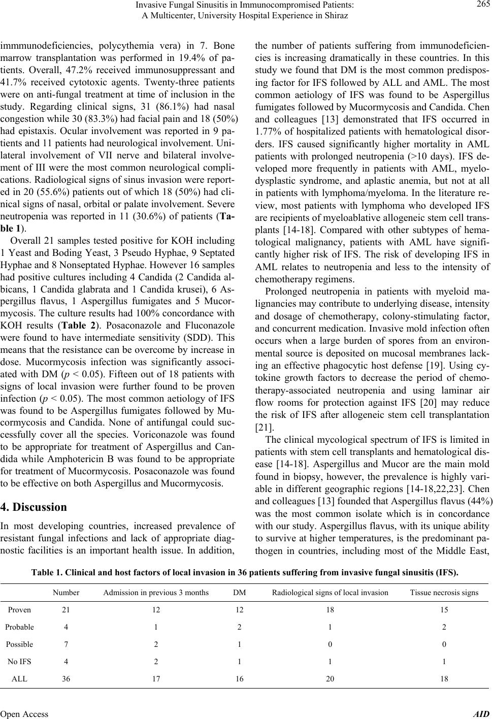 Invasive Fungal Sinusitis in Immunocompromised Patients: A Multicenter, University Hospital Experience in Shiraz Open Access AID 265 immmunodeficiencies, polycythemia vera) in 7. Bone marrow transplantation was performed in 19.4% of pa- tients. Overall, 47.2% received immunosuppressant and 41.7% received cytotoxic agents. Twenty-three patients were on anti-fungal treatment at time of inclusion in the study. Regarding clinical signs, 31 (86.1%) had nasal congestion while 30 (83.3%) had facial pain and 18 (50%) had epistaxis. Ocular involvement was reported in 9 pa- tients and 11 patients had neurological involvement. Uni- lateral involvement of VII nerve and bilateral involve- ment of III were the most common neurological compli- cations. Radiological signs of sinus invasion were report- ed in 20 (55.6%) patients out of which 18 (50%) h ad cli- nical signs of nasal, orbital or palate involvement. Severe neutropenia was reported in 11 (30.6%) of patients (Ta- ble 1). Overall 21 samples tested positive for KOH including 1 Yeast and Boding Yeast, 3 Pseudo Hyphae, 9 Septated Hyphae and 8 Non sep tated Hyph ae. Howe ver 16 samples had positive cultures including 4 Candida (2 Candida al- bicans, 1 Candida glabrata and 1 Candida krusei), 6 As- pergillus flavus, 1 Aspergillus fumigates and 5 Mucor- mycosis. The culture results had 100% concordance with KOH results (Table 2). Posaconazole and Fluconazole were found to have intermediate sensitivity (SDD). This means that the resistance can be overcome by increase in dose. Mucormycosis infection was significantly associ- ated with DM (p < 0.05). Fifteen out of 18 patients with signs of local invasion were further found to be proven infection (p < 0.05). The most common aetiology of IFS was found to be Aspergillus fumigates followed by Mu- cormycosis and Candida. None of antifungal could suc- cessfully cover all the species. Voriconazole was found to be appropriate for treatment of Aspergillus and Can- dida while Amphotericin B was found to be appropriate for treatment of Mucormycosis. Posaconazole was found to be effective on both Aspergillus and Mucormycosis. 4. Discussion In most developing countries, increased prevalence of resistant fungal infections and lack of appropriate diag- nostic facilities is an important health issue. In addition, the number of patients suffering from immunodeficien- cies is increasing dramatically in these countries. In this study we found that DM is the most common predispos- ing factor for IFS followed by A LL and AML. The most common aetiology of IFS was found to be Aspergillus fumigates followed by Mucormycosis and Candida. Chen and colleagues [13] demonstrated that IFS occurred in 1.77% of hospitalized patients with hematological disor- ders. IFS caused significantly higher mortality in AML patients with prolonged neutropenia (>10 days). IFS de- veloped more frequently in patients with AML, myelo- dysplastic syndrome, and aplastic anemia, but not at all in patients with lymphoma/myeloma. In the literature re- view, most patients with lymphoma who developed IFS are recipients of myeloablative allogeneic stem cell trans- plants [14-18]. Compared with other subtypes of hema- tological malignancy, patients with AML have signifi- cantly higher risk of IFS. The risk of developing IFS in AML relates to neutropenia and less to the intensity of chemotherapy regi mens. Prolonged neutropenia in patients with myeloid ma- lignancies may contribute to underlying disease, intensity and dosage of chemotherapy, colony-stimulating factor, and concurrent medication. Invasive mold infection often occurs when a large burden of spores from an environ- mental source is deposited on mucosal membranes lack- ing an effective phagocytic host defense [19]. Using cy- tokine growth factors to decrease the period of chemo- therapy-associated neutropenia and using laminar air flow rooms for protection against IFS [20] may reduce the risk of IFS after allogeneic stem cell transplantation [21]. The clinical mycological spectrum of IFS is limited in patients with stem cell transplants and hematological dis- ease [14-18]. Aspergillus and Mucor are the main mold found in biopsy, however, the prevalence is highly vari- able in different geographic regions [14-18,22,23]. Chen and colleagues [13 ] founded that Aspergillus flavus (44%) was the most common isolate which is in concordance with our study. Aspergillus flavus, with its uniqu e ability to survive at higher temperatures, is the predominant pa- thogen in countries, including most of the Middle East, Table 1. Clinical and host factors of local invasion in 36 patients suffering from invasive fungal sinusitis (IFS). Number Admission in pr evious 3 months DM Radiological signs of local invasion Tissue necrosis signs Proven 21 12 12 18 15 Probable 4 1 2 1 2 Possible 7 2 1 0 0 No IFS 4 2 1 1 1 ALL 36 17 16 20 18 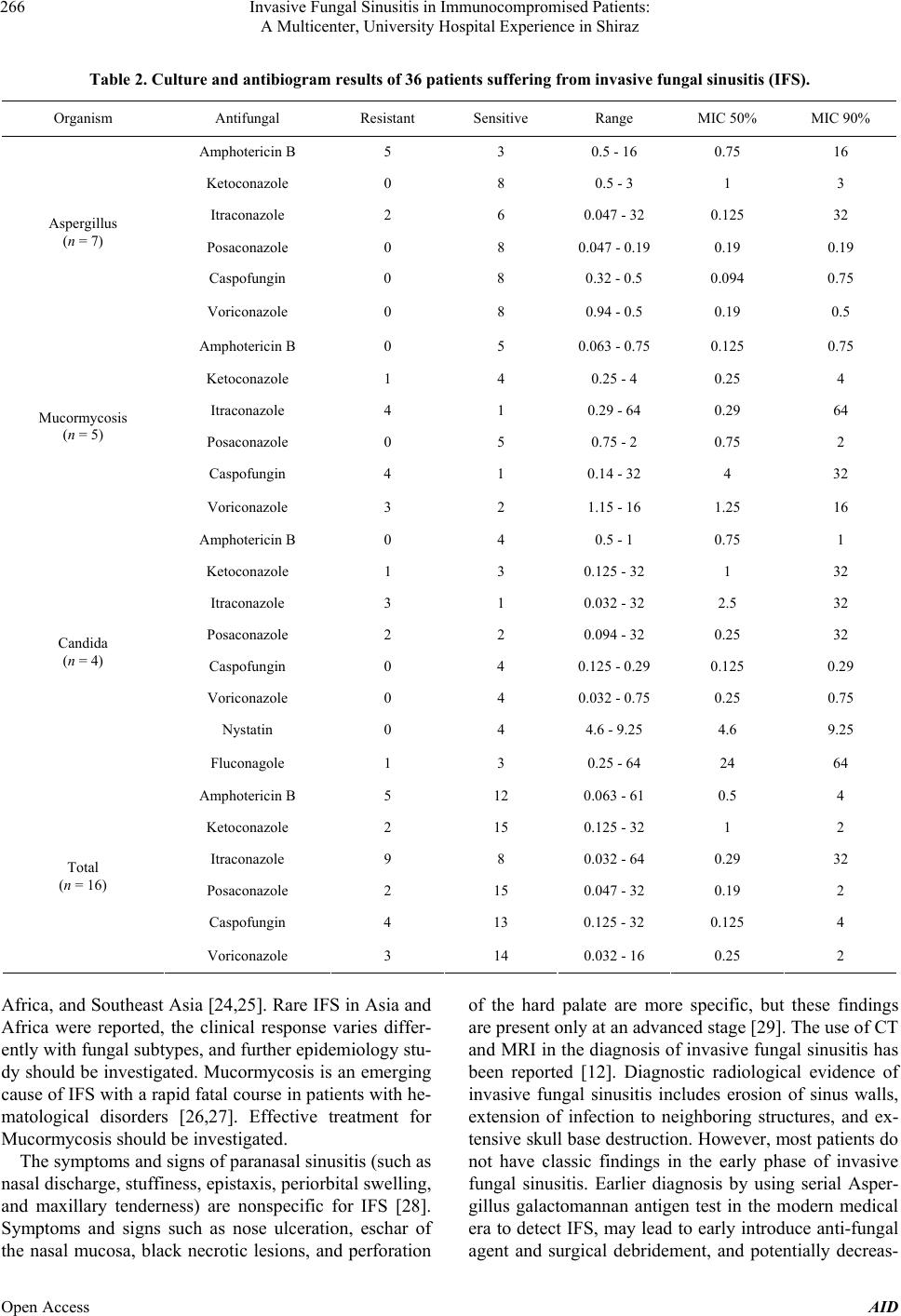 Invasive Fungal Sinusitis in Immunocompromised Patients: A Multicenter, University Hospital Experience in Shiraz 266 Table 2. Culture and antibiogram results of 36 patients suffering from invasive fungal sinusitis (IFS). Organism Antifungal Resistant Sensitive Range MIC 50% MIC 90% Amphotericin B 5 3 0.5 - 16 0.75 16 Ketoconazole 0 8 0.5 - 3 1 3 Itraconazole 2 6 0.047 - 32 0.125 32 Posaconazole 0 8 0.047 - 0.19 0.19 0.19 Caspofungin 0 8 0.32 - 0.5 0.094 0.75 Aspergillus (n = 7) Voriconazole 0 8 0.94 - 0. 5 0.19 0.5 Amphotericin B 0 5 0.063 - 0.75 0.125 0.75 Ketoconazole 1 4 0.25 - 4 0.25 4 Itraconazole 4 1 0.29 - 6 4 0.29 64 Posaconazole 0 5 0.75 - 2 0.75 2 Caspofungin 4 1 0.14 - 32 4 32 Mucormycosis (n = 5) Voriconazole 3 2 1.15 - 16 1.25 16 Amphotericin B 0 4 0.5 - 1 0.75 1 Ketoconazole 1 3 0.125 - 32 1 32 Itraconazole 3 1 0.032 - 32 2.5 32 Posaconazole 2 2 0.094 - 32 0.25 32 Caspofungin 0 4 0.125 - 0.29 0.125 0.29 Voriconazole 0 4 0.032 - 0.75 0.25 0.75 Nystatin 0 4 4.6 - 9.25 4.6 9.25 Candida (n = 4) Fluconagole 1 3 0.25 - 64 24 64 Amphotericin B 5 12 0.063 - 61 0.5 4 Ketoconazole 2 15 0.125 - 32 1 2 Itraconazole 9 8 0.032 - 64 0.29 32 Posaconazole 2 15 0.047 - 32 0.19 2 Caspofungin 4 13 0.125 - 32 0.125 4 Total (n = 16) Voriconazole 3 14 0.032 - 16 0.25 2 Africa, and Southeast Asia [24,25]. Rare IFS in Asia and Africa were reported, the clinical response varies differ- ently with fungal sub types, and fu rther epidemio logy stu - dy should be investigated. Mucormycosis is an emerging cause of IFS with a rapid fatal course in patients with he- matological disorders [26,27]. Effective treatment for Mucormycosis should be investigated. The symptoms and signs of paranasal sinusitis (such as nasal discharge, stuffiness, epistaxis, periorbital swelling, and maxillary tenderness) are nonspecific for IFS [28]. Symptoms and signs such as nose ulceration, eschar of the nasal mucosa, black necrotic lesions, and perforation of the hard palate are more specific, but these findings are present only at an advanced stage [29]. The use of CT and MRI in the diagnos is of invasive fungal sinusitis has been reported [12]. Diagnostic radiological evidence of invasive fungal sinusitis includes erosion of sinus walls, extension of infection to neighboring structures, and ex- tensive skull base destruction. However, most patients do not have classic findings in the early phase of invasive fungal sinusitis. Earlier diagnosis by using serial Asper- gillus galactomannan antigen test in the modern medical era to detect IFS, may lead to early introduce anti-fungal agent and surgical debridement, and potentially decreas- Open Access AID  Invasive Fungal Sinusitis in Immunocompromised Patients: A Multicenter, University Hospital Experience in Shiraz 267 ed morbidity and mortality in high risk patients. 5. Conclusion In conclusion, DM is the most common predisposing fac- tor for IFS followed by ALL and AML. The most com- mon aetiology of IFS was found to be Aspergillus fumi- gant followed by M ucorm y c osi s and Candida. None of an- tifungal agents could successfully cover all the species. 6. Acknowledgments This research project was financially supported by Health Policy Research Center affiliated with Shiraz University of Medical Sciences. Also, there is no conflict of interest to be declared regarding this manuscript. REFERENCES [1] J. Crawford, D. C. Dale and G. H. Lyman, “Chemothera- py-Induced Neutropenia: Risks, Consequences, and New Directions for Its Management,” Cancer, Vol. 100, 2004, pp. 228-237. http://dx.doi.org/10.1002/cncr.11882 [2] S. Vento and F. Cainelli, “Infections in Patients with Can- cer Undergoing Chemotherapy: Etiology, Prevention, and Treatment,” Lancet Oncology, Vol. 4, 2003, pp. 595-604. http://dx.doi.org/10.1016/S1470-2045(03)01218-X [3] E. J. Bow, “Management of the Febrile Neutropenic Can- cer Patient: Lessons from 40 Years of Study,” Clin Mi- crobiol Infect, Vol. 11, No. S5, 2005, pp. 24-29. http://dx.doi.org/10.1111/j.1469-0691.2005.01240.x [4] N. G. Almyroudis and B. H. Segal, “Prevention and Treat- ment of Invasive Fungal Diseases in Neutropenic Pa- tients,” Current Opinion in Infectious Diseases, Vol. 22, 2009, pp. 385-393. http://dx.doi.org/10.1097/QCO.0b013e32832e074d [5] S. Ascioglu, J. H. Rex, B. de Pauw, J. E. Bennett, J. Bille, F. Crokaert, et al., “Invasive Fungal Infections Coopera- tive Group of the European Organization for Research and Treatment of Cancer; Mycoses Study Group of the National Institute of Allergy and Infectious Diseases. De- fining Opportunistic Invasive Fungal Infections in Immu- nocompromised Patients with Cancer and Hematopoietic Stem Cell Transplants: An International Consensus,” Cli- nical Infectious Diseases, Vol. 34, 2002, pp. 7-14. http://dx.doi.org/10.1086/323335 [6] B. De Pauw, T. J. Walsh, J. P. Donnelly, D. A. Stevens, J. E. Edwards, T. Calandra, et al., “European Organization for Research and Treatment of Cancer/Invasive Fungal Infections Cooperative Group; National Institute of Aller- gy and Infect ious Diseases Mycose s Study Group (EORTC/ MSG) Consensus Group. Revised Definitions of Invasive Fungal Disease from the European Organization for Re- search and Treatment of Cancer/Invasive Fungal Infec- tions Cooperative Group and the National Institute of Al- lergy and Infectious Diseases Mycoses Study Group (EORTC/MSG) Consensus Group,” Clinical Infectious Diseases, Vol. 46, 2008, pp. 1813-1821. http://dx.doi.org/10.1086/588660 [7] R. Martino and M. Subira, “Invasive Fungal Infections in Hematology: New Trends,” Annals of Hematology, Vol. 81, 2002, pp. 233-243. http://dx.doi.org/10.1007/s00277-002-0466-3 [8] E. J. Anaissie, “Diagnosis and Therapy of Fungal Infec- tion in Patients with Leukemia—New Drugs and Immu- notherapy,” Best Practice & Research Clinical Haemato- logy, Vol. 21, 2008, pp. 683-690. http://dx.doi.org/10.1016/j.beha.2008.10.001 [9] R. D. deShazo, K. Chapin and R. E. Swain, “Fungal Sinu- sitis,” The New England Journal of Medicine, Vol. 337, 1997, pp. 254-259. http://dx.doi.org/10.1056/NEJM199707243370407 [10] S. L. Parikh, G. Venkatraman and J. M. DelGaudio, “In- vasive Fungal Sinusitis: A 15-Year Review from a Single Institution,” American Journal of Rhinology, Vol. 18, 2004, pp. 75-81. [11] W. T. Anselmo-Lima, R. P. Lopes, F. C. Valera and R. C. Demarco, “Invasive Fungal Rhinosinusitis in Immuno- compromised Patients,” Rhinology, Vol. 42, 2004, pp. 141-144. [12] R. C. Howells and H. H. Ramadan, “Usefulness of Com- puted Tomography and Magnetic Resonance in Fulmi- nant Invasive Fungal Rhinosinusitis,” American Journal of Rhinology, Vol. 15, 2001, pp. 255-261. [13] C. Y. Chen, W. H. Shen g, A. Ch eng, Y. C. Chen, W. Tsay , J. L. Tang, et al., “Invasive Fungal Sinusitis in Patients with Hematological Malignancy: 15 Years Experience in a Single University Hospital in Taiwan,” BMC Infect Di- sease, Vol. 11, 2011, p. 250. http://dx.doi.org/10.1186/1471-2334-11-250 [14] P. C. Iwen, M. E. Rupp and S. H. Hinric hs, “I nvasi ve Mold Sinusitis: 17 Cases in Immunocompromised Patients and Review of the Literature,” Clinical Infectious Diseases, Vol. 24, 1997, pp. 1178-1184. http://dx.doi.org/10.1086/513662 [15] P. E. Drakos, A. Nagler, R. Or, E. Naparstek, J. Kapelu- shnik, D. Engelhard, G. Rahav, D. Ne’emean and S. Sla- vin, “Invasive Fungal Sinusitis in Patients Undergoing Bone Marrow Transplantation,” Bone Marrow Transplant, Vol. 12, 1993, pp. 203-208. [16] D. Saah, P. E. Drakos, J. Elidan, I. Braverman, R. Or and A. Nagler, “Rhinocerebral Aspergillosis in Patients Un- dergoing Bone Marrow Transplantation,” Annals of Oto- logy, Rhinology, and Laryngology, Vol. 103, 1994, pp. 306-310. [17] G. H. Talbot, A. Huang and M. Provencher, “Invasive Aspergillus Rhinosinusitis in Patients with Acute Leuke- mia,” Reviews of Infectious Diseases, Vol. 13, 1991, pp. 219-232. http://dx.doi.org/10.1093/clinids/13.2.219 [18] R. S. Weber and G. Lopez-Berestein, “Treatment of Inva- sive Aspergillus Sinusitis with Liposomal-Amphotericin B,” Laryngoscope, Vol. 97, 1987, pp. 937-941. http://dx.doi.org/10.1288/00005537-198708000-00010 [19] A. Wald, W. Leisen ring, J. A. van Burik and R. A. Bow- den, “Epidemiology of Aspergillus Infections in a Large Open Access AID  Invasive Fungal Sinusitis in Immunocompromised Patients: A Multicenter, University Hospital Experience in Shiraz Open Access AID 268 Cohort of Patients Undergoing Bone Marrow Transplan- tation,” The Journal of Infectious Diseases, Vol. 175, 1997, pp. 1459-1466. http://dx.doi.org/10.1086/516480 [20] M. Ruhnke, A. Böhme, D. Buchheidt, K. Donhuijsen, H. Einsele, R. Enzensberger, et al., “Infectious Diseases Working Party (AGIHO) of the German Society of He- matology and Oncology (DGHO) Diagnosis of Invasive Fungal Infections in Hematology and Oncology—Guide- lines of the Infectious Diseases Working Party (AGIHO) of the German Society of Hematology and Oncology (DGHO),” Annals of Hematology, Vol. 82, No. S2, 2003, pp. S141-S148. http://dx.doi.org/10.1007/s00277-003-0768-0 [21] D. C. Lanza, H. J. Dhong, P. Tantilipikorn, J. Tanabodee, D. M. Nadel and D. W. Kennedy, “Fungus and Chronic Rhinosinusitis: From Bench to Clinical Understanding,” Annals of Otology, Rhinology, and Laryngology, Vol. 196, No. S1, 2006, pp. 27-34. [22] Y. Li, Y. Li, P. Li and G. Zhang, “Diagnosis and Endo- scopic Surgery of Chronic Invasive Fungal Rhinosinusi- tis,” American Journal of Rhinology and Allergy, Vol. 23, 2009, pp. 622-625. http://dx.doi.org/10.2500/ajra.2009.23.3361 [23] A. P. Ingley, S. L. Parikh and J. M. DelGaudio, “Orbital and Cranial Nerve Presentations and Sequelae Are Hall- marks of Invasive Fungal Sinusitis Caused by Mucor in Contrast to Aspergillus,” American Journal of Rhinology, Vol. 22, 2008, pp. 155-158. http://dx.doi.org/10.2500/ajr.2008.22.3141 [24] S. Krishnan, E. K. Manavathu and P. H. Chandrasekar, “Aspergillus Flavus: An Emerging Non-Fumigatus As- pergillus Species of Significance,” Mycoses, Vol. 52, 2009, pp. 206-222. http://dx.doi.org/10.1111/j.1439-0507.2008.01642.x [25] G. Surya Prakash Rao, S. B. Mann, P. Ta lwar and M. M. Arora, “Primary Mycotic Infection of Paranasal Sinuses,” Mycopathologia, Vol. 84, 1984, pp. 73-76. http://dx.doi.org/10.1007/BF00436515 [26] W. A. Bethge, M. Schmalzing, G. Stuhler, U. Schuma- cher, S. M. Kröber, M. Horger, H. Einsele, L. Kanz and H. Hebart, “Mucormycoses in Patients with Hematologic Malignancies: An Emerging Fungal Infection,” Haema- tologica, Vol. 90, No. S1, 2005, p. ECR22. [27] A. Nosari, P. Oreste, M. Montillo, G. Carrafiello, M. Draisci, G. Muti, et al., “Mucormycosis in Hematologic Malignancies: An Emerging Fungal Infection,” Haema- tologica, Vol. 85, 2000, pp. 1068-1071. [28] A. E. Suslu, O. Ogretmenoglu, N. Suslu, O. T. Yucel and T. M. Onerci, “Acute Invasive Fungal Rhinosinusitis: Our Experience with 19 Patients,” European Archives of Otor- hinolaryngology, Vol. 266, 2009, pp. 77-82. http://dx.doi.org/10.1007/s00405-008-0694-9 [29] R. Y. Hachem, M. R. Boktour, H. A. Hanna, R. Husni, E. Y. Hanna, X. Keutgen, et al., “Sinus Surgery Combined with Antifungal Therapy Is Effective in the Treatment of Invasive Aspergillus Sinusiti s in Neutropenic Patients with Cancer,” Infection, Vol. 36, 2008, pp. 539-542. http://dx.doi.org/10.1007/s15010-008-7388-5 |