

Paper Menu >>
Journal Menu >>
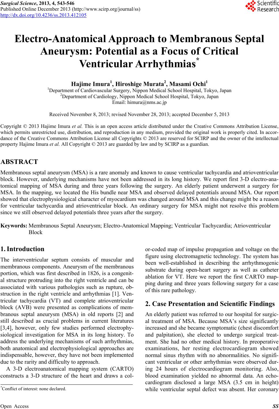 Surgical Science, 2013, 4, 543-546 Published Online December 2013 (http://www.scirp.org/journal/ss) http://dx.doi.org/10.4236/ss.2013.412105 Open Access SS Electro-Anatomical Approach to Membranous Septal Aneurysm: Potential as a Focus of Critical Ventricular Arrhythmias* Hajime Imura1, Hiroshige Murata2, Masami Ochi1 1Department of Cardiovascular Surgery, Nippon Medical School Hospital, Tokyo, Japan 2Department of Cardiology, Nippon Medical School Hospital, Tokyo, Japan Email: [email protected] Received November 8, 2013; revised November 28, 2013; accepted December 5, 2013 Copyright © 2013 Hajime Imura et al. This is an open access article distributed under the Creative Commons Attribution License, which permits unrestricted use, distribution, and reproduction in any medium, provided the original work is properly cited. In accor- dance of the Creative Commons Attribution License all Copyrights © 2013 are reserved for SCIRP and the owner of the intellectual property Hajime Imura et al. All Copyright © 2013 are guarded by law and by SCIRP as a guardian. ABSTRACT Membranous septal aneurysm (MSA) is a rare anomaly and known to cause ventricular tachycardia and atrioventricular block. However, underlying mechanisms have not been addressed in its long history. We report first 3-D electro-ana- tomical mapping of MSA during and three years following the surgery. An elderly patient underwent a surgery for MSA. In the map ping, we located the His b undle near MSA and observed delayed potentials arou nd MSA. Our report showed that electrophysiological character of myocardium was changed around MSA and this change might be a reason for ventricular tachycardia and atrioventricular block. An ordinary surgery for MSA might not resolve this problem since we still observed delayed potentials three years after the surgery. Keywords: Membranous Septal Aneurysm; Electro-Anatomical Mapping; Ventricular Tachycardia; Atrioventricular Block 1. Introduction The interventricular septum consists of muscular and membranous components. Aneurysm of the membranous portion, which was first described in 1826, is a cong enit- al structure protruding into the right ventricle and can be associated with various pathologies such as rupture, ob- struction in the right ventricle and arrhythmias [1]. Ven- tricular tachycardia (VT) and complete atrioventricular block (AVB) were presented as complications of mem- branous septal aneurysm (MSA) in old reports [2] and still described as crucial problems in current literatures [3,4], however, only few studies performed electrophy- siological investigation for MSA in its long history. To address the underlying mechanisms of such arrhythmias, both anatomical and electrophysiological approaches are indispensable, however, they have not been implemented due to the rarity and difficulty to approach. A 3-D electroanatomical mapping system (CARTO) constructs a 3-D structure of the heart and draws a col- or-coded map of impulse propagation and voltage on the figure using electromagnetic technology. The system has been well-established in describing the arrhythmogenic substrate during open-heart surgery as well as catheter ablation for VT. Here we report the first CARTO map- ping during and three years following surgery for a case of this rare pathology. 2. Case Presentation and Scientific Findings An elderly patient was referred to our hosp ital for surgic- al treatment of MSA. Because MSA’s size significantly increased and she became symptomatic (chest discomfort and palpitation), she elected to undergo surgical treat- ment. She had no other medical history. In preoperative examinations, her resting electrocardiogram showed normal sinus rhythm with no abnormalities. No signifi- cant ventricular or other arrhythmias were observed dur- ing 24 hours of electrocardiogram monitoring. Also, blood examination yielded no abnormal data. An echo- cardiogram disclosed a large MSA (3.5 cm in height) while ventricular septal defect was absent. Her coronary *Conflict of interest: none declared. 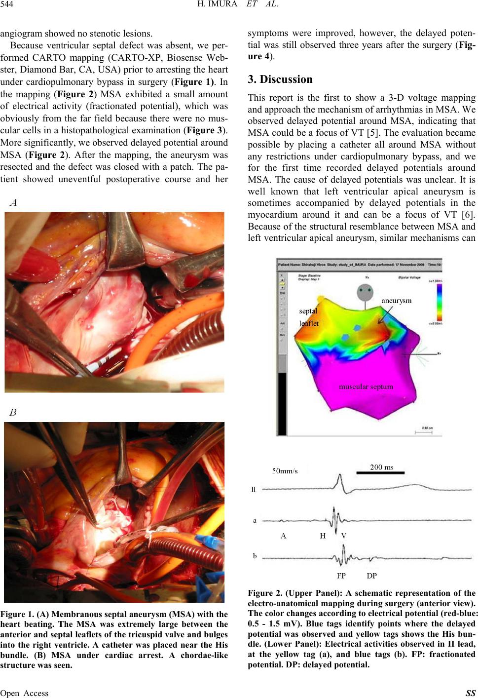 H. IMURA ET AL. 544 angiogram showed no stenotic lesions. Because ventricular septal defect was absent, we per- formed CARTO mapping (CARTO-XP, Biosense Web- ster, Diamond Bar, CA, USA) prior to arresting the heart under cardiopulmonary bypass in surgery (Figure 1). In the mapping (Figure 2) MSA exhibited a small amount of electrical activity (fractionated potential), which was obviously from the far field because there were no mus- cular cells in a histopathological examination (Figure 3). More significantly, we observed delayed potential around MSA (Figure 2). After the mapping, the aneurysm was resected and the defect was closed with a patch. The pa- tient showed uneventful postoperative course and her Figure 1. (A) Membranous septal aneurysm (MSA) with the heart beating. The MSA was extremely large between the anterior and septal leaflets of the tricuspid valve and bulges into the right ventricle. A catheter was placed near the His bundle. (B) MSA under cardiac arrest. A chordae-like structure was seen. symptoms were improved, however, the delayed poten- tial was still observed three years after the surgery (Fig- ure 4). 3. Discussion This report is the first to show a 3-D voltage mapping and approach the mechanism of arrhythmias in MSA. We observed delayed potential around MSA, indicating that MSA could be a focus of VT [5]. The evaluation became possible by placing a catheter all around MSA without any restrictions under cardiopulmonary bypass, and we for the first time recorded delayed potentials around MSA. The cause of delayed potentials was unclear. It is well known that left ventricular apical aneurysm is sometimes accompanied by delayed potentials in the myocardium around it and can be a focus of VT [6]. Because of the structural resemblance between MSA and left ventricular apical aneurysm, similar mechanisms can Figure 2. (Upper Panel): A schematic representation of the electro-anatomical mapping during surgery (anterior view). The color changes according to electrical potential (red-blue: 0.5 - 1.5 mV). Blue tags identify points where the delayed potential was observed and yellow tags shows the His bun- dle. (Lower Panel): Electrical activities observed in II lead, at the yellow tag (a), and blue tags (b). FP: fractionated potential. DP: delayed potential. Open Access SS 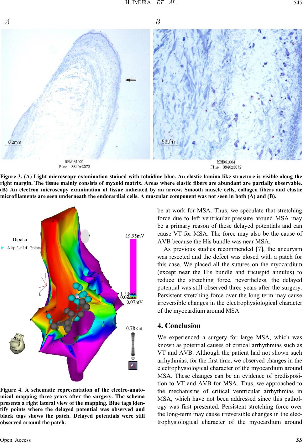 H. IMURA ET AL. Open Access SS 545 Figure 3. (A) Light microscopy examination stained with toluidine blue. An elastic lamina-like structure is visible along the right margin. The tissue mainly consists of myxoid matrix. Areas where elastic fibers are abundant are partially observable. (B) An electron microscopy examination of tissue indicated by an arrow. Smooth muscle cells, collagen fibers and elastic microfilaments are seen underneath the e ndocar dial cells. A muscular c omponent was not seen in both (A) and (B). Figure 4. A schematic representation of the electro-anato- mical mapping three years after the surgery. The schema presents a right lateral view of the mapping. Blue tags iden- tify points where the delayed potential was observed and black tags shows the patch. Delayed potentials were still be at work for MSA. Thus observed around the patch. , we speculate that stretching urysm w rgery for large MSA, which was force due to left ventricular pressure around MSA may be a primary reason of these delayed potentials and can cause VT for MSA. The force may also be the cause of AVB because the His bundle was near MSA. As previous studies recommended [7], the ane as resected and the defect was closed with a patch for this case. We placed all the sutures on the myocardium (except near the His bundle and tricuspid annulus) to reduce the stretching force, nevertheless, the delayed potential was still observed three years after the surgery. Persistent stretching force over the long term may cause irreversible changes in the electrophysiological character of the myocardium around MSA 4. Conclusion We experienced a su known as potential causes of critical arrhythmias such as VT and AVB. Although the patient had not shown such arrhythmias, for the first time, we observed changes in the electrophysiological character of the myocardium around MSA. These changes can be an evidence of predisposi- tion to VT and AVB for MSA. Thus, we approached to the mechanisms of critical ventricular arrhythmias in MSA, which have not been addressed since this pathol- ogy was first presented. Persistent stretching force over the long-term may cause irreversible changes in the elec- trophysiological character of the myocardium around 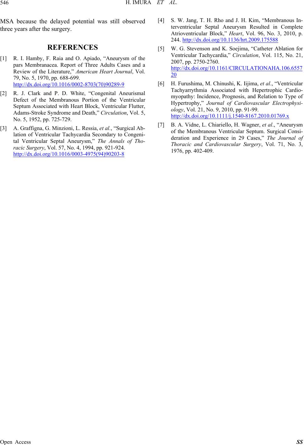 H. IMURA ET AL. 546 MSA because the delayed potential was still observed three years after the surgery. REFERENCES [1] R. I. Hamby, “Aneurysm of the F. Raia and O. Apiado, pars Membranacea. Report of Three Adults Cases and a Review of the Literature,” American Heart Journal, Vol. 79, No. 5, 1970, pp. 688-699. http://dx.doi.org/10.1016/0002-8703(70)90289-9 urisma . Ressia, et al., “Surgical Ab- [2] R. J. Clark and P. D. White, “Congenital Anel Defect of the Membranous Portion of the Ventricular Septum Associated with Heart Block, Ventricular Flutter, Adams-Stroke Syndrome and Death,” Circulation, Vol. 5, No. 5, 1952, pp. 725-729. [3] A. Graffigna, G. Minzioni, L lation of Ventricular Tachycardia Secondary to Congeni- tal Ventricular Septal Aneurysm,” The Annals of Tho- racic Surgery, Vol. 57, No. 4, 1994, pp. 921-924. http://dx.doi.org/10.1016/0003-4975(94)90203-8 [4] S. W. Jang, T. H. Rho and J. H. Kim, “Membranous In- terventricular Septal Aneurysm Resulted in Complete Atrioventricular Block,” Heart, Vol. 96, No. 3, 2010, p. 244. http://dx.doi.org/10.1136/hrt.2009.175588 [5] W. G. Stevenson and K. Soejima, “Catheter Ablation for Ventricular Tachycardia,” Circulation, Vol. 115, No. 21, 2007, pp. 2750-2760. http://dx.doi.org/10.1161/CIRCULATIONAHA.106.6557 20 [6] H. Furushima, M. Chinushi, K. Iijima, et al., “Ventricular Tachyarrythmia Associated with Hepertrophic Cardio- myopathy: Incidence, Prognosis, and Relation to Type of Hypertrophy,” Journal of Cardiovascular Electrophysi- ology, Vol. 21, No. 9, 2010, pp. 91-99. http://dx.doi.org/10.1111/j.1540-8167.2010.01769.x [7] B. A. Vidne, L. Chiariello, H. Wagner, et al., “Aneurysm of the Membranous Ventricular Septum. Surgical Consi- deration and Experience in 29 Cases,” The Journal of Thoracic and Cardiovascular Surgery, Vol. 71, No. 3, 1976, pp. 402-409. Open Access SS |