

Paper Menu >>
Journal Menu >>
 Advances in Infectious Diseases, 2013, 3, 248-252 Published Online December 2013 (http://www.scirp.org/journal/aid) http://dx.doi.org/10.4236/aid.2013.34037 Open Access AID Rhino-Orbito-Cerebral Mucormycosis in a Diabetic Patient with Idiopathic CD4 + Lymphocytopenia Nuno Rocha Pereira1, Paulo Andrade1, Laura Sá1, Cândida Abreu1, Rita Figueiredo2, Roberto Silva3, António Sarmento1 1Infectious Diseases Department, Centro Hospitalar São João and Faculty of Medicine of Porto University, Porto, Portugal; 2Neuro- radiology Department, Centro Hospitalar São João, Porto, Portugal; 3Pathology Department, Centro Hospitalar São João and Faculty of Medicine of Porto University, Porto, Portugal. Email: [email protected] Received September 28th, 2013; revised October 26th, 2013; accepted November 1st, 2013 Copyright © 2013 Nuno Rocha Pereira et al. This is an open access article distributed under the Creative Commons Attribution Li- cense, which permits unrestricted use, distribution, and reproduction in any medium, provided the original work is properly cited. ABSTRACT A 54-year-old diabetic male, with idiopathic CD4 + lymphocytopenia, was presented with a two-month history of head- ache and periocular pain followed by vertigo, left hypoacusia and left peripheral facial palsy and hypoesthesia. More than a month after admission, a palate ulcer appeared and Infectious Diseases consultation was required. Mucormycosis was suspected and the diagnosis was confirmed by histologic examination. Despite early surg ery and combination anti- fungal therapy, the patien t did not survive. This case report illustrates the difficulty in diagnosing a rare condition with non-specific clinical manifestations and underlines the importance of a timely multidisciplinary approach in order to recognise this highly fatal disease earlier. It also describes a previously non-reported situation of mucormycosis in a patient with idiopathic CD4 + lymphocytopenia. Keywords: Mucormycosis; Idiopathic CD4 + Lymphocytopenia; Diabetes; Amphotericin B 1. Introduction Mucormycosis is a life threatening fungal infection caus- ed by Mucorales fungi. There are six major clinical forms of the disease: cutaneous, pulmonary, gastrointestinal, disseminated, uncommon rare forms (endocarditis, oste- omyelitis, peritonitis and renal infection) and rhino-orbi- to-cerebral (ROCM) [1]. The latter is the most common form and is associated with diabetes in approximately two-thirds of cases [2]. Other risk factors for mucormy- cosis include haematological malignancies, hematopoi- etic stem cell and solid organ transplantation, imunoss- upresssive drugs, deferoxamine therapy and prolonged use of voriconazole [1]. We present a case of ROCM in a diabetic patient with idiopathic CD4 + lymphocytopenia (ICL), to our knowl- edge, a previously non-described association. 2. Case Presentation An Infectious Diseases consultation was required for a 54-year-old male who had developed an hard palate ul- ceration. The patient had been admitted 6 weeks before to the Ne urolog y w ard w ith a th ree-d ay h isto r y of lef t pe- ripheral facial palsy and left facial hypoesthesia. A brain computed tomography (CT) scan showed a right cerebel- lar lesion. His past medical history was remarkable for ICL, di- agnosed 5 years earlier after etiologic workup of crypto- coccal meningitis; arterial hypertension, obesity, dislipi- demia and a poorly controlled type 2 diabetes mellitus were also elicited. For the last five years he was under suppressive therapy with fluconazole and trimethoprim/ sulfamethoxazole plus azithromycin for opportunistic in- fection prophylaxis. Two months before admission he started complaining of headache and right periocular pain. One month later, he developed left hypoacusia and vertigo and three days before admission left peripheral facial palsy and hypoes- thesia. On admission at the emergency room (D1) he com- plained of bilateral frontal headache, bilateral periorbital pain, vertigo, nausea and vomiting. Neurological exami- nation revealed bilateral temporal optic margin blurring, left face hypoesthesia, left peripheral facial palsy, later- 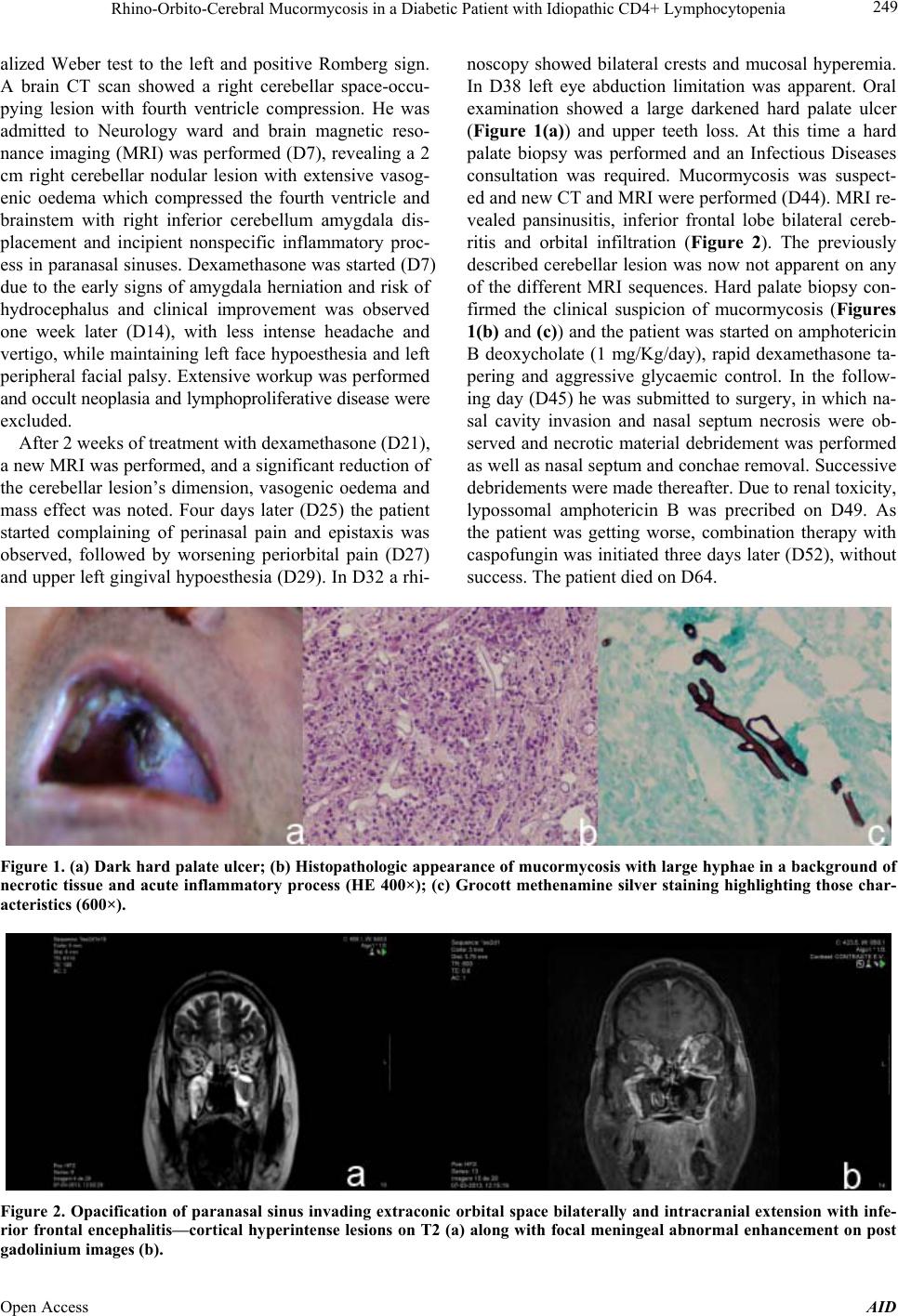 Rhino-Orbito-Cerebral Mucormycosis in a Diabetic Patient with Idiopathic CD4+ Lymphocytopenia 249 alized Weber test to the left and positive Romberg sign. A brain CT scan showed a right cerebellar space-occu- pying lesion with fourth ventricle compression. He was admitted to Neurology ward and brain magnetic reso- nance imaging (MRI) was performed (D7), revealing a 2 cm right cerebellar nodular lesion with extensive vasog- enic oedema which compressed the fourth ventricle and brainstem with right inferior cerebellum amygdala dis- placement and incipient nonspecific inflammatory proc- ess in paranasal sinuses. Dexamethasone was started (D7) due to the early signs of amygdala herniation and risk of hydrocephalus and clinical improvement was observed one week later (D14), with less intense headache and vertigo, while maintaining left face hypoesthesia and left peripheral facial palsy. Extensive workup was performed and occult neoplasia and lymphoproliferative disease were excluded. After 2 weeks of treatment with dexamethasone (D21), a new MRI was performed, and a significant reduction of the cerebellar lesion’s dimension, vasogenic oedema and mass effect was noted. Four days later (D25) the patient started complaining of perinasal pain and epistaxis was observed, followed by worsening periorbital pain (D27) and upper left gingival hypo esthesia (D29). In D32 a rhi- noscopy showed bilateral crests and mucosal hyperemia. In D38 left eye abduction limitation was apparent. Oral examination showed a large darkened hard palate ulcer (Figure 1(a)) and upper teeth loss. At this time a hard palate biopsy was performed and an Infectious Diseases consultation was required. Mucormycosis was suspect- ed and new CT and MRI were performed (D44). MRI re- vealed pansinusitis, inferior frontal lobe bilateral cereb- ritis and orbital infiltration (Figure 2). The previously described cerebellar lesion was now not apparent on any of the different MRI sequences. Hard palate biopsy con- firmed the clinical suspicion of mucormycosis (Figures 1(b) and (c)) and the patient was started on amphotericin B deoxycholate (1 mg/Kg/day), rapid dexamethasone ta- pering and aggressive glycaemic control. In the follow- ing day (D45) he was submitted to surgery, in which na- sal cavity invasion and nasal septum necrosis were ob- served and necrotic material debridement was performed as well as nasal septum and conchae removal. Successive debridements were made thereafter. Due to renal toxicity, lypossomal amphotericin B was precribed on D49. As the patient was getting worse, combination therapy with caspofungin was initiated three days later (D52), withou t success. The patient died on D64. Figure 1. (a) Dark hard palate ulcer; (b) Histopathologic appearance of mucormycosis with large hyphae in a background of necrotic tissue and acute inflammatory process (HE 400×); (c) Grocott methenamine silver staining highlighting those char- acteristics (600×). Figure 2. Opacification of paranasal sinus invading extraconic orbital space bilaterally and intracranial extension with infe- rior frontal encephalitis—cortical hyperintense lesions on T2 (a) along with focal meningeal abnormal enhancement on post adolinium images (b). g Open Access AID 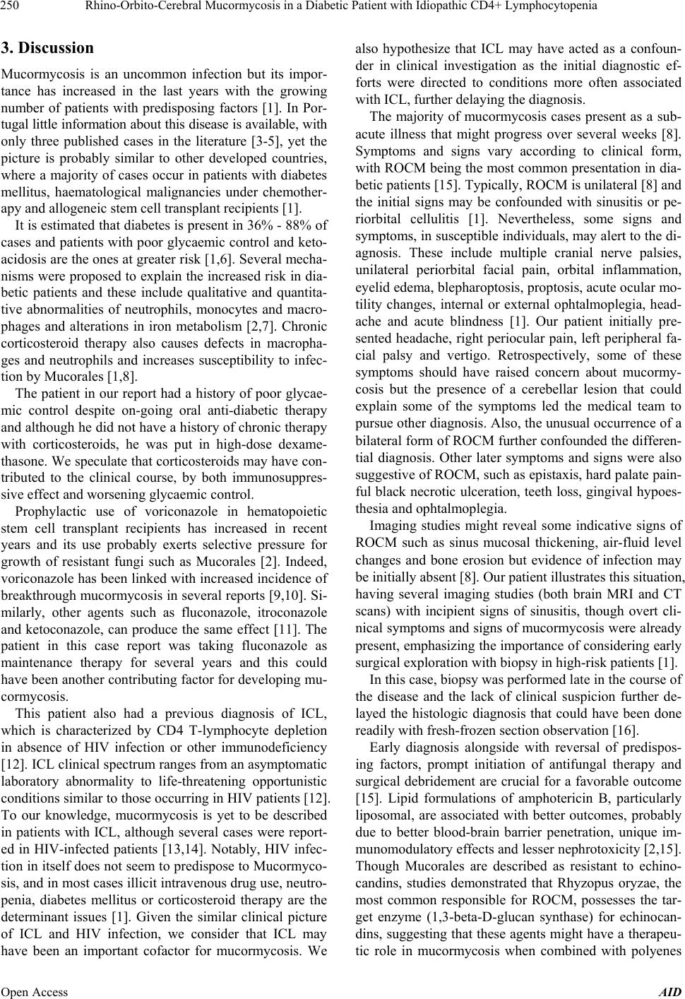 Rhino-Orbito-Cerebral Mucormycosis in a Diabetic Patient with Idiopathic CD4+ Lymphocytopenia 250 3. Discussion Mucormycosis is an uncommon infection but its impor- tance has increased in the last years with the growing number of patients with predisposing factors [1]. In Por- tugal little information about this disease is available, with only three published cases in the literature [3-5], yet the picture is probably similar to other developed countries, where a majority of cases occur in patients with diabetes mellitus, haematological malignancies under chemother- apy and allogeneic stem cell transplant recipients [1]. It is estimated that diabetes is p resent in 36% - 88% of cases and patients with poor glycaemic control and keto- acidosis are the ones at greater risk [1,6]. Several mecha- nisms were proposed to explain the increased risk in dia- betic patients and these include qualitative and quantita- tive abnormalities of neutrophils, monocytes and macro- phages and alterations in iron metabolism [2,7]. Chronic corticosteroid therapy also causes defects in macropha- ges and neutrophils and increases susceptibility to infec- tion by Mucorales [1,8]. The patient in our report had a history of poor glycae- mic control despite on-going oral anti-diabetic therapy and although he did not have a history of chronic therapy with corticosteroids, he was put in high-dose dexame- thasone. We speculate that corticostero ids may have con- tributed to the clinical course, by both immunosuppres- sive effect and worsening glycaemic control. Prophylactic use of voriconazole in hematopoietic stem cell transplant recipients has increased in recent years and its use probably exerts selective pressure for growth of resistant fungi such as Mucorales [2]. Indeed, voriconazole has been linked with increased incidence of breakthrough mucormycosis in several reports [9,10]. Si- milarly, other agents such as fluconazole, itroconazole and ketoconazole, can produce the same effect [11]. The patient in this case report was taking fluconazole as maintenance therapy for several years and this could have been another contributing factor for developing mu- cormycosis. This patient also had a previous diagnosis of ICL, which is characterized by CD4 T-lymphocyte depletion in absence of HIV infection or other immunodeficiency [12]. ICL clinical spectrum ranges from an asymptomatic laboratory abnormality to life-threatening opportunistic conditions similar to tho se occurring in HIV patien ts [12]. To our knowledge, mucormycosis is yet to be described in patients with ICL, although several cases were report- ed in HIV-infected patients [13,14]. Notably, HIV infec- tion in itself does not seem to predispo se to Mucormyco- sis, and in most cases illicit intravenou s drug use, neutro- penia, diabetes mellitus or corticosteroid therapy are the determinant issues [1]. Given the similar clinical picture of ICL and HIV infection, we consider that ICL may have been an important cofactor for mucormycosis. We also hypothesize that ICL may have acted as a confoun- der in clinical investigation as the initial diagnostic ef- forts were directed to conditions more often associated with ICL, further dela y i ng t he diagnosis. The majority of mucormycosis cases present as a sub- acute illness that might progress over several weeks [8]. Symptoms and signs vary according to clinical form, with ROCM being the most common presentation in dia- betic patients [15 ]. Typically, ROCM is un ilateral [8] and the initial signs may be confounded with sinusitis or pe- riorbital cellulitis [1]. Nevertheless, some signs and symptoms, in susceptible individuals, may alert to the di- agnosis. These include multiple cranial nerve palsies, unilateral periorbital facial pain, orbital inflammation, eyelid edema, blepharoptosis, proptosis, acute ocular mo- tility changes, internal or external ophtalmoplegia, head- ache and acute blindness [1]. Our patient initially pre- sented headache, right periocular pain, left peripheral fa- cial palsy and vertigo. Retrospectively, some of these symptoms should have raised concern about mucormy- cosis but the presence of a cerebellar lesion that could explain some of the symptoms led the medical team to pursue other diagnosis. Also, the unusual occurrence of a bilateral form of ROCM further confounded the differen- tial diagnosis. Other later symptoms and signs were also suggestive of ROCM, such as epistaxis, hard p alate pain- ful black necrotic ulceration, teeth loss, gingival hypoes- thesia and ophtalmoplegia. Imaging studies might reveal some indicative signs of ROCM such as sinus mucosal thickening, air-fluid level changes and bone erosion but evidence of infection may be initially absent [8]. Our patient illustrates th is situation, having several imaging studies (both brain MRI and CT scans) with incipient signs of sinusitis, though overt cli- nical symptoms and signs of mucormycosis wer e already present, emphasizing the importance of considering early surgical explor at i on wit h bi o p s y in hig h- ri sk pat i ent s [1] . In this case, biopsy was performed late in the course of the disease and the lack of clinical suspicion further de- layed the histologic diagnosis that could have been done readily with fresh-frozen section observation [16]. Early diagnosis alongside with reversal of predispos- ing factors, prompt initiation of antifungal therapy and surgical debridement are crucial for a favorable outcome [15]. Lipid formulations of amphotericin B, particularly liposomal, are associated with better outcomes, probably due to better blood-brain barrier penetration, unique im- munomodulatory effects and lesser nephrotoxicity [2,15]. Though Mucorales are described as resistant to echino- candins, studies demonstrated that Rhyzopus oryzae, the most common responsible for ROCM, possesses the tar- get enzyme (1,3-beta-D-glucan synthase) for echinocan- dins, suggesting that these agents might have a therapeu- tic role in mucormycosis when combined with polyenes Open Access AID  Rhino-Orbito-Cerebral Mucormycosis in a Diabetic Patient with Idiopathic CD4+ Lymphocytopenia 251 [2,17]. This theoretical benefit was confirmed in a retro- spective study comparing amphotericin B plus caspofun- gin with amphotericin B alone and the benefit was par- ticularly evident in patients with cerebral involvement [18]. In spite of the critical role of early anti-fungal ther- apy, surgery still constitutes the cornerstone for success- ful treatment of ROCM, since this infection is associated with angioinvasion and extensive necrosis that can im- pair adequate antifungal therapy to affected tissues [15]. The extent of surgery is dictated by the patient’s indivi- dual presentation but for maxillofacial infection increas- ing emphasis is put in less disfiguring surgical proce- dures [2]. Other therapeutic options for mucormycosis include posaconazole rescue therapy [1], hyperbaric oxy- gen [19], iron chelation with deferasirox [1] and the use of several kinds of immunotherapy [20]. Our patient was not diagnosed early in the clinical course, yet antifung al ther apy was quickly initiated, rapid tapering of corticosteroid and aggressive glycaemic con- trol were performed and surgery was done one day after diagnosis. Due to finantial restrains amphotericin B de- oxycolate was the initial therapeutic choice however, once nephrotoxicity was present, liposomal amphotericin B was started and caspofungin was later added, due to poor therapeutic resp onse. Due to d isease ex tension, with brain and bilateral orbital involvement, a choice was made not to perform aggressive desfigurative surgery and a mi- nimally invasive approach was done. 4. Conclusion This case illustrates the difficulty in diagnosing a rare con- dition with non-specific clinical manifestations. It also underlines the importance of awareness for risk factors and clinical features of mucormycosis. High index of sus- picion, multidisciplinary approach, newer diagnostic and treatment tools altogether should p ermit, in the future, an improvement in the outcome of this devastating disease. 5. Acknowledgements The authors greet all the professionals involved in man- agement of the patient and thank Dr. Henrique Costa for his article review. REFERENCES [1] G. Petrikkos, A. Skiada, O. Lortholary, E. Roilides, T. J. Walsh and D. P. Kontoyiannis, “Epidemiology and Clini- cal Manifestations of Mucormycosis,” Clinical Infectious Diseases, Vol. 54, No. S1, 2012, pp. S23-S34. http://dx.doi.org/10.1093/cid/cir866 [2] M. N. Gamaletsou, N. V. Sipsas, E. Roilides and T. J. Walsh, “Rhino-Orbital-Cerebral Mucormycosis,” Current Infectious Disease Reports, Vol. 14, No. 4, 2012, pp. 423- 434. http://dx.doi.org/10.1007/s11908-012-0272-6 [3] J. G. Cortez, A. Speidel, C. Peixoto, E. Selicka, C. Va- lente, P. Figueiredo and A. Vieira, “Mind the Gap: Man- agement of an Emergent and Threatening Invasive Fungal Infection—A Case Report of Rhino-Orbital-Cerebral and Pulmonary Mucormycosis,” Medical Mycology Case Re- ports, Vol. 2, 2013, pp. 79-84. http://dx.doi.org/10.1016/j.mmcr.2013.02.008 [4] V. Oliveira and A. Costa, “Cerebral Hematoma Caused by Mucormycosis,” Revista de Neurología, Vol. 33, No. 10, 2001, pp. 951-953. [5] D. S. Marques, C. Pinho Vaz, R. Branca, et al., “Rhizo- mucor and Scedosporium Infection Post Hematopoietic Stem-Cell Transplant,” Case Reports in Medicine, Vol. 2011, 2011, Article ID: 830769. [6] M. M. Roden, T. E. Zaoutis, W. L. Buchanan, et al., “Epi- demiology and Outcome of Zygomycosis: A Review of 929 Reported Cases,” Clinical Infectious Diseases, Vol. 41, No. 5, 2005, pp. 634-653. http://dx.doi.org/10.1086/432579 [7] B. Rammaert, F. Lanternier, S. Poiree, R. Kania and O. Lortholary, “Diabetes and Mucormycosis: A Complex In- terplay,” Diabetes & Metabolism, Vol. 38, No. 3, 2012, pp. 193-204. http://dx.doi.org/10.1016/j.diabet.2012.01.002 [8] U. Wali, A. Balkhair and A. Al-Mujaini, “Cerebro-Rhino Orbital Mucormycosis: An Update,” The Journal of In- fection and Public Health, Vol. 5, No. 2, 2012, pp. 116- 126. http://dx.doi.org/10.1016/j.jiph.2012.01.003 [9] S. M. Trifilio, C. L. Bennett, P. R . Yarnold, et al., “Break- through Zygomycosis after Voriconazole Administration among Patients with Hematologic Malignancies Who Re- ceive Hematopoietic Stem-Cell Transplants or Intensive Chemotherapy,” Bone Marrow Transplant, Vol. 39, No. 7, 2007, pp. 425-429. http://dx.doi.org/10.1038/sj.bmt.1705614 [10] D. P. Kontoyiannis, M. S. Lionakis, R. E. Lewis, et al., “Zygomycosis in a Tertiary-Care Cancer Center in the Era of Aspergillus-Active Antifungal Therapy: A Case- Control Observational Study of 27 Recent Cases,” The Journal of Infectious Diseases, Vol. 191, No. 8, 2005, pp. 1350-1360. http://dx.doi.org/10.1086/428780 [11] L. Pagano, B. Gleissner, L. Fianchi, “Breakthrough Zy- gomycosis and Voriconazole,” The Journal of Infectious Diseases, Vol. 192, No. 8, 2005, pp. 1496-1497. http://dx.doi.org/10.1086/444433 [12] L. Luo and T. Li, “Idiopathic CD4 Lymphocytopenia and Opportunistic Infection—An Update,” FEMS Immunol- ogy and Medical Microbiology, Vol. 54, No. 3, 2008, pp. 283-289. http://dx.doi.org/10.1111/j.1574-695X.2008.00490.x [13] S. Antinori, M. Nebuloni, C. Magni, et al., “Trends in the Postmortem Diagnosis of Opportunistic Invasive Fungal Infections in Patients with AIDS: A Retrospective Study of 1630 Autopsies Performed between 1984 and 2002,” American Journal of Clinical Pathology, Vol. 132, No. 2, 2009, pp. 221-227. http://dx.doi.org/10.1309/AJCPRAAE8LZ7DTNE [14] L. Nichols, R. Z. Ocque and I. Daly, “Zygomycosis Asso- Open Access AID  Rhino-Orbito-Cerebral Mucormycosis in a Diabetic Patient with Idiopathic CD4+ Lymphocytopenia Open Access AID 252 ciated with HIV Infection and Liver Transplantation,” Pathology Research International, Vol. 2011, 2011, Arti- cle ID: 545981. [15] H. Y. Sun and N. Singh, “Mucormycosis: Its Contempo- rary Face and Management Strategies,” The Lancet Infec- tious Diseases, Vol. 11, No. 4, 2011, pp. 301-311. http://dx.doi.org/10.1016/S1473-3099(10)70316-9 [16] T. J. Walsh, M. N. Gamaletsou, M. R. McGinnis, R. T. Hayden and D. P. Kontoyiannis, “Early Clinical and La- boratory Diagnosis of Invasive Pulmonary, Extrapulmon- ary, and Disseminated Mucormycosis (Zygomycosis),” Clinical Infectious Diseases, Vol. 54, No. S1, 2012, pp. S55-S60. http://dx.doi.org/10.1093/cid/cir868 [17] B. Spellberg, A. Ibrahim, E. Roilides, et al., “Combina- tion Therapy for Mucormycosis: Why, What, and How?” Clinical Infectious Diseases, Vol. 54, No. S1, 2012, pp. S73-S78. http://dx.doi.org/10.1093/cid/cir885 [18] C. Reed, R. Bryant, A. S. Ibrahim, et al., “Combination Polyene-Caspofungin Treatment of Rhino-Orbital-Cere- bral Mucormycosis,” Clinical Infectious Diseases, Vol. 47, No. 3, 2008, pp. 364-371. http://dx.doi.org/10.1086/589857 [19] A. Tragiannidis and A. H. Groll, “Hyperbaric Oxygen The- rapy and Other Adjunctive Treatments for Zygomycosis,” Clinical Microbiology and Infection, Vol. 15, No. S5, 2009, pp. 82-86. http://dx.doi.org/10.1111/j.1469-0691.2009.02986.x [20] A. Safdar, “Immunotherapy for Invasive Mold Disease in Severely Immunosuppressed Patients,” Clinical Infectious Diseases, Vol. 57, No. 1, 2013, pp. 94-100. http://dx.doi.org/10.1093/cid/cit187 |