

Paper Menu >>
Journal Menu >>
 Advances in Infectious Diseases, 2013, 3, 231-237 Published Online December 2013 (http://www.scirp.org/journal/aid) http://dx.doi.org/10.4236/aid.2013.34034 Open Access AID 231 Development of New Strategy for Non-Antibiotic Therapy: Dromedary Camel Lactoferrin Has a Potent Antimicrobial and Immunomodulator Effects* Alaa B. Ismael1,2#, Salama M. Abd El Hafez3,4, Manal B. Mahmoud4, Abdel-Kader A. Elaraby5, Hany M. Hassan4 1Department of Medical Biotechnology, Faculty of Applied Medical Sciences, Taif University, Turaba, KSA; 2Department of Animal Medicine, Faculty of Veterinary Medicine, Zagazig University, Zagazig, Egypt; 3Department of Medical Microbiology, Faculty of Applied Medical Sciences, Taif University, Turaba, KSA; 4Immunobiology and Immunopharmacology Unit, Animal Reproduction Research Institute (ARRI), Giza, Egypt; 5Holding Company for Biological Products & Vaccines VACSERA, Giza, Egypt. Email: #[email protected] Received August 27th, 2013; revised September 26th, 2013; accepted October 3rd, 2013 Copyright © 2013 Alaa B. Ismael et al. This is an open access article distributed under the Creative Commons Attribution License, which permits unrestricted use, distribution, and reproduction in any medium, provided the original work is properly cited. ABSTRACT The human and bovine lactoferrin have been studied extensively, but very few reports have been published concerning camel lactoferrin (cLf). The present study aimed to isolate cLf and evaluate its efficiency including antimicrobial activ- ity and immunomodulator effects. cLf isolation was attempted from camel milk whey using a cation exchange chroma- tography by SP-Sepharose. The antimicrobial activity of the isolated cLf was investigated against Staphylococcus au- reus (S. aureus), Streptococcus agalactiae (S. agalactiae), Escherichia coli (E. coli) and Pseudomonas aeruginosa (P. aerogenosa) strains. The immune effect of cLf was studied by lymphocyte transformation test. It was found that cLf was separated around molecular weight of 80 kDa and showed significant inhibitory effect against E. coli followed by P. aeruginosa, S. agalactiae and S. aureus. cLf increased lymphocyte transformations mean values in a dose dependant manner. The highest transformations mean value was determined at 50 µg/mL. In conclusion, these results suggest that cLf is a potent natural antimicrobial and novel immunomodulator agent. Keywords: Dromedary Camel Lactoferrin; Isolation; Antimicrobial and Immunomodulator Effects 1. Introduction Few studies have been reported on camels and camel milk [1]. Dromedary camel milk and their products are a good nutritional source for the people living in the arid and urban areas. In addition, fresh and fermented camel milk were reported to provide particular health benefits to the consumer depending on the bioactive substances in milk [2]. Antibiotics are commonly used for both pro- phylaxis and treatment of various bacterial infections in human and farm animals. In recent years, antibiotics re- sistance in bacteria of animal origin and its impact on human health have drawn much attention worldwide [3]. Bovine mastitis is the most common cause for the use of antibiotics agents in lactating dairy cattle [4] and the de- tection of antibiotics residues in milk poses health haz- ards to consumers, and the cause of high economic impor- tance because such milk is unfit for processing and subse- quent consumption [5]. Moreover, the antibiotic therapy has many complications as hypersensitivity, direct toxic- ity, antibiotic-induced immunosuppresion and super-infec- tions. This is highlighting the need for a new strategy for non-antibiotic therapy using novel immunomodulators as naturally released immunomodulators (Lactoferrin (Lf), cathelicidins and defenses) or bacterial products (Perip- lasmic proteins and lipopolysaccharides). Lactoferrin (Lf), is an iron-binding glycoprotein found in a variety of body secretions including tears, bronchial mucus, and saliva and it is found in high concentrations in the mammary secretions of nonlactating dairy animals. It is important in regulation of iron metabolism [6]. This natural antimicrobial agent is a multifunctional bioactive molecule with a critical role in many important physio- *No conflict of interest to declare. #Corresponding author.  Development of New Strategy for Non-Antibiotic Therapy: Dromedary Camel Lactoferrin Has a Potent Antimicrobial and Immunomodulator Effects 232 logical pathways. Lf could elicit a variety of inhibitory effects against microorganisms, comprising stasis, cidal, adhesion-blockade, cationic, synergistic, and opsonic me- chanisms. Broad-spectrum activities against different bac- teria, viruses, fungi, and parasites, in combination with anti-inflammatory and immunomodulatory properties, make Lf a potent innate host defense mechanism [7]. The large potential applications of Lf have led scientists to develop this nutraceutical protein for use in feed, food and pharmaceutical applications. Camel lactoferrin (cLf) purification, biochemical, and immunological characterization have shown its similarity to human and bovine Lf, as well as the cross-react with the anti-human Lf antibodies [8-10]. The amounts of lactoferrin and immunoglobulins were found to be great- er in dromedary camel milk than bovine or buffalo milk [8,10,11]. Incubation of human leukocytes with cLf leads to a complete virus entry inhibition after seven days’ incubation. Thus, cLf markedly inhibits hepatitis C virus genotype 4 infection of human peripheral blood leuko- cytes [12]. The miR-214 is directly involved in Lf ex- pression and Lf mediated cancer susceptibility (proapop- totic activities) in mammary epithelial cells [13]. Many processing technologies have been developed to isolate the high purity fraction of Lf. And most of the te- chnologies use a cation exchange chromatography on SP-Sepha-rose [14,15]. The aim of this investigation was mainly to isolate cLf from camel milk whey and evaluate its efficacy in vitro including antimicrobial and immunomodulator effects. We use cLf but not bovine Lf because cLf is more bioac- tive [16]. 2. Materials and Methods 2.1. Isolation of Lactoferrin from Camel Milk Whey Lactoferrin (Lf) isolation was attempted from camel milk whey. It was purified using a cation exchange chroma- tography on SP-Sepharose following the procedure that previously described [14]. Briefly, milk whey was ob- tained from camel milk using ultra speed centrifuge, 15000 × g at 4˚C for 30 min. Skimmed milk was then di- luted 1:1 with the dilution buffer (0.04 M NaH2PO4, 0.8 M NaCl, 0.04% (v/v) Tween 20, pH 7.4) and it was in- cubated with SP-Sepharose at 4˚C overnight. Afterwards, the SP-Sepharose was washed with the washing buffer (0.02 M NaH2PO4, 0.4 M NaCl, 0.02% (v/v) Tween 20, pH 7.4) to elude the unbound proteins. The gel then packed into a column (5 × 30 cm or 3 × 30 cm, depend- ing on the milk volume) and lactoferrin was eluted with the elution buffer (0.02 M NaH2PO4, 1 M NaCl, pH 7.4). The column was run at a flow rate of 3 mL/min. 2.2. Electrophoresis of Milk and Fractions Containing Lactoferrin Purity control and characterization of camel Lf (cLf) was done using sodium dodecyl sulfate-polyacrylamide gel electrophoresis (SDS-PAGE). Collected fractions of ca- mel milk whey and broad range protein ladder (Fermen- tra SM1841) were resolved in 12% polyacrylamide mi- nigel-protein II electrophoresis cell (Bio-Rad). Samples were diluted in sample buffer 2-mercaptoethanol (Sigma Chemical Co.), boiled for 5 minutes before being loaded in the gels and run at 70 volts for 3 hours. Gels were stained with 1% Coomassie blue R-250 (Sigma Chemical Co.), then distained at room temperature in 5% methanol and 7.5% acetic acid with shaking for 30 minutes. The different fractions were quantified using Bio-Rad GS 700 imaging densitometer molecular analysis software against broad range marker [17]. 2.3. Antimicrobial Activity Assays Escherichia coli (E. coli), Pseudomonas aeruginosa (P. aerogenosa), Staphylococcus aureus (S. aureus) and Streptococcus agalactiae (S. agalactiae) isolates were used to study the antimicrobial activity of cLf. The tested microorganisms were kept in their specific soft agar. Working cultures were obtained by growing the tested isolates on their specific media. After an overnight incu- bation, an isolated colony was transferred to 10 mL of Mueller-Hinton broth (MHB, Difco Laboratories, Detroit, MI) and incubated at 37˚C for 16 - 20 h. Final concentra- tion of 1 × 10⁶ CFU/mL was used. A volume of 1 mL of cLf solution in different concentrations (1 and 3 mg/mL) was added to 4 wells of tissue culture plates (NUNC. A/S, Roskilde, Denmark) for each of tested microorganisms as previously described [18]. The tested microorganisms in phosphate buffer saline (PBS, 10 mM, pH 7.4) was used a control. Plates were incubated at 37˚C. Aliquots were removed after 1, 3, 6, 12, 24 hours and ten serially di- luted, then plated at 37˚C on Mueller Hinton agar (MHA, Difco Laboratories, Detroit, MI) to be counted after 48 h incubation. Total aerobic bacterial count (TBC) of tested microorganisms was done in which viable aerobic me- sophlic bacteria were determined as previously described [19]. All equipments used were either sterile new glass or plastic to avoid iron contamination. All experiments were repeated at least two times. 2.4. In Vitro Lymphocyte Proliferation Studies Lymphocyte proliferation test using MTT (3-(4, 5-di- methyl thiazol-2-yl) 2, 5-diphenyl tetrazolium bromide) was performed [20] with modification. Briefly, heaprini- zed calf blood samples were aseptically collected Open Access AID 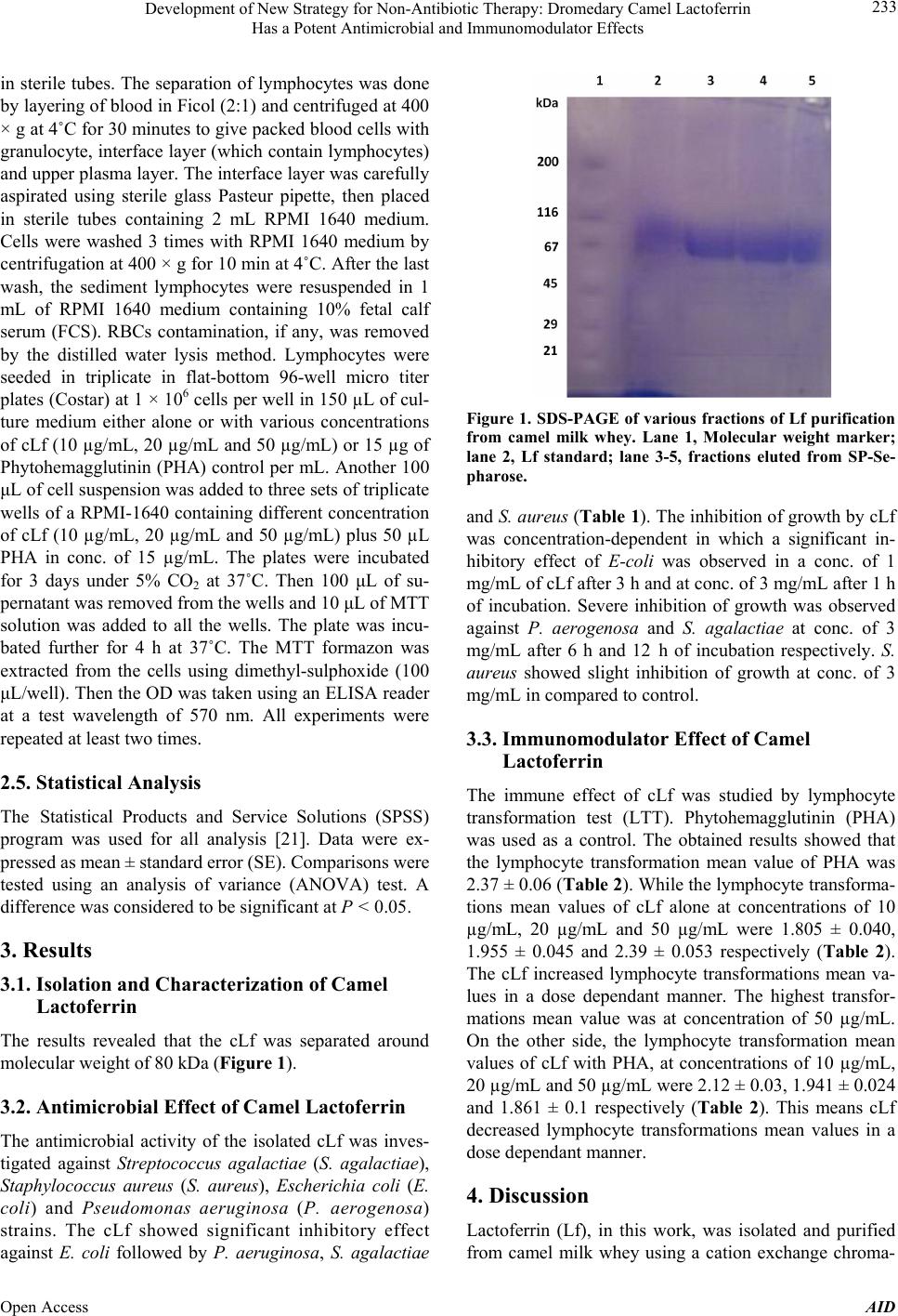 Development of New Strategy for Non-Antibiotic Therapy: Dromedary Camel Lactoferrin Has a Potent Antimicrobial and Immunomodulator Effects 233 in sterile tubes. The separation of lymphocytes was done by layering of blood in Ficol (2:1) and centrifuged at 400 × g at 4˚C for 30 minutes to give packed blood cells with granulocyte, interface layer (which contain lymphocytes) and upper plasma layer. The interface layer was carefully aspirated using sterile glass Pasteur pipette, then placed in sterile tubes containing 2 mL RPMI 1640 medium. Cells were washed 3 times with RPMI 1640 medium by centrifugation at 400 × g for 10 min at 4˚C. After the last wash, the sediment lymphocytes were resuspended in 1 mL of RPMI 1640 medium containing 10% fetal calf serum (FCS). RBCs contamination, if any, was removed by the distilled water lysis method. Lymphocytes were seeded in triplicate in flat-bottom 96-well micro titer plates (Costar) at 1 × 10⁶ cells per well in 150 µL of cul- ture medium either alone or with various concentrations of cLf (10 µg/mL, 20 µg/mL and 50 µg/mL) or 15 µg of Phytohemagglutinin (PHA) control per mL. Another 100 μL of cell suspension was added to three sets of triplicate wells of a RPMI-1640 containing different concentration of cLf (10 µg/mL, 20 µg/mL and 50 µg/mL) plus 50 µL PHA in conc. of 15 µg/mL. The plates were incubated for 3 days under 5% CO2 at 37˚C. Then 100 μL of su- pernatant was removed from the wells and 10 μL of MTT solution was added to all the wells. The plate was incu- bated further for 4 h at 37˚C. The MTT formazon was extracted from the cells using dimethyl-sulphoxide (100 μL/well). Then the OD was taken using an ELISA reader at a test wavelength of 570 nm. All experiments were repeated at least two times. 2.5. Statistical Analysis The Statistical Products and Service Solutions (SPSS) program was used for all analysis [21]. Data were ex- pressed as mean ± standard error (SE). Comparisons were tested using an analysis of variance (ANOVA) test. A difference was considered to be significant at P < 0.05. 3. Results 3.1. Isolation and Characterization of Camel Lactoferrin The results revealed that the cLf was separated around molecular weight of 80 kDa (Figure 1). 3.2. Antimicrobial Effect of Camel Lactoferrin The antimicrobial activity of the isolated cLf was inves- tigated against Streptococcus agalactiae (S. agalactiae), Staphylococcus aureus (S. aureus), Escherichia coli (E. coli) and Pseudomonas aeruginosa (P. aerogenosa) strains. The cLf showed significant inhibitory effect against E. coli followed by P. aeruginosa, S. agalactiae Figure 1. SDS-PAGE of various fractions of Lf purification from camel milk whey. Lane 1, Molecular weight marker; lane 2, Lf standard; lane 3-5, fractions eluted from SP-Se- pharose. and S. aureus (Table 1). The inhibition of growth by cLf was concentration-dependent in which a significant in- hibitory effect of E-coli was observed in a conc. of 1 mg/mL of cLf after 3 h and at conc. of 3 mg/mL after 1 h of incubation. Severe inhibition of growth was observed against P. aerogenosa and S. agalactiae at conc. of 3 mg/mL after 6 h and 12 h of incubation respectively. S. aureus showed slight inhibition of growth at conc. of 3 mg/mL in compared to control. 3.3. Immunomodulator Effect of Camel Lactoferrin The immune effect of cLf was studied by lymphocyte transformation test (LTT). Phytohemagglutinin (PHA) was used as a control. The obtained results showed that the lymphocyte transformation mean value of PHA was 2.37 ± 0.06 (Table 2 ). While the lymphocyte transforma- tions mean values of cLf alone at concentrations of 10 µg/mL, 20 µg/mL and 50 µg/mL were 1.805 ± 0.040, 1.955 ± 0.045 and 2.39 ± 0.053 respectively (Table 2). The cLf increased lymphocyte transformations mean va- lues in a dose dependant manner. The highest transfor- mations mean value was at concentration of 50 µg/mL. On the other side, the lymphocyte transformation mean values of cLf with PHA, at concentrations of 10 µg/mL, 20 µg/mL and 50 µg/mL were 2.12 ± 0.03, 1.941 ± 0.024 and 1.861 ± 0.1 respectively (Table 2). This means cLf decreased lymphocyte transformations mean values in a dose dependant manner. 4. Discussion Lactoferrin (Lf), in this work, was isolated and purified from camel milk whey using a cation exchange chroma- Open Access AID 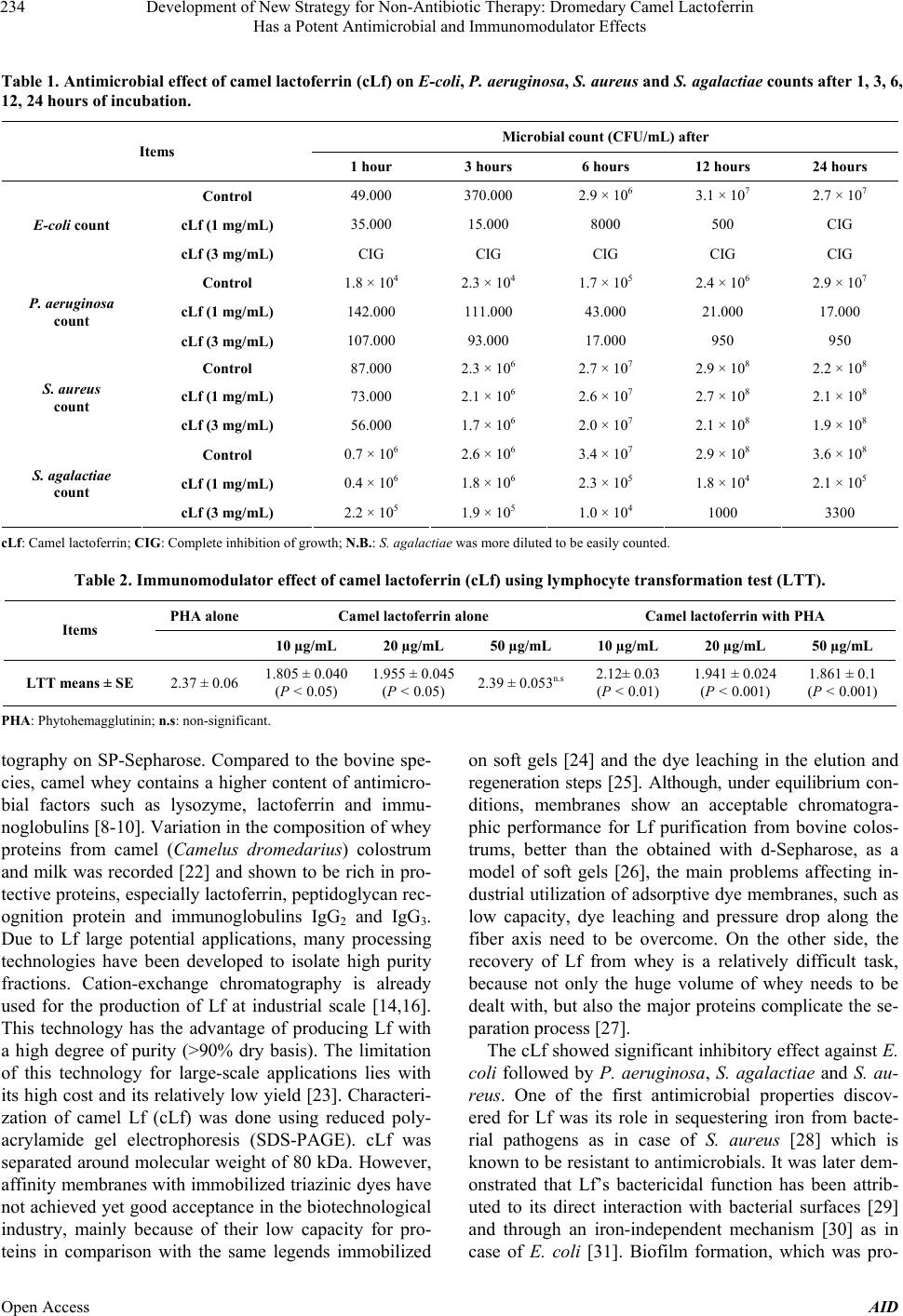 Development of New Strategy for Non-Antibiotic Therapy: Dromedary Camel Lactoferrin Has a Potent Antimicrobial and Immunomodulator Effects Open Access AID 234 Table 1. Antimicrobial effect of camel lactoferrin (cLf) on E-coli, P. aeruginosa, S. aureus and S. agalactiae counts after 1, 3, 6, 12, 24 hours of incubation. Microbial count (CFU/mL) after Items 1 hour 3 hours 6 hours 12 hours 24 hours Control 49.000 370.000 2.9 × 10⁶ 3.1 × 10⁷ 2.7 × 10⁷ cLf (1 mg/mL) 35.000 15.000 8000 500 CIG E-coli count cLf (3 mg/mL) CIG CIG CIG CIG CIG Control 1.8 × 10⁴ 2.3 × 10⁴ 1.7 × 10⁵ 2.4 × 10⁶ 2.9 × 10⁷ cLf (1 mg/mL) 142.000 111.000 43.000 21.000 17.000 P. aeruginosa count cLf (3 mg/mL) 107.000 93.000 17.000 950 950 Control 87.000 2.3 × 10⁶ 2.7 × 10⁷ 2.9 × 10⁸ 2.2 × 10⁸ cLf (1 mg/mL) 73.000 2.1 × 10⁶ 2.6 × 10⁷ 2.7 × 10⁸ 2.1 × 10⁸ S. aureus count cLf (3 mg/mL) 56.000 1.7 × 10⁶ 2.0 × 10⁷ 2.1 × 10⁸ 1.9 × 10⁸ Control 0.7 × 10⁶ 2.6 × 10⁶ 3.4 × 10⁷ 2.9 × 10⁸ 3.6 × 10⁸ cLf (1 mg/mL) 0.4 × 10⁶ 1.8 × 10⁶ 2.3 × 10⁵ 1.8 × 10⁴ 2.1 × 10⁵ S. agalactiae count cLf (3 mg/mL) 2.2 × 10⁵ 1.9 × 10⁵ 1.0 × 10⁴ 1000 3300 cLf: Camel lactoferrin; CIG: Complete inhibition of growth; N.B.: S. agalactiae was more diluted to be easily counted. Table 2. Immunomodulator effect of camel lactoferrin (cLf) using lymphocyte transformation test (LTT). PHA alone Camel lactoferrin alone Camel lactoferrin with PHA Items 10 µg/mL 20 µg/mL 50 µg/mL 10 µg/mL 20 µg/mL 50 µg/mL LTT means ± SE 2.37 ± 0.061.805 ± 0.040 (P < 0.05) 1.955 ± 0.045 (P < 0.05) 2.39 ± 0.053n.s 2.12± 0.03 (P < 0.01) 1.941 ± 0.024 (P < 0.001) 1.861 ± 0.1 (P < 0.001) PHA: Phytohemagglutinin; n.s: non-significant. tography on SP-Sepharose. Compared to the bovine spe- cies, camel whey contains a higher content of antimicro- bial factors such as lysozyme, lactoferrin and immu- noglobulins [8-10]. Variation in the composition of whey proteins from camel (Camelus dromedarius) colostrum and milk was recorded [22] and shown to be rich in pro- tective proteins, especially lactoferrin, peptidoglycan rec- ognition protein and immunoglobulins IgG2 and IgG3. Due to Lf large potential applications, many processing technologies have been developed to isolate high purity fractions. Cation-exchange chromatography is already used for the production of Lf at industrial scale [14,16]. This technology has the advantage of producing Lf with a high degree of purity (>90% dry basis). The limitation of this technology for large-scale applications lies with its high cost and its relatively low yield [23]. Characteri- zation of camel Lf (cLf) was done using reduced poly- acrylamide gel electrophoresis (SDS-PAGE). cLf was separated around molecular weight of 80 kDa. However, affinity membranes with immobilized triazinic dyes have not achieved yet good acceptance in the biotechnological industry, mainly because of their low capacity for pro- teins in comparison with the same legends immobilized on soft gels [24] and the dye leaching in the elution and regeneration steps [25]. Although, under equilibrium con- ditions, membranes show an acceptable chromatogra- phic performance for Lf purification from bovine colos- trums, better than the obtained with d-Sepharose, as a model of soft gels [26], the main problems affecting in- dustrial utilization of adsorptive dye membranes, such as low capacity, dye leaching and pressure drop along the fiber axis need to be overcome. On the other side, the recovery of Lf from whey is a relatively difficult task, because not only the huge volume of whey needs to be dealt with, but also the major proteins complicate the se- paration process [27]. The cLf showed significant inhibitory effect against E. coli followed by P. aeruginosa, S. agalactiae and S. au- reus. One of the first antimicrobial properties discov- ered for Lf was its role in sequestering iron from bacte- rial pathogens as in case of S. aureus [28] which is known to be resistant to antimicrobials. It was later dem- onstrated that Lf’s bactericidal function has been attrib- uted to its direct interaction with bacterial surfaces [29] and through an iron-independent mechanism [30] as in case of E. coli [31]. Biofilm formation, which was pro-  Development of New Strategy for Non-Antibiotic Therapy: Dromedary Camel Lactoferrin Has a Potent Antimicrobial and Immunomodulator Effects 235 posed as a colonial organization adhesion method for P. aeruginosa, is a well-studied phenomenon. Through bio- film formation, bacteria become highly resistant to host cell defense mechanisms and antibiotic treatment [32]. It is well known that some bacterial strains require high le- vels of iron to form biofilms. Thus, Lf’s function as an iron chelator has been hypothesized to effectively inhibit biofilm formation through iron sequestration [33]. Occur- rence in various milieus strongly emphasizes the signifi- cance of the structure-function relationship in the multi- functionality of the Lf [7]. Regarding the immune effect of cLf, the cLf increased lymphocyte transformations mean values in a dose de- pendant manner. The highest transformations mean value was of lactoferrin in conc. of 50 µg/mL. This finding was agreed with [34] who reported that the addition of re- combinant human lactoferrin (Talactoferrin Alfa (TLf)) to human peripheral blood or monocyte-derived dendritic cell cultures resulted in cell maturation, as evidenced by up-regulated expression of CD80, CD83, and CD86, pro- duction of proinflammatory cytokines, and increased ca- pacity to stimulate the proliferation of allogeneic lym- phocytes. In addition, this finding was agreed to some extend with [35] who found that the effects of Lf in ex- perimental models were differential and dependent on an individual PBMC reactivity, mitogen or alloantigen and Lf concentration. Generally, lymphocytes from donors responsive to Lf exhibited higher proliferation indices to PHA when compared with non-responsive individuals, suggest that the differential action of Lf might be due to its ability to sense the activation status of lymphocytes, although he mentioned that data on Lf effects on mito- gen-induced proliferation are scarce, though fairly con- sistent both in the mouse and human systems. It has been demonstrated that human and bovine lactoferrin inhibit proliferative responses in vitro. In addition, the cLf decreased lymphocyte transforma- tions mean values in a dose dependant manner when combined with PHA. This opinion goes hand in hand with [36] who reported that purified lactoferrin, isolated from human milk, was tested for its effect on human T- lymphocyte proliferative responses to Phytohemagglu- tinin (PHA) and to alloantigen in mixed lymphocyte cul- ture. Lf inhibited proliferation in both assays in a dose- dependent manner. The suppressive effect was not due to Lf mediated cytotoxicity since washing cells that had been pre-incubated with Lf restored their proliferative ac- tivity. Lf was most effective in suppressing the PHA re- sponse when added within 24 h of culture initiation. Iron saturated Lf failed to inhibit PHA-induced proliferation, suggesting that the mechanisms of suppression involve the chelating property of Lf. The suppressive effect of Lf on T-lymphocyte proliferative response in vitro supports the notion that Lf has significant immunoregulatory po- tential in vivo. The same agreement was concluded by [35] that the effects of Lf on the proliferative response of lymphocytes to PHA were generally stimulatory at lower and inhibitory at higher concentrations of Lf. The increas- ed production of cytokines may play a significant role in the down-regulation of mitogen-induced lymphocyte pro- liferation in the presence of Lf. 5. Conclusion and Recommendation In conclusion, these results suggest that cLf is a potent natural antimicrobial and novel immunomodulator agent. The extensive uses of Lf in the treatment of various in- fectious diseases in animals and humans have been the driving force in Lf research, however, a lot of work is required to obtain a better understanding of its activity. Further studies will be needed for molecular cloning, pro- moter analysis and identification of camel lactoferrin gene. 6. Acknowledgements This research was financially supported by the dean of Scientific Research (Project No.1-433-1582), Faculty of Applied Medical Sciences (Turaba), Taif University, KSA. REFERENCES [1] K. I. Ereifej, M. H. Alu’datt, H. A. AlKhalidy, I. Alli and T. Rababah, “Comparison and Characterization of Fat and Protein Composition for Camel Milk from Eight Jorda- nian Locations,” Food Chemistry, Vol. 127, No. 1, 2011, pp. 282-289. http://dx.doi.org/10.1016/j.foodchem.2010.12.112 [2] O. A. Al Haj and H. A. Al Kanhal, “Compositional, Te- chnological and Nutritional Aspects of Dromedary Camel Milk,” International Dairy Journal, Vol. 20, No. 12, 2010, pp. 811-821. http://dx.doi.org/10.1016/j.idairyj.2010.04.003 [3] I. Phillips, M. Casewell, T. Cox, B. De Groot, C. Friis, R. Jones, C. Nightingale, R. Preston and J. Waddell, “Does the Use of Antibiotics in Food Animals Pose a Risk to Human Health? A Critical Review of Published Data,” Journal of Antimicrobial Chemotherapy, Vol. 53, No. 1, 2004, pp. 28-52. http://dx.doi.org/10.1093/jac/dkg483 [4] P. J. Rajala-Schultz, K. L. Smith, J. S. Hogan and B. C. Love, “Antimicrobial Susceptibility of Mastitis Pathogens from First Lactation and Older Cows,” Veterinary Micro- biology, Vol. 102, No. 1-2, 2004, pp. 33-42. http://dx.doi.org/10.1016/j.vetmic.2004.04.010 [5] A. A. Adesiyun, S. Stoute and B. David, “Pre-Processed Bovine Milk Quality in Trinidad: Prevalence and Charac- teristics of Bacterial Pathogens and Occurrence of Anti- microbial Residues in Milk from Collection Centresm,” Food Control, Vol. 18, No. 4, 2007, pp. 312-320. Open Access AID  Development of New Strategy for Non-Antibiotic Therapy: Dromedary Camel Lactoferrin Has a Potent Antimicrobial and Immunomodulator Effects 236 http://dx.doi.org/10.1016/j.foodcont.2005.10.012 [6] P. P. Ward, E. Paz and O. M. Conneely, “Multifunctional Roles of Lactoferrin: A Critical Overview,” Cellular and Molecular Life Sciences, Vol. 62, No. 22, 2005, pp. 2540- 2548. http://dx.doi.org/10.1007/s00018-005-5369-8 [7] A. S. Naidu, “Lactoferrin: Natural, Multifunctional, Anti- microbial,” CRC Press LLC, Florida, 2000. http://dx.doi.org/10.1201/9781420041439 [8] E. I. El-Agamy, “Effect of Heat Treatment on Camel Milk Proteins with Respect to Antimicrobal Factors: A Com- parison with Cows’ and Buffalo Milk Protein,” Food Chemistry, Vol. 68, No. 2, 2000, pp. 227-232. http://dx.doi.org/10.1016/S0308-8146(99)00199-5 [9] D. Levieux, A. Levieux, H. El-Hatmi and J. P. Rigaudière, “Immunochemical Quantification of Heat Denaturation of Camel (Camelus Dromedarius) Whey Proteins,” Journal of Dairy Research, Vol. 72, No. 1, 2006, pp. 1-9. [10] G. Konuspayeva, B. Faye, G. Loiseau and D. Levieux, “Lactoferrin and Immunoglobulin Contents in Camel’s Milk (Camelus bactrianus, Camel us dromedarius, and Hy- brids) from Kazakhstan,” Journal of Dairy Science, Vol. 90, No. 1, 2007, pp. 38-46. http://dx.doi.org/10.3168/jds.S0022-0302(07)72606-1 [11] S. Kappeler, Z. Farah and Z. Puhan, “Alternative Splicing of Lactophorin mRNA from Lactating Mammary Gland of the Camel (Camelus dromedarius),” Journal of Dairy Science, Vol. 82, No. 10, 1999, pp. 2084-2093. http://dx.doi.org/10.3168/jds.S0022-0302(99)75450-0 [12] El-RM Redwan and Tabll, “Camel Lactoferrin Markedly Inhibits Hepatitis C Virus Genotype 4 Infection of Hu- man Peripheral Blood Leukocytes,” Journal of Immuno- assay Immunochemistry, Vol. 28, No. 3, 2007, pp. 267- 277. http://dx.doi.org/10.1080/15321810701454839 [13] Y. Liao, X. Du and B. Lönnerdal, “miR-214 Regulates Lactoferrin Expression and Pro-Apoptotic Function in Mammary Epithelial Cells,” Journal of Nutrition, Vol. 140, No. 9, 2010, pp. 1552-1556. http://dx.doi.org/10.3945/jn.110.124289 [14] P. H. C. Van Berkel, M. E. J. Geerts, H. A. van Veen, P. M. Kooiman, H. A. Pieper, A. de Boer and J. H. Nuijens, “Glycosylated and Unglycosylated Human Lactoferrins Both Bind Iron and Show Identical Affinities towards Human Lysozyme and Bacterial Lipopoly-Saccharide, but Differ in Their Susceptibilities towards Tryptic Proteoly- sis,” Biochemical Journal, Vol. 312, No. 1, 1995, pp. 107-114. [15] E. I. El-Agamy, R. Ruppanner, A. Ismail, C. P. Champag- ne and R. Assaf, “Purification and Characterization of La- ctoferrin, Lactoperoxydase, Lysozyme and Immunoglo- bulins from Camel’s Milk,” International Dairy Journal, Vol. 6, No. 2, 1996, pp. 129-145. http://dx.doi.org/10.1016/0958-6946(94)00055-7 [16] C. Conesa, L. Sanchez, C. Rota, M. D. Pérez, M. Calvo, S. Farnaud and R. W. Evans, “Isolation of Lactoferrin from Milk of Different Species: Calorimetric and Antimicro- bial Studies,” Comparative Biochemistry and Physiology, Part B, Vol. 150, 2008, pp. 131-139. [17] U. K. Laemmli, “Cleavage of Structural Proteins during the Assembly of the Head of Bacteriophage T4,” Nature, Vol. 227, 1970, pp. 680-685. http://dx.doi.org/10.1038/227680a0 [18] H. A. E. Asfour, M. H. Yassin and A. M. Gomaa, “Anti- bacterial Activity of Bovine Milk Lactoferrin against Some Mastitis Causative Pathogens with Special Regards to Mycoplasmas,” International Journal of Microbiologi- cal Research, Vol. 1, No. 3, 2010, pp. 97-105. [19] N. Richardson, “Standard Methods for the Examination of Dairy Product,” 15th Edition, APHA, Washington DC, 1985. [20] G. Rai-Elbalhaa, J. L. Pellerin, G. Bodin, H. A. Abdullah and H. Hiron, “Lymphoblastic Transformation Assay of Sheep Peripheral Blood Lymphocytes: A New Rapid and Easy-to-Read Technique,” Comparative Immunology, Mi- crobiology & Infectious Diseases, Vol. 8, No. 3-4, 1985, pp. 311-318. http://dx.doi.org/10.1016/0147-9571(85)90010-4 [21] M. Borenstein, H. Rothstein and J. Cohen, “Sample Po- wer Statistics 10,” SPSS Inc., Chicago, 1997. [22] H. El-Hatmi, J.-M. Girardet, J.-L. Gaillard, M. H. Yahya- oui and H. Attia, “Characterisation of Whey Proteins of Camel (Camelus dromedarius) Milk and Colostrum,” Small Ruminant Research, Vol. 70, No. 2-3, 2007, pp. 267-271. http://dx.doi.org/10.1016/j.smallrumres.2006.04.001 [23] E. Fuda, P. Jauregi and D. L. Pyle, “Recovery of Lactofer- rin and Lactoperoxidase from Sweet Whey Using Colloi- dal Gas Aphrons (CGAs) Generated from an Anionic Sur- factant,” AOT Biotechnological Progress, Vol. 20, No. 2, 2004, pp. 514-525. http://dx.doi.org/10.1021/bp034198d [24] R. Ghosh, “Protein Separation Using Membrane Chroma- tography: Opportunities and Challenges,” Journal of Chro- matograph, Vol. A952, No. 1-2, 2002, p. 13. [25] D. L. Stewart, D. Purvis and C. R. Lowe, “Affinity Chro- matography on Novel Perfluorocarbon Supports: Immo- bilisation of C.I. Reactive Blue 2 on a Polyvinyl Alcohol- Coated Perfluoropolymer Support and Its Application in Affinity Chromatography,” Journal of Chromatograph, Vol. A510, No. 27, 1990, p. 177. [26] F. J. Wolman, D. Gonzalez Maglio, M. Grasselli and O. Cascone, “One-Step Lactoferrin Purification from Bovine Whey and Colostrum by Affinity Membrane Chromatog- raphy,” Journal of Membrane Science, Vol. 288, No. 1-2, 2007, pp. 132-138. http://dx.doi.org/10.1016/j.memsci.2006.11.011 [27] S. Yoshida and X. Y. Ye, “Isolation of Lactoperoxidase and Lactoferrin from Bovine Milk Acid Whey by Carbo- xymethyl Cation Exchange Chromatography,” Journal of Dairy Science, Vol. 74, No. 5, 1991, pp. 1439-1444. http://dx.doi.org/10.3168/jds.S0022-0302(91)78301-X [28] R. S. Bhimani, Y. Vendrov and P. Furmanski, “Influence of Lactoferrin Feeding and Injection against Systemic Staphylococcal Infections in Mice,” Journal of Applied Microbiology, Vol. 86, No. 1, 1999, pp. 135-144. http://dx.doi.org/10.1046/j.1365-2672.1999.00644.x [29] S. Farnaud and R. W. Evans, “Lactoferrin: A Multifunc- tional Protein with Antimicrobial Properties,” Molecular Open Access AID  Development of New Strategy for Non-Antibiotic Therapy: Dromedary Camel Lactoferrin Has a Potent Antimicrobial and Immunomodulator Effects Open Access AID 237 Immunology, Vol. 40, No. 7, 2005, pp. 395-405. http://dx.doi.org/10.1016/S0161-5890(03)00152-4 [30] P. Valenti and G. Antonini, “Lactoferrin: An Important Host Defense against Microbial and Viral Attack,” Cellu- lar and Molecular Life Sciences, Vol. 62, No. 22, 2005, pp. 2576-2587. http://dx.doi.org/10.1007/s00018-005-5372-0 [31] A. Nacimiento and L. O. Giugliano, “Human Milk Frac- tions Inhibit the Adherence of Diffusely Adherent Esche- richia coli (DAEC) and Enteroaggregative E. coli (EAEC) to HeLa Cells,” FEMS Microbiological Letters, Vol. 184, No. 1, 2000, pp. 91-94. [32] P. K. Singh, M. R. Parsek, E. P. Greenberg and M. J. Welsh, “A Component of Innate Immunity Prevents Bac- terial Biofilm Development,” Nature, Vol. 417, No. 6888, 2002, pp. 552-555. http://dx.doi.org/10.1038/417552a [33] E. D. Weinberg, “Suppression of Bacterial Biofilm For- mation by Iron Limitation,” Medical Hypotheses, Vol. 63, No. 5, 2004, pp. 863-865. http://dx.doi.org/10.1016/j.mehy.2004.04.010 [34] R. Gonzalo, D. Yang, P. Tewary, A. Varadhachary and J. J. Oppenheim, “Lactoferrin Acts as an Alarmin to Promote the Recruitment and Activation of APCs and Antigen- Specific Immune Responses,” Journal of Immunology, Vol. 180, No. 10, 2008, pp. 6868-6876. [35] M. Zimecki, S. Dariusz, A. S. Pniak and L. K. Marian, “Lactoferrin Regulates Proliferative Response of Human Peripheral Blood Mononuclear Cells to Phytohaemagluti- nin and Mixed Lymphocyte Reaction,” Archivum Immu- nologiae et Therapiae Experimentalis, Vol. 49, No. 2, 2001, pp. 147-154. [36] E. R. Richie, J. K. Hilliard, R. Gilmore and D. J. Gillespie, “Human Milk Derived Lactoferrin Inhibits Mitogen and Alloantigen Induced Human Lymphocyte Proliferation,” Journal of Reproductive Immunology, Vol. 12, No. 2, 1987, pp. 137-148. http://dx.doi.org/10.1016/0165-0378(87)90041-6 |