

Paper Menu >>
Journal Menu >>
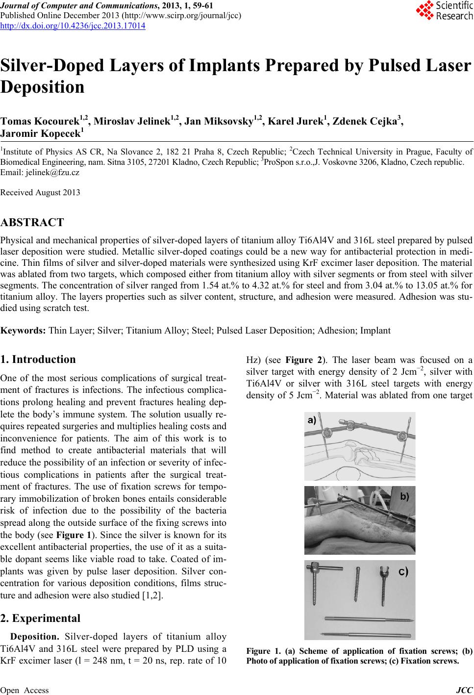 Journal of Computer and Communications, 2013, 1, 59-61 Published Online December 2013 (http://www.scirp.org/journal/jcc) http://dx.doi.org/10.4236/jcc.2013.17014 Open Access JCC 59 Silver-Doped Layers of Implants Prepared by Pulsed Laser Deposition Tomas Kocourek1,2, Miroslav Jelinek1,2, Jan Miksovsky1,2, Karel Jurek1, Zdenek Cejka3, Jaromir Kopecek1 1Institute of Physics AS CR, Na Slovance 2, 182 21 Praha 8, Czech Republic; 2Czech Technical University in Prague, Faculty of Biomedical Engineering, nam. Sitna 3105, 27201 Kladno, Czech Republic; 3ProSpon s.r.o.,J. Voskovne 3206, Kladno, Czech republic. Email: [email protected] Received August 2013 ABSTRACT Physical and mechanical properties of silver-doped layers of titanium alloy Ti6Al4V and 316L steel prepared by pulsed laser deposition were studied. Metallic silver-doped coatings could be a new way for antibacterial protection in medi- cine. Thin films of s ilver and silver-doped materials were synthesized using KrF excimer laser deposition. The material was ablated from two targets, which composed either from titanium alloy with silver segments or from steel with silver segments. The concentration of silver ranged from 1.54 at.% to 4.32 at.% for steel and from 3.04 at.% to 13.05 at.% for titanium alloy. The layers properties such as silver content, structure, and adhesion were measured. Adhesion was stu- died using scratch test. Keywords: Thin Layer; Silver; Titanium Alloy; Steel; Pulsed Laser Deposition; Adhes ion; Implant 1. Introduction One of the most serious complications of surgical treat- ment of fractures is infections. The infectious complica- tions prolong healing and prevent fractures healing dep- lete the body’s immune system. The solution usually re- quires repeated surgeries and multiplies healin g costs and inconvenience for patients. The aim of this work is to find method to create antibacterial materials that will reduce the possibility of an infection or severity of infec- tious complications in patients after the surgical treat- ment of fractures. The use of fixation screws for tempo- rary immobilization of broken bones entails considerable risk of infection due to the possibility of the bacteria spread along the outside surface of the fixing screws into the body (see Figu re 1). Since the silver is known for its excellent antibacterial properties, the use of it as a suita- ble dopant seems like viable road to take. Coated of im- plants was given by pulse laser deposition. Silver con- centration for various deposition conditions, films struc- ture and adhesion were also studied [1,2]. 2. Experimental Depo sit ion. S ilv er-doped layers of titanium alloy Ti6Al4V and 316L steel were prepared by PLD using a KrF excimer laser (l = 248 nm, t = 20 ns, rep. rate of 10 Hz) (see Figure 2). The laser beam was focused on a silver target with energy density of 2 Jcm−2, silver with Ti6Al4V or silver with 316L steel targets with energy density of 5 Jcm−2. Material was ablated from one target Figure 1. (a) Scheme of application of fixation screws; (b) Photo of application of fixation screws; (c) Fixation screws. 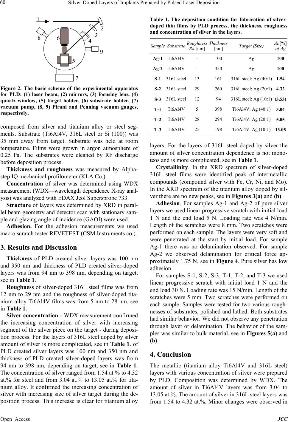 Silver-Doped Layers of Implants Prepared by Pulsed Laser Deposition Open Access JCC 60 Figure 2. The basic scheme of the experimental apparatus for PLD: (1) laser beam, (2) mirrors, (3) focusing lens, (4) quartz window, (5) target holder, (6) substrate holder, (7) vacuum pump, (8, 9) Pirani and Penning vacuum gauges, respectively. composed from silver and titanium alloy or steel seg- ments. Substrate (Ti6Al4V, 316L steel or Si (100)) was 35 mm away from target. Substrate was held at room temperature. Films were grown in argon atmosphere of 0.25 Pa. The substrates were cleaned by RF discharge before deposition process. Thickness and roughness was measured by Alpha- step IQ mechanical profilometer (KLA Co.). Concentration of silver was determined using WDX measurement (WDX—wavelength dependence X-ray anal- ysis) was analyzed with EDAX Jeol Supersprobe 733. Structure of layers was determined by XRD in paral- lel beam geometry and detector scan with stationar y sam- ple and glazing angle of incidence (GAOI) were used. Adhesion. For the adhesion measurements we used macro scratch tester REVETEST (CSM Instruments co.). 3. Results and Discussion Thickness of PLD created silver layers was 100 nm and 350 nm and thickness of PLD created silver-doped layers was from 94 nm to 398 nm, depending on target, see in Table 1. Roughness of silver-doped 316L steel films was from 12 nm to 29 nm and the roughness of silver-doped tita- nium alloy Ti6Al4V films was from 5 nm to 28 nm, see in Table 1. Silver concentration - WDX measurement confirmed the increasing concentration of silver with increasing segment of the silver piece on the target - during deposi- tion process. For the layers of 316L steel doped by silver amount of silver is more complicated, see in Table 1 . of PLD created silver layers was 100 nm and 350 nm and thickness of PLD created silver-doped layers was from 94 nm to 398 nm, depending on target, see in Table 1. The concentration of silv er rang ed from 1.54 at. % to 4.32 at.% for steel and from 3.04 at.% to 13.05 at.% for tita- nium alloy. It confirmed the increasing concentration of silver with increasing size of silver target during the de- position process. This increase is clear for titanium alloy Table 1. The deposition condition for fabrication of silver- doped thin films by PLD process, the thickness, roughness and concentration of silver in the layers. Sample Substrate Roughness Ra [nm] Thickness [nm] Target (Size) At.[%] of Ag Ag-1 Ti6Al4V - 100 Ag 100 Ag-2 Ti6Al4V - 350 Ag 100 S-1 316L steel 13 161 316L steel: Ag (40:1) 1.54 S-2 316L steel 29 260 316L steel: Ag (20:1) 4.32 S-3 316L steel 12 94 316L steel: Ag (10:1) (3.53) T-1 Ti6Al4V 5 398 Ti6Al4V: Ag (40:1) 3.04 T-2 Ti6Al4V 28 294 Ti6Al4V: Ag (20:1) 5.05 T-3 Ti6Al4V 25 198 Ti6Al4V: Ag (10:1) 13.05 layers. For the layers of 316L steel doped by silver the amount of silver concentration dependence is not mono- teos and is more complicated, see in Table 1. Crystallinity. In the XRD spectrum of silver-doped 316L steel films were identified peak of intermetallic compounds (compound silver with Fe, Cr, Ni, and Mo). In the XRD spectrum of the titanium alloy doped by sil- ver there are no new peaks, see in Figures 3(a) and (b). Adhesion. For samples Ag-1 and Ag-2 of pure silver layers we used linear progressiv e scratch with initial load 1 N and the end load 5 N. Loading rate was 4 N/min. Length of the scratches were 8 mm. Two scratches were performed on each sample. The layers were very soft and were penetrated at the start by initial load. For sample Ag-1 there was no delamination observed. For sample Ag-2 we observed delamination for critical force ap- proximately 1.75 N, see in Fig ure 4. Pure silver has low adhesion. For samples S-1, S-2, S-3, T-1, T-2, and T-3 we used linear progressive scratch with initial load 1 N and the end load 30 N. Loading rate was 15 N/min. Length of the scratches were 5 mm. Two scratches were performed on each sample. Samples were tested for two various rough- nesses of substrates, polished and lathed. Both substrates had similar behavior. We did not observe any penetration through layer or delamination. The behavior of the sam- ples was similar to bulk material, see in Figures 5(a) and (b). 4. Conclusion The metallic (titanium alloy Ti6Al4V and 316L steel) layers with various concentration of silver were prepared by PLD. Composition was determined by WDX. The amount of silver in Ti6Al4V layers was from 3.04 to 13.05 at.%. The amount of silver in 316L steel layers was from 1.54 to 4.32 at.%. Minor changes were observed in 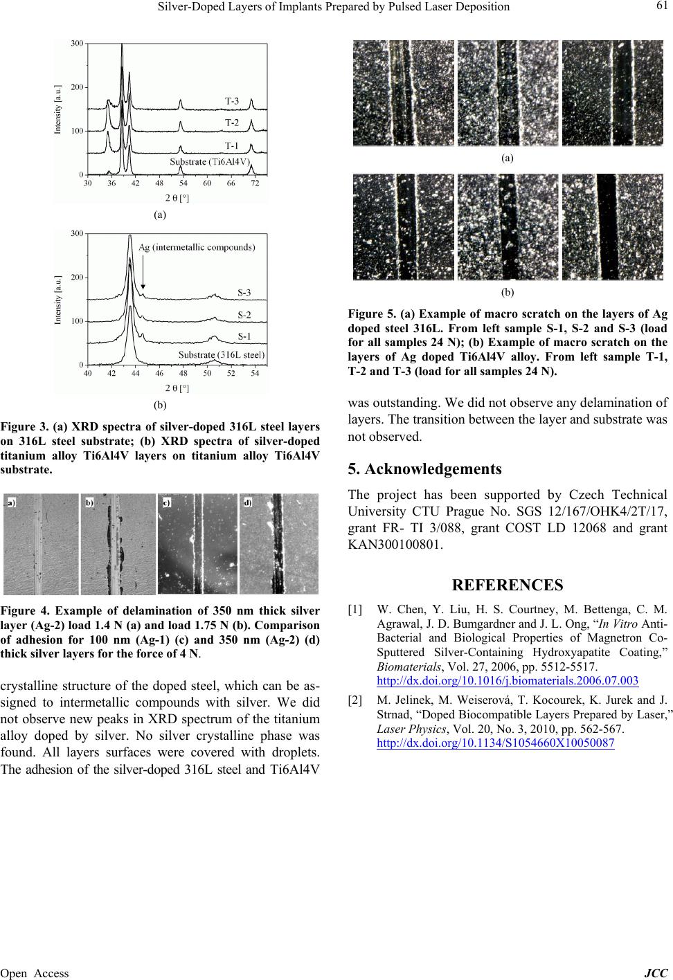 Silver-Doped Layers of Implants Prepared by Pulsed Laser Deposition Open Access JCC 61 (a) (b) Figure 3. (a) XRD spectra of silver-doped 316L steel layers on 316L steel substrate; (b) XRD spectra of silver-doped titanium alloy Ti6Al4V layers on titanium alloy Ti6Al4V substrate. Figure 4. Example of delamination of 350 nm thick silver layer (Ag-2) load 1.4 N (a) and load 1.75 N (b). Comparison of adhesion for 100 nm (Ag-1) (c) and 350 nm (Ag-2) (d) thick silver layers for the force of 4 N. crystalline structure of the doped steel, which can be as- signed to intermetallic compounds with silver. We did not observe new peaks in XRD spectrum of the titanium alloy doped by silver. No silver crystalline phase was found. All layers surfaces were covered with droplets. The adhesion of the silver-doped 316L steel and Ti6Al4V (a) (b) Figure 5. (a) Example of macro scratch on the layers of Ag doped steel 316L. From left sample S-1, S-2 and S-3 (load for all samples 24 N); (b) Example of macro scratch on the layers of Ag doped Ti6Al4V alloy. From left sample T-1, T-2 and T-3 (load for all samples 24 N). was outstanding. We did not observe any delamination of layers. The transition between the layer and substrate was not obser ved. 5. Acknowledgements The project has been supported by Czech Technical University CTU Prague No. SGS 12/167/OHK4/2T/17, grant FR- TI 3/088, grant COST LD 12068 and grant KAN300100801. REFERENCES [1] W. Chen, Y. Liu, H. S. Courtney, M. Bettenga, C. M. Agrawal, J. D. Bumgardner and J. L. Ong, “In Vitro Anti- Bacterial and Biological Properties of Magnetron Co- Sputtered Silver-Containing Hydroxyapatite Coating,” Biomaterials, Vol. 27, 2006, pp. 5512-5517. http://dx.doi.org/10.1016/j.biomaterials.2006.07.003 [2] M. Jelinek, M. Weiserová, T. Kocourek, K. Jurek and J. Strnad, “Doped Biocompatible Layers Prepared by Laser,” Laser Physics, Vol. 20, No. 3, 2010, pp. 562-567. http://dx.doi.org/10.1134/S1054660X10050087 |