

Paper Menu >>
Journal Menu >>
 Advances in Bioscience and Biotechnology, 2013, 4, 1007-1012 ABB http://dx.doi.org/10.4236/abb.2013.411134 Published Online November 2013 (http://www.scirp.org/journal/abb/) A proteomic analysis of the effect of radiation therapy on wound healing in women reconstructed with the TRAM flap Bekka O. Christensen1,2, Jens Overgaard2, Henrik Vorum4, Bent Honore3, Tine E. Damsgaard1 1Department of Plastic Surgery, Aarhus University Hospital, Aarhus, Denmark 2Department of Experimental Clinical Oncology, Aarhus University Hospital, Aarhus, Denmark, 3Department of Medical Biochemistry, University of Aarhus, Aarhus, Denmark 4Department of Ophthalmology, Aalborg University Hospital, Aalborg, Denmark Email: [email protected] Received 10 July 2013; revised 20 September 2013; accepted 13 October 2013 Copyright © 2013 Bekka O. Christensen et al. This is an open access article distributed under the Creative Commons Attribution License, which permits unrestricted use, distribution, and reproduction in any medium, provided the original work is properly cited. ABSTRACT The incidence of breast cancer is still increasing, and with improved cancer treatment, more women live longer with the side effects of their treatment. The response of normal tissue to radiation continues for years after the treatment is completed. The influence of radiotherapy on the outcome of breast reconstruct- tive surgery remains unpredictable. The combination of two surgical sites of which o ne is previously irradi- ated, is rarely encountered in humans and thus com- piles a unique opportunity to study the implications of irradiation followed by surgery. The aim of this study was to examine the long-term effect of radiation therapy on the proteins expressed in the wound tissue after a breast reconstruction. Ten patients were in- cluded in the study, all treated with radiotherapy af- ter a mastectomy and breast reconstruction with a contralateral pedicled TRAM flap. Expanded poly- tetrafluoretylene polymer tubes were implanted for 10 days, subcutaneously, below the inframammary fold and below the donor site. The protein from the newly synthesized granulation tissue in the tubes was extracted and analyzed for differences in protein ex- pression with 2D gel electrophoresis and mass spec- trometry. A total of 676 proteins were detected; of these, 4 proteins changed significantly and were suc- cessfully identified. TPM4 and APOA4 from the ra- diation treated tissue were shown to be significantly decreased, whereas IGKC and VDAC1 were found to be significantly increased. The proteomic technique combined with the ePTFE tube wound model can elucidate some of the molecular alterations in the wound healing induced by radiation therapy. The protein modifications of TPM4, APOA4, IGKC and VDAC1 may influence the cell proliferation, apop- tosis and the inflammation of the tissue repair proc- ess. Keywords: TRAM Flap; Radiation Therapy; Reconstructive Surgery; Human; Proteomics; TPM; VDAC; APOA4 1. INTRODUCTION The healing process is the basis of all surgeries. There- fore, it is crucial to gain knowledge of the biology of the tissue repair process and the factors influencing it: how to optimize the healing process, diminish the postopera- tive complications. The incidence of breast cancer is still increasing, and with improved cancer treatment, more women live lon- ger with the side-effects of their cancer treatment. The destruction or damage of healthy tissue/cells outside the tumour has both acute and long-term side-effects. Late sequelae from radiation therapy (RT) include skin atro- phy, pigmentation changes, soft tissue fibrosis and mi- cro-vascular damage [1]. The combination of RT and breast reconstruction has generated significant discussion. Several studies have retrospectively evaluated aesthetic outcome and compli- cations after mastectomy, radiation therapy and subse- quent breast reconstruction. The overall conclusion is that radiation therapy increases morbidity and decreases the aesthetic outcome; patients will contract more com- plications and an inferior outcome is unpredictable [2-6]. In 1982, Hartrampf et al. introduced the pedicled transverse abdominal muscle flap (TRAM flap) for re- construction of the breast. Variations of this versatile flap OPEN ACCESS  B. O. Christensen et al. / Advances in Bioscience and Biotechnology 4 (2013) 1007-1012 1008 are common methods for autologous breast reconstruct- tion, and often recommended for women previously treated with radiation therapy, as it transposes non radi- ated tissue into the irradiated area. Radiation therapy is confined to the irradiated tissue volume and therefore, enables examination of both irra- diated and non irradiated tissue from the same individual. The combination of two surgical sites, one of which is previously irradiated, is rarely encountered in humans. Thus, the TRAM flap procedure compiles a unique op- portunity to study the implications of irradiation fol- lowed by surgery. Previous studies have reported impaired wound heal- ing and radiation induced tissue changes [1,7-9]. The pathogenesis of the radiation induced tissue alterations are complex, involving a cascade of molecular and cel- lular events. The time span in which such alterations occur is uncertain. The proteome is the entire complement of proteins, in- cluding modifications that vary with time and require- ments. It is well known that mRNA is not always trans- lated into protein; the amount of protein produced for a given amount of mRNA depends on both the gene. It is transcribed from the current physiological state of the cell. These (post-translational) modifications affect the function of the proteins, and are therefore important in the understanding of the patophysiological process caus- ing the changes in the tissue repair process. Proteo- mic analysis can determine the expression levels of pro- teins, including the modifications [10]. Proteomic analysis, based on two dimensional elec- trophoresis (2-DE), has primarily been conducted to study carcinogenesis, signal pathway profiling, and mo- lecular markers [11]. To our knowledge, no study has used proteomics to examine the changes in the tissue repair response, induced by previous radiation therapy, in newly synthesized granulation tissue from humans. Most studies working with radiation therapy and proteins use techniques involving specific protein antibodies. They require a beforehand knowledge of which proteins are in- fluenced by RT. Other studies focus on cell cultures or wound fluids [12,13]. The expanded polytetrafluorethylene polymer tube (ePTFE tube) is a model for studying wound healing in humans [14]. The proteomic analysis provides the possibility of examining proteins without a priori knowledge of which protein properties are altered by radiation therapy and subsequent surgery. 2. MATERIALS AND METHODS 2.1. Patient After a modified radical mastectomy for breast cancer, ten women were consecutively included in the study. The women were reconstructed with a contralateral pedicled TRAM flap at the Department of Plastic Surgery, Aarhus University Hospital, Denmark. All had been treated with radiation therapy according to the national guidelines from the Danish Breast Cancer Cooperative Group [15]. Data were obtained from personal communication, physical examination and charts including information regarding BMI (body mass index), tobacco smoking and adjuvant therapy. 2.2. Surgery The TRAM flap was harvested with a horizontal ellipse of tissue, incorporating the full width and a variable length of the rectus abdominis muscle. The musculocu- taneous flap was tensionlessly transposed into the recre- ated mastectomy defect, supplied by the superior epigas- tric vessels. The transposed tissue was partly de-epithel- ized and shaped into a new breast mound. Supra umbili- cal fascial sheath defects were closed primarily and in- fraubillical fascial defects were reconstructed using a 15 × 15 cm on-lay polypropylene mesh. The umbilicus was brought out through a central opening in the midline of the abdominal flap and sutured in place. Closure was performed using a layered closure of Scarpa’s fascia, subcutaneous tissue and skin. 2.3. The ePTFE Tube The ePTFE tubes (International Polymer Enginering, Tempe, Arizona, USA.) have a 0.12 cm inner diameter, a 0.25 cm outer diameter and a pore size of 90 - 120 µm. The 9 cm ePTFE tube was threaded on a 3-0 non-ab- sorbable nylon suture and sterilized before implantation. The ePTFE tubes were implanted during general an- esthesia, in the subcutaneous tissue at the intrammamary fold and at the donor site, using the Seldinger technique. Approximately 1cm of the ePTFE tube was left protrude- ing outside the skin at the lateral perforation. The ePTFE tube was sutured to the skin with a single nylon 4-0 su- ture and covered with Tegaderm®. The ePTFE tubes were removed on postoperative day 10 - 12. The external part of the ePTFE tube was discarded and the former subcutaneous parts were immediately divided and snap frozen in liquid nitrogen and stored at −140 ˚C. 2.4. Proteomic Analysis The ePTFE tubes were homogenized in lysis buffer and the supernatants were removed. Two-dimensional gel electrophoresis, image analysis and identification of dif- ferentially expressed protein spots by mass spectrometry were essentially performed as previously described [16]. The dry and transparent gels were scanned in the transmissive mode on a GS-710 Imaging Densitometer Copyright © 2013 SciRes. OPEN ACCESS 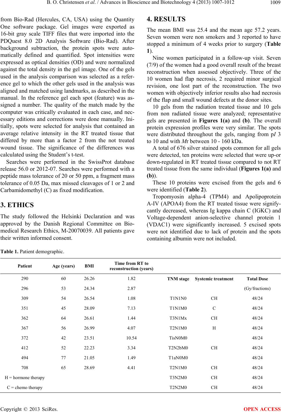 B. O. Christensen et al. / Advances in Bioscience and Biotechnology 4 (2013) 1007-1012 Copyright © 2013 SciRes. 1009 OPEN ACCESS from Bio-Rad (Hercules, CA, USA) using the Quantity One software package. Gel images were exported as 16-bit gray scale TIFF files that were imported into the PDQuest 8.0 2D Analysis Software (Bio-Rad). After background subtraction, the protein spots were auto- matically defined and quantified. Spot intensities were expressed as optical densities (OD) and were normalized against the total density in the gel image. One of the gels used in the analysis comparison was selected as a refer- ence gel to which the other gels used in the analysis was aligned and matched using landmarks, as described in the manual. In the reference gel each spot (feature) was as- signed a number. The quality of the match made by the computer was critically evaluated in each case, and nec- essary editions and corrections were done manually. Ini- tially, spots were selected for analysis that contained an average relative intensity in the RT treated tissue that differed by more than a factor 2 from the not treated wound tissue. The significance of the differences was calculated using the Student’s t-test. Searches were performed in the SwissProt database release 56.0 or 2012-07. Searches were performed with a peptide mass tolerance of 20 or 50 ppm, a fragment mass tolerance of 0.05 Da, max missed cleavages of 1 or 2 and Carbamidomethyl (C) as fixed modification. 3. ETHICS The study followed the Helsinki Declaration and was approved by the Danish Regional Committee on Bio- medical Research Ethics, M-20070039. All patients gave their written informed consent. 4. RESULTS The mean BMI was 25.4 and the mean age 57.2 years. Seven women were non smokers and 3 reported to have stopped a minimum of 4 weeks prior to surgery (Table 1). Nine women participated in a follow-up visit. Seven (7/9) of the women had a good overall result of the breast reconstruction when assessed objectively. Three of the 10 women had flap necrosis, 2 required minor surgical revision, one lost part of the reconstruction. The two women with objectively inferior results also had necrosis of the flap and small wound defects at the donor sites. 10 gels from the radiation treated tissue and 10 gels from non radiated tissue were analyzed; representative gels are presented in Figures 1(a) and (b). The overall protein expression profiles were very similar. The spots were distributed throughout the gels, ranging from pI 3 to 10 and with Mr between 10 - 160 kDa. A total of 676 silver stained spots common for all gels were detected, ten proteins were selected that were up-or down-regulated in RT treated tissue compared to not RT treated tissue from the same individual (Figures 1(a) and (b)). These 10 proteins were excised from the gels and 6 were identified (Table 2). Tropomyosin alpha-4 (TPM4) and Apolipoprotein A-IV (APOA4) from the RT treated tissue were signify- cantly decreased, whereas Ig kappa chain C (IGKC) and Voltage-dependent anion-selective channel protein 1 (VDAC1) were significantly increased. 5 excised spots were not identified due to lack of protein and the spots containing albumin were not included. Table 1. Patient demographic. Patient Age (years) BMI Time from RT to reconstruction (years) 290 60 26.26 1.82 TNM stage Systemic treatment Total Dose 296 53 24.34 2.87 (Gy/fractions) 309 54 26.54 1.08 T1N1N0 CH 48/24 351 45 28.09 7.13 T1N1M0 C 48/24 362 64 26.61 1.44 T3N1Mx CH 48/24 367 56 26.99 4.07 T2N1M0 H 48/24 372 42 23.51 10.54 TisN0M0 48/24 412 52 22.23 3.34 T2N2bM0 CH 48/24 494 77 21.05 1.49 T1aN0M0 48/24 708 65 28.69 4.41 T2N1M0 CH 48/24 H = hormone therapy T3N2M0 CH 48/24 C = chemo therapy T2N2M0 CH 48/24 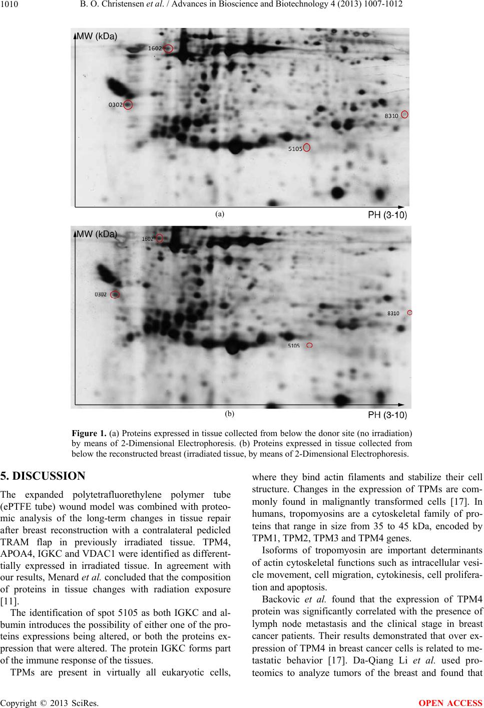 B. O. Christensen et al. / Advances in Bioscience and Biotechnology 4 (2013) 1007-1012 1010 (a) (b) Figure 1. (a) Proteins expressed in tissue collected from below the donor site (no irradiation) by means of 2-Dimensional Electrophoresis. (b) Proteins expressed in tissue collected from below the reconstructed breast (irradiated tissue, by means of 2-Dimensional Electrophoresis. 5. DISCUSSION The expanded polytetrafluorethylene polymer tube (ePTFE tube) wound model was combined with proteo- mic analysis of the long-term changes in tissue repair after breast reconstruction with a contralateral pedicled TRAM flap in previously irradiated tissue. TPM4, APOA4, IGKC and VDAC1 were identified as different- tially expressed in irradiated tissue. In agreement with our results, Menard et al. concluded that the composition of proteins in tissue changes with radiation exposure [11]. The identification of spot 5105 as both IGKC and al- bumin introduces the possibility of either one of the pro- teins expressions being altered, or both the proteins ex- pression that were altered. The protein IGKC forms part of the immune response of the tissues. TPMs are present in virtually all eukaryotic cells, where they bind actin filaments and stabilize their cell structure. Changes in the expression of TPMs are com- monly found in malignantly transformed cells [17]. In humans, tropomyosins are a cytoskeletal family of pro- teins that range in size from 35 to 45 kDa, encoded by TPM1, TPM2, TPM3 and TPM4 genes. Isoforms of tropomyosin are important determinants of actin cytoskeletal functions such as intracellular vesi- cle movement, cell migration, cytokinesis, cell prolifera- tion and apoptosis. Backovic et al. found that the expression of TPM4 protein was significantly correlated with the presence of lymph node metastasis and the clinical stage in breast cancer patients. Their results demonstrated that over ex- pression of TPM4 in breast cancer cells is related to me- tastatic behavior [17]. Da-Qiang Li et al. used pro- eomics to analyze tumors of the breast and found that t Copyright © 2013 SciRes. OPEN ACCESS 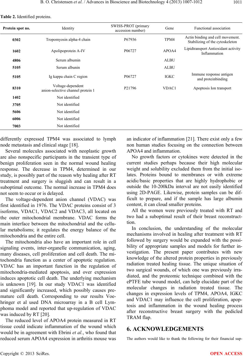 B. O. Christensen et al. / Advances in Bioscience and Biotechnology 4 (2013) 1007-1012 1011 Table 2. Identified proteins. Protein spot no. Identity SWISS-PROT (primary accession number) Gene Functional association 0302 Tropomyosin alpha-4 chain P67936 TPM4 Actin binding and cell movement. Stabilizing of the cytoskeleton 1602 Apolipoprotein A-IV P06727 APOA4 Lipidtransport Antioxidant activity Inflammation 4806 Serum albumin ALBU 5105 Serum albunin ALBU 5105 Ig kappa chain C region P06727 IGKC Immune response antigen and proteinbinding 8310 Voltage-dependent anion-selective channel protein 1 P21796 VDAC1 Apoptosis Ion transport 1402 Not identified 3705 Not identified 5606 Not identified 6006 Not identified 7003 Not identified differently expressed TPM4 was associated to lymph node metastasis and clinical stage [18]. Several molecules associated with neoplastic growth are also nonspecific participants in the transient type of benign proliferation seen in the normal wound healing response. The decrease in TPM4, determined in our study, is possibly part of the reason why healing after RT treatment and surgery is sluggish and can result in a suboptimal outcome. The normal increase in TPM4 does not seem to occur or is delayed. The voltage-dependent anion channel (VDAC) was first identified in 1976. The VDAC proteins consist of 3 isoforms, VDAC1, VDAC2 and VDAC3, all located on the outer mitochondrial membrane. VDAC forms the main interface between the mitochondrial and the cellu- lar metabolisms; it regulates the energy balance of the mitochondria and the entire cell. The mitochondria also have an important role in cell signaling events, inter-organelle communication, aging, many diseases, cell proliferation and cell death. The mi- tochondria function as a center of apoptotic regulation. VDAC has an important function in the regulation of mitochondria-mediated apoptosis, and over expression induces apoptotic cell death. The underlying mechanism is unknown [19]. In our study VDAC1 was identified and significantly increased, which possibly causes pre- mature cell death. Corresponding to our results Voe- hringer et al used DNA microarray in a B cell Lym- phoma model and reported that up-regulation of VDAC was induced by RT [20]. The reduced level of APOA4 protein measured in RT tissue could indicate inflammation of the wound which would be in agreement with Ebrini et al., who found that reduced serum APOA4 expression in arthritis mouse was an indicator of inflammation [21]. There exist only a few non human studies focusing on the connection between APOA4 and inflammation. No growth factors or cytokines were detected in the current studies perhaps because their high molecular weight and solubility excluded them from the initial iso- lates. Proteins bound to membranes or with extreme acidic/basic properties that are highly hydrophobic or outside the 10-200kDa interval are not easily identified using 2D-PAGE. Likewise, protein samples can be dif- ficult to prepare, and if the sample has large albumin content, it can cloud smaller proteins. All the women were previously treated with RT and two had a suboptimal result of their breast reconstruct- tion. In conclusion, the understanding of the molecular mechanisms involved in healing after treatment with RT followed by surgery would be expanded with the possi- bility of appropriate samples and models for further in- vestigation. The present paper contributes with new knowledge of the altered protein properties in previously radiation treated healing tissue. The unique situation of two surgical wounds, of which one was previously irra- diated, and the proteomic technique combined with the ePTFE tube wound model, can help elucidate part of the molecular changes in radiation treated tissue. The changes in expression levels of TPM4, APOA4, IGKC and VDAC1 may influence the cell proliferation, apop- tosis and inflammation in the wound healing process after reconstructive breast surgery with the pedicled TRAM flap. 6. ACKNOWLEDGEMENTS The authors would like to thank the following for their financial sup- Copyright © 2013 SciRes. OPEN ACCESS 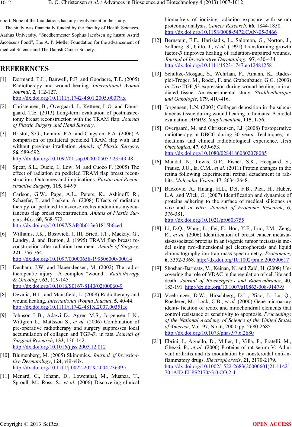 B. O. Christensen et al. / Advances in Bioscience and Biotechnology 4 (2013) 1007-1012 1012 nport. None of the foundations had any involvement in the study. The study was financially funded by the Faculty of Health Sciences, Aarhus University, “Snedkermester Sophus Jacobsen og hustru Astrid Jacobsens Fond”, The A. P. Møller Foundation for the advancement of medical Science and The Danish Cancer Society. REFERENCES [1] Dormand, E.L., Banwell, P.E. and Goodacre, T.E. (2005) Radiotherapy and wound healing. International Wound Journal, 2, 112-127. http://dx.doi.org/10.1111/j.1742-4801.2005.00079.x [2] Christensen, B., Overgaard, J., Kettner, L.O. and Dams- gaard, T.E. (2013) Long-term evaluation of postmastec- tomy breast reconstruction with the TRAM flap. Journal of Plastic Surgery and Hand Surgery. [3] Bristol, S.G., Lennox, P.A. and Clugston, P.A. (2006) A comparison of ipsilateral pedicled TRAM flap with and without previous irradiation. Annals of Plastic Surgery, 56, 589-592. http://dx.doi.org/10.1097/01.sap.0000205057.23543.48 [4] Spear, S.L., Ducic, I., Low, M. and Cuoco F. (2005) The effect of radiation on pedicled TRAM flap breast recon- struction: Outcomes and implications. Plastic and Recon- stractive Surgery, 115, 84-95. [5] Carlson, G.W., Page, A.L., Peters, K., Ashinoff, R., Schaefer, T. and Losken, A. (2008) Effects of radiation therapy on pedicled transverse rectus abdominis myocu- taneous flap breast reconstruction. Annals of Plastic Sur- gery May, 60, 568-572. http://dx.doi.org/10.1097/SAP.0b013e31815b6ced [6] Williams, J.K., Bostwick, J. III, Bried, J.T., Mackay, G., Landry, J. and Benton, J. (1995) TRAM flap breast re- construction after radiation treatment. Annals of Surgery, 221, 756-764. http://dx.doi.org/10.1097/00000658-199506000-00014 [7] Denham, J.W. and Hauer-Jensen, M. (2002) The radio- therapeutic injury—A complex “wound”. Radiotherapy & Oncology, 63, 129-145. http://dx.doi.org/10.1016/S0167-8140(02)00060-9 [8] Devalia, H.L. and Mansfield, L. (2008) Radiotherapy and wound healing. International Wound Journal, 5, 40-44. http://dx.doi.org/10.1111/j.1742-481X.2007.00351.x [9] Johnson L.B., Adawi D., Agren M.S., Jorgensen L.N., Wittgren L., Mattsson S., et al. (2006) Combination of pre-operative radiotherapy and surgery suppresses local accumulation of collagen and TGF-β1 in rats. Journal of Surgical Research, 133, 136-142. http://dx.doi.org/10.1016/j.jss.2005.12.012 [10] Blumenberg, M. (2005) Skinomics. Journal of Investiga- tive Dermatology, 124, viii-viix. http://dx.doi.org/10.1111/j.0022-202X.2004.23639.x [11] Menard, C., Johann, D., Lowenthal, M., Muanza, T., Sproull, M., Ross, S., et al. (2006) Discovering clinical biomarkers of ionizing radiation exposure with serum proteomic analysis. Cancer Research, 66, 1844-1850. http://dx.doi.org/10.1158/0008-5472.CAN-05-3466 [12] Bernstein, E.F., Harisiadis, L., Salomon, G., Norton, J., Sollberg, S., Uitto, J., et al. (1991) Transforming growth factor-β improves healing of radiation-impaired wounds. Journal of Investigative Dermatology, 97, 430-434. http://dx.doi.org/10.1111/1523-1747.ep12481258 [13] Schultze-Mosgau, S., Wehrhan, F., Amann, K., Rades- piel-Troger, M., Rodel, F. and Grabenbauer, G.G. (2003) In Vivo TGF-β3 expression during wound healing in irra- diated tissue. An experimental study. Strahlentherapie und Onkologie, 179, 410-416. [14] Jorgensen, L.N. (2003) Collagen deposition in the subcu- taneous tissue during wound healing in humans: A model evaluation. APMIS. Supplementum, 115, 1-56. [15] Overgaard, M. and Christensen, J.J. (2008) Postoperative radiotherapy in DBCG during 30 years. Techniques, in- dications and clinical radiobiological experience. Acta Oncologica, 47, 639-653. http://dx.doi.org/10.1080/02841860802078085 [16] Mandal, N., Lewis, G.P., Fisher, S.K., Heegaard, S., Prause, J.U., la, C.M., et al. (2011) Protein changes in the retina following experimental retinal detachment in rab- bits. Molecular Vision, 17, 2634-2648. [17] Backovic, A., Huang, H.L., Del, F.B., Piza, H., Huber, L.A. and Wick, G. (2007) Identification and dynamics of proteins adhering to the surface of medical silicones in vivo and in vitro. Journal of Proteome Research, 6, 376-381. http://dx.doi.org/10.1021/pr0603755 [18] Li, D.Q., Wang, L., Fei, F., Hou, Y.F., Luo, J.M., Zeng, R., et al. (2006) Identification of breast cancer metasta- sis-associated proteins in an isogenic tumor metastasis mo- del using two-dimensional gel electrophoresis and liquid chromatography-ion trap-mass spectrometry. Proteomics, 6, 3352-3368. http://dx.doi.org/10.1002/pmic.200500617 [19] Shoshan-Barmatz, V., Keinan, N. and Zaid, H. (2008) Un- covering the role of VDAC in the regulation of cell life and death. Journal of Bioenergetics and Biomembranes, 40, 183-191. http://dx.doi.org/10.1007/s10863-008-9147-9 [20] Voehringer, D.W., Hirschberg, D.L., Xiao, J., Lu, Q., Roederer, M., Lock, C.B., et al. (2000) Gene microarray identi- fication of redox and mitochondrial elements that control resistance or sensitivity to apoptosis. Proceedings of the National Academy of Science of the United States of America, Vol. 97, No. 6, 2000, pp. 2680-2685. http://dx.doi.org/10.1073/pnas.97.6.2680 [21] Ebrini, I., Agnello, D., Miller, I., Villa, P., Fratelli, M., Ghezzi, P., et al. (2000) Proteins of rat serum V: Adju- vant arthritis and its modulation by nonsteroidal anti-in- flammatory drugs. Electrophoresis, 21, 2170-2179. http://dx.doi.org/10.1002/1522-2683(20000601)21:11<21 70::AID-ELPS2170>3.0.CO;2-1 Copyright © 2013 SciRes. OPEN ACCESS |