

Paper Menu >>
Journal Menu >>
 J. Biomedical Science and Engineering, 2011, 4, 119-121 doi:10.4236/jbise.2011.42017 Published Online February 2011 (http://www.SciRP.org/journal/jbise/ JBiSE ). Published Online February 2011 in SciRes. http://www.scirp.org/journal/JBiSE Computer aided modeling and analysis of a new biomedical and surgical instrument Zheng L i University of Bridgeport, Bridgeport, USA. Email: [email protected] Received 12 November 2010; revised 15 November 2010; accepted 19 November 2010. ABSTRACT This paper describes the recent research and devel- opment of an endo surgical/biomedical instrument in surgical suture applications for minimally invasive therapy procedure. The newly developed instruments can not only protect the wound during the surgical procedure but also actively help the healing process. The new mechanism design of the surgical instrument aids in better ergonomic design, reliable functionality, and continuous cost reduction in product manufac- turing. 3-D modeling technique, func tionality an alysis, kinematical simulation and computer aided solution have been applied to the instrument design, devel- opment and future improvement to meet the specific requirements of minimally invasive surgery proce- dure. The improved new endo surgical/biomedical instrument can prevent patient’s vessels and tissues from being damaging because the distal move of clips are well controlled without clip drop-off incident. Plus the operational force to form the clip is lower than regular surgical/biomedical instruments due to this special new mechanism design. In addition to the above, the manufacturing and product cost can be decreased because the dimensional tolerance of com- ponents, such as clip channel and jaw guide track, can be loose due to this new instrument design. The prototypes of this new endo surgical/biomedical in- strument design are analyzed through computer aided modeling and simulation, in order to prove its feasible functionality, reliable performance, and me- chanical advantage. All these improved features have also been tested and verified through the prototypes. Keywords: Hemostasis; Endoscopic Device; Computational Simulation; 3-D Modeling; Mechanical Advantage 1. INTRODUCTION The newly developed technologies have directed mini- mally invasive surgeries [1]. The positive and feasible changes in surgical instruments have led to the new de- velopment of surgical techniques [2] and [3]. The bio- medical and surgical instrument market is always ad- justed and controlled for its functionality, performance, feasibility, quality, safety, and manufacturing cost. The surgical instrument market is very competitive, price sensitive and depicted by advanced technologies [4] and [5]. Biomedical and surgical instrument is technology based product and normally advanced techniques are especially required to develop special technology to compete the products in today’s challenging market [6]. The applications include the closure of tissue defects, perforations, and anastomotic leakage in the esophagus and stomach. The endo surgical instrument has also been used to prevent post-polypectomy bleeding, placement of enteral feeding tubes. The recent studies show the versatility of endo surgical clips in therapeutic and en- doscopic applications. This endo surgical instrument is the innovative product that will allow for greater ease of use for surgeons and help to improve patient outcomes. Based on the field and clinical feedback, the new technology that simultaneously opens and aligns the jaws has been implemented, allow- ing well controlled surgical clip feeding and closure. This new surgical instrument design can provide more con- sistent and reliable mechanism to protect the clip from external and unanticipated disturbance while the surgical clip sits in the jaw track. Endo surgical instrument has been widely used in hemostasis during endoscopy of the upper and lower gastrointestinal tract in which the bleeding lesions can be successfully clipped. The alternatives to endoscopic clipping of peptic ulcers are thermal therapy (such as electrocautery to burn the vessel causing the bleeding), or injection of epinephrine to constrict the blood vessel. Comparative studies between endo surgical clips and thermal therapy verify that endo surgical clips cause less trauma to the mucosa around the ulcer than electrocau- tery. 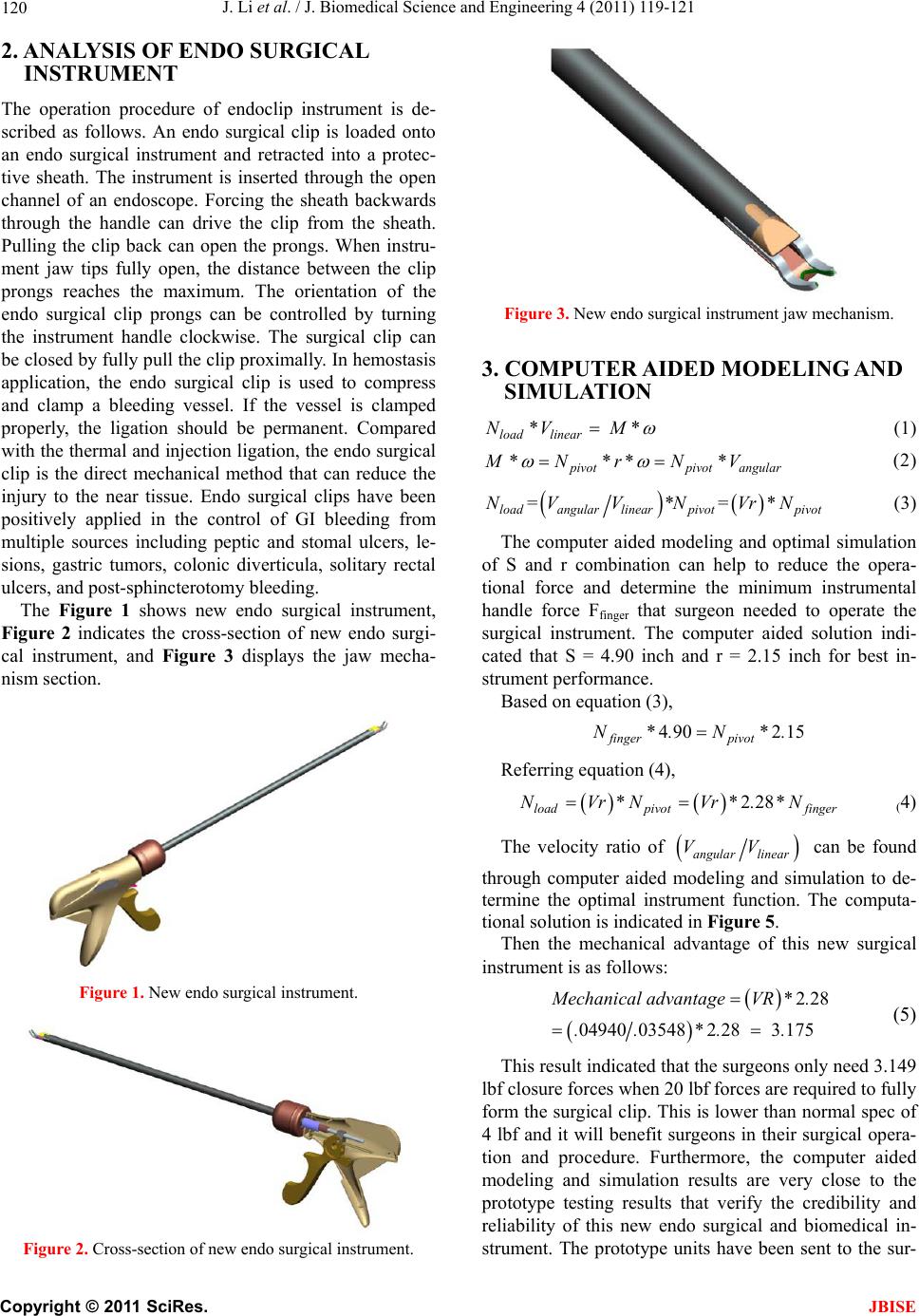 J. Li et al. / J. Biomedical Science and Engineering 4 (2011) 119-121 120 2. ANALYSIS OF ENDO SURGICAL INSTRUMENT The operation procedure of endoclip instrument is de- scribed as follows. An endo surgical clip is loaded onto an endo surgical instrument and retracted into a protec- tive sheath. The instrument is inserted through the open channel of an endoscope. Forcing the sheath backwards through the handle can drive the clip from the sheath. Pulling the clip back can open the prongs. When instru- ment jaw tips fully open, the distance between the clip prongs reaches the maximum. The orientation of the endo surgical clip prongs can be controlled by turning the instrument handle clockwise. The surgical clip can be closed by fully pull the clip proximally. In hemostasis application, the endo surgical clip is used to compress and clamp a bleeding vessel. If the vessel is clamped properly, the ligation should be permanent. Compared with the thermal and injection ligation, the endo surgical clip is the direct mechanical method that can reduce the injury to the near tissue. Endo surgical clips have been positively applied in the control of GI bleeding from multiple sources including peptic and stomal ulcers, le- sions, gastric tumors, colonic diverticula, solitary rectal ulcers, and post-sphincterotomy bleeding. The Figure 1 shows new endo surgical instrument, Figure 2 indicates the cross-section of new endo surgi- cal instrument, and Figure 3 displays the jaw mecha- nism section. Figure 1. New endo surgical instrument. Figure 2. Cross-section of new endo surgical instrument. Figure 3. New endo surgical instrument jaw mechanism. 3. COMPUTER AIDED MODELING AND SIMULATION load linear N*V M* (1) pivotpivot angular M *N*r*N*V (2) loadangular linearpivotpivot N=VV*N =Vr*N (3) The computer aided modeling and optimal simulation of S and r combination can help to reduce the opera- tional force and determine the minimum instrumental handle force Ffinger that surgeon needed to operate the surgical instrument. The computer aided solution indi- cated that S = 4.90 inch and r = 2.15 inch for best in- strument performance. Based on equation (3), 490215 finger pivot N*.N*. Referring equation (4), 228 load pivotfinger NVr*NVr* .*N ( 4) The velocity ratio of angular linear VV can be found through computer aided modeling and simulation to de- termine the optimal instrument function. The computa- tional solution is indicated in Figure 5. Then the mechanical advantage of this new surgical instrument is as follows: 228 04940035482 283 175 M echanical ad vant ageVR*. ..*. . (5) This result indicated that the surgeons only need 3.149 lbf closure forces when 20 lbf forces are required to fully form the surgical clip. This is lower than normal spec of 4 lbf and it will benefit surgeons in their surgical opera- tion and procedure. Furthermore, the computer aided modeling and simulation results are very close to the prototype testing results that verify the credibility and reliability of this new endo surgical and biomedical in- strument. The prototype units have been sent to the sur- C opyright © 2011 SciRes. JBISE 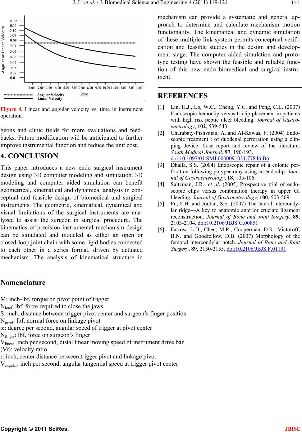 J. Li et al. / J. Biomedical Science and Engineering 4 (2011) 119-121 Copyright © 2011 SciRes. 121 JBISE Figure 4. Linear and angular velocity vs. time in instrument operation. geons and clinic fields for more evaluations and feed- backs. Future modification will be anticipated to further improve instrumental function and reduce the unit cost. 4. CONCLUSION This paper introduces a new endo surgical instrument design using 3D computer modeling and simulation. 3D modeling and computer aided simulation can benefit geometrical, kinematical and dynamical analysis in con- ceptual and feasible design of biomedical and surgical instruments. The geometric, kinematical, dynamical and visual limitations of the surgical instruments are ana- lyzed to assist the surgeon in surgical procedure. The kinematics of precision instrumental mechanism design can be simulated and modeled as either an open or closed-loop joint chain with some rigid bodies connected to each other in a series format, driven by actuated mechanism. The analysis of kinematical structure in mechanism can provide a systematic and general ap- proach to determine and calculate mechanism motion functionality. The kinematical and dynamic simulation of these multiple link system permits conceptual verifi- cation and feasible studies in the design and develop- ment stage. The computer aided simulation and proto- type testing have shown the feasible and reliable func- tion of this new endo biomedical and surgical instru- ment. REFERENCES [1] Lin, H.J., Lo, W.C., Cheng, Y.C. and Peng, C.L. (2007) Endoscopic hemoclip versus triclip placement in patients with high risk peptic ulcer bleeding. Journal of Gastro- enterology, 102, 539-543. [2] Charabaty-Pishvaian, A. and Al-Kawas, F. (2004) Endo- scopic treatment t of duodenal perforation using a clip- ping device: Case report and review of the literature. South Medical Journal, 97, 190-193. doi:10.1097/01.SMJ.0000091031.77846.B6 [3] Dhalla, S.S. (2004) Endoscopic repair of a colonic per- foration following polypectomy using an endoclip. Jour- nal of Gastroenterology, 18, 105-106. [4] Saltzman, J.R., et al. (2005) Prospective trial of endo- scopic clips versus combination therapy in upper GI bleeding. Journal of Gastroenterology, 100, 503-509. [5] Fu, F.H. and Jordan, S.S. (2007) The lateral intercondy- lar ridge—A key to anatomic anterior cruciate ligament reconstruction. Journal of Bone and Joint Surgery, 89, 2103-2104. doi:10.2106/JBJS.G.00851 [6] Farrow, L.D., Chen, M.R., Cooperman, D.R., Victoroff, B.N. and Goodfellow, D.B. (2007) Morphology of the femoral intercondylar notch. Journal of Bone and Joint Surgery, 89, 2150-2155. doi:10.2106/JBJS.F.01191 Nomenclature M: inch-lbf, torque on pivot point of trigger Nload: lbf, force required to close the jaws S: inch, distance between trigger pivot center and surgeon’s finger position Npivot: lbf, normal force on linkage pivot ω: degree per second, angular speed of trigger at pivot center Nfinger: lbf, force on surgeon’s finger Vlinear: inch per second, distal linear moving speed of instrument drive bar (Vr): velocity ratio r: inch, center distance between trigger pivot and linkage pivot Vangular: inch per second, angular tangential speed at trigger pivot center |