

Paper Menu >>
Journal Menu >>
 Open Journal of Urology, 2013, 3, 287-292 Published Online November 2013 (http://www.scirp.org/journal/oju) http://dx.doi.org/10.4236/oju.2013.37054 Open Access OJU RNase L Variants Do Not Appear to Impact on Clinical Features of Sporadic Prostate Cancer Patients* Frank T. D’Arcy1,2, Ruth Foley1, Thomas H. Lynch2 1Institute of Molecular Medicine, Trinity College, Dublin, Ireland 2Department of Urology, St. James Hospital, Dublin, Ireland Email: [email protected] Received November 4, 2013; revised November 23, 2013; accepted November 28, 2013 Copyright © 2013 Frank T. D’Arcy et al. This is an open access article distributed under the Creative Commons Attribution License, which permits unrestricted use, distribution, and reproduction in any medium, provided the original work is properly cited. ABSTRACT Introduction: Prostate cancer is the most common non-cutaneous male cancers, contributing to significant mortality rates globally. Mutations of RNase L, an enzyme involved in inflammatory and immunological pathways, have been speculated to predispose to cancer. This study assesses three different mutations of the RNase L gene in Irish prostate cancer patients, including one linked with general cancer susceptibility never investigated before in prostate cancer (rs3738579), and reports on links with aggressive cancer. Methods: 134 patients had their RNase L mutation status determined by polymerase chain reaction (PCR) of serum DNA. Complementary clinical details for each patient are statistically analysed. Results: No link to age of diagnosis, high grade disease or prostate specific antigen (PSA) level at diagnosis was demonstrated with any of the studied single nucleotide polymorphisms (SNP). The SNP variation was consistent with that of published international series. Conclusion: SNP genotypic frequencies in Ireland are consistent with international findings. The studied RNase L mutations including rs3738579 do not appear to have a significant impact on our patient population. Keywords: RNase L; R462Q; D541E; Prostate Cancer 1. Introduction RNase L is a gene found on the hereditary prostate can- cer (HPC) locus of chromosome 1 that codes for a latent endoribonuclease. This enzyme participates in an interferon inducible RNA decay pathway that has a role in inflam- mation and cellular immunity against viral infection. Sus- tained activation will lead to apoptosis. The gene is 741 amino acids long with 8 exons and is roughly 13 kilo- bases. It is converted from an inactive monomeric form to a potent dimeric structure by the action of a series of 2’ to 5’ linked oligodenylates, commonly known as 2 - 5 A. RNase L has been a candidate gene for prostate cancer researchers for a number of years and many variants in- cluding R462Q [1], E265X [1], M1I [2] and 471ΔAAAG [3] have been described. The R462Q RNase L mis-sense mutation (rs486927) arises when a substitution of the “G” to “A” base along exon 1 at mRNA position 1552 gives an amino acid change of argenine to glutamine at amino acid position 462. The consequence of this is a variant of the gene that produces an enzyme functioning at a third of the wild- type enzyme’s efficiency [4]. It has been of interest to researchers since it was linked to prostate cancer in 2002 [2]. Subsequent studies have been mixed with some show- ing a link with this variant and familial prostate cancer [5,6], whilst others have not [7]. A meta-analysis of 7 papers has failed to show a significant link between this mutation and prostate cancer regardless of ethnicity or family history [8]. D541E (rs627928) comes about following a substitution of the “T” base at mRNA position 1790 with “G”, caus- ing a change in the protein at amino acid position 541 from aspartate to glutamic acid. This area of the gene codes for a region within the protein kinase domain of the enzyme, an area with an important role in dimeriza- tion of the protein into its active state. Despite the muta- tion, this enzyme has comparable catalytic activity with the wildtype variant [4]. Its significance is the source of some debate within the literature. Studies have linked *All funding for described work came via Trinity College Dublin and The Irish Cancer Society.  F. T. D’ARCY ET AL. 288 this change to an increased risk of familial prostate can- cer [7] and sporadic, metastatic disease [9], but in a Japa- nese study [10] the wildtype variant was actually linked to an increased risk of familial prostate cancer. Other investigators have found no significant associations with it [1,5,11]. An undescribed mutation (rs3738579) located in the 5’ untranscribed region (5’-UTR) has been identified as a general indicator of increased cancer susceptibility [12]. The exact role of this variant on expression is unclear but mutations are seen disproportionally in cervical as well as head and neck squamous cell cancers. Work in the fields of cervical cancer (human papillo- mavirus) [13], gastric cancer (helicobacter pylori) [14], B-cell lymphomas (Epstein-Barr virus) [15] and osteo- genic sarcomas (chronic osteomyelitis) [16] have all found definite links between chronic infection and neoplasia. Whilst no such links have yet been established in prostate cancer, there are inconsistent reports in the literature of positive associations of the disease with sexually trans- mitted infections (STIs) [17-19], and one study finding men with 25 or more sexual partners being 2.8 times more likely to be diagnosed with prostate cancer than a man with 5 or less partners [20]. If inflammation or viral infection plays a role in prostate cancer development, it is speculated that polymorphisms in genes involved in the inflammatory and infectious disease pathways like RNase L could be important. This study aims to sequence RNase L single nucleo- tide polymorphisms (SNPs) in men with known cases of sporadic prostate cancer and correlate to the clinical as- pects of the cancer. 2. Methods and Materials Patient samples were obtained from the Prostate Cancer Research Consortium (PCRC) bio-repository. This is a collaboration of three University institutions affiliated with five major hospitals. Complementary clinical details relating to each patient are also logged into a password protected, secure database. All volunteers were pre-op- erative radical prostatectomy patients with localised pros- tate cancer, enrolled with full consent in line with ethical advice. All cases of prostate cancer analysed were spo- radic. Using the database of known SNPs published online at the National Centre for Biotechnology Information (NCBI), the sequence of code surrounding the SNPs of interest were obtained. Primers were designed using Primer Express (Applied Biosystems, USA). Details of the actual sequence of in- terest and the primer set are given in Table 1. Primers were supplied by MWG and were purchased at 0.01 mol scale, HPS purification. DNA was extracted from peripheral blood DNA by the Autopure automated system (Qiagen, USA) which uses puregene chemistry. The product was purified (Qiaquick PCR purification kit from Qiagen, USA) and sequencing reactions with a fluorescent dye terminator (BigDye v3.1, ABI) were performed. Results were interpreted with Se- quencing Analysis v5.1 from Applied Biosystems. Polymerase chain reaction (PCR) was carried out with the product initially being exposed to 94˚C for thirty seconds, followed by 35 cycles where the temperature alternated from 94˚C for 30 more seconds, then [53.1˚C (R462Q)/53˚C (D541E)/55˚C (5’-UTR)] for 30 seconds followed by a minute at 72˚C. The reaction culminated in 30 minutes exposure to 72˚C. 3. Results Table 2 illustrates the frequencies of each RNase L SNP studied in the prostate cancer population. These figures are in keeping with those published on the NCBI database. R462Q 9% of patients had the homozygous AA genotype (R462Q), which corresponds to the less efficient variant of the RNase L enzyme, while 52.2% were heterozygous for this SNP. Table 3 outlines R462Q status versus histo- logical disease aggression. On initial inspection it would appear that a disproportionate amount of men with the AA (i.e. the R462Q) genotype have tumours that are of Gleason score 7 or greater (7 out of 11). This association, whilst a trend did not demonstrate statistical significance (p = 0.258). No association was seen between this SNP and either percentage gland involvement by tumour (p = 0.57) or number of affected first-degree relatives (p = 0.69). Furthermore, anova 1-way p values for age and prostate specific antigen (PSA) levels at diagnosis also failed to demonstrate any statistical significance (p = Table 1. Description of primers selected for each SNP studied. DbSNP ID Base Amino acid Primer Sequence Forward 5’-TGGAAGCGTGTTTGGATGTG-3’ rs486907 G/A R462Q Reverse 5’-TGCAGATCCTGGTGGGTGTA -3’ Forward 5’-TTGATTTATGGCTTTTGTGCAGG-3’ rs627928 T/G D541E Reverse 5’-TGAGGTCCTTAGTTTCCTCATCT-3’ Forward 5’-GTGGAAT GTCAGAAGAC TGAGAAC-3’ rs3738579 C/T - Reverse 5’-AATGCCACCTGCTACCACTT-3’ Open Access OJU 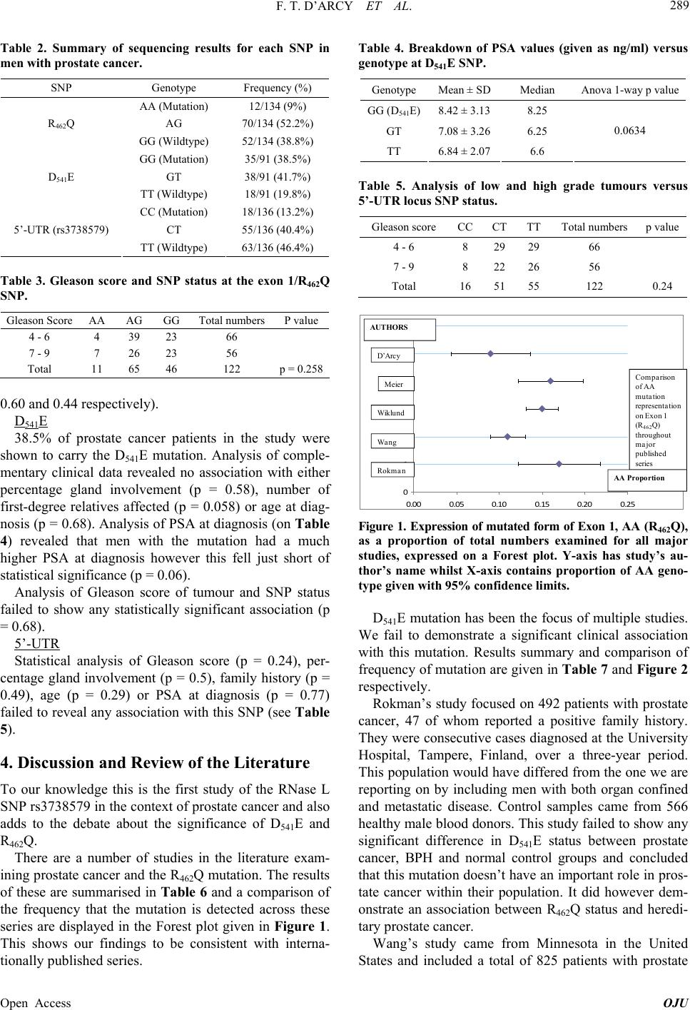 F. T. D’ARCY ET AL. 289 Table 2. Summary of sequencing results for each SNP in men with prostate cancer. SNP Genotype Frequency (%) AA (Mutation) 12/134 (9%) AG 70/134 (52.2%) R462Q GG (Wildtype) 52/134 (38.8%) GG (Mutation) 35/91 (38.5%) GT 38/91 (41.7%) D541E TT (Wildtype) 18/91 (19.8%) CC (Mutation) 18/136 (13.2%) CT 55/136 (40.4%) 5’-UTR (rs3738579) TT (Wildtype) 63/136 (46.4%) Table 3. Gleason score and SNP status at the exon 1/R462Q SNP. Gleason Score AA AG GGTotal numbers P value 4 - 6 4 39 23 66 7 - 9 7 26 23 56 Total 11 65 46 122 p = 0.258 0.60 and 0.44 respectively). D541E 38.5% of prostate cancer patients in the study were shown to carry the D541E mutation. Analysis of comple- mentary clinical data revealed no association with either percentage gland involvement (p = 0.58), number of first-degree relatives affected (p = 0.058) or age at diag- nosis (p = 0.68). Analysis of PSA at diagnosis (on Table 4) revealed that men with the mutation had a much higher PSA at diagnosis however this fell just short of statistical significance (p = 0.06). Analysis of Gleason score of tumour and SNP status failed to show any statistically significant association (p = 0.68). 5’-UTR Statistical analysis of Gleason score (p = 0.24), per- centage gland involvement (p = 0.5), family history (p = 0.49), age (p = 0.29) or PSA at diagnosis (p = 0.77) failed to reveal any association with this SNP (see Table 5). 4. Discussion and Review of the Literature To our knowledge this is the first study of the RNase L SNP rs3738579 in the context of prostate cancer and also adds to the debate about the significance of D541E and R462Q. There are a number of studies in the literature exam- ining prostate cancer and the R462Q mutation. The results of these are summarised in Table 6 and a comparison of the frequency that the mutation is detected across these series are displayed in the Forest plot given in Figure 1. This shows our findings to be consistent with interna- tionally published series. Table 4. Breakdown of PSA values (given as ng/ml) versus genotype at D541E SNP. Genotype Mean ± SDMedian Anova 1-way p value GG (D541E)8.42 ± 3.138.25 GT 7.08 ± 3.266.25 TT 6.84 ± 2.076.6 0.0634 Table 5. Analysis of low and high grade tumours versus 5’-UTR locus SNP status. Gleason score CCCTTT Total numbersp value 4 - 6 8 29 29 66 7 - 9 8 22 26 56 Total 16 51 55 122 0.24 0 1 2 3 4 5 6 0.000.05 0.100.15 0.20 0.25 系列1 Rokman Wa ng Wiklund Meier D’Arcy AUTHORS Comparison of AA mutation representation on Exon 1 (R 462 Q) throughout major published series AA Proportion Figure 1. Expression of mutated form of Exon 1, AA (R 462Q), as a proportion of total numbers examined for all major studies, expressed on a Forest plot. Y-axis has study’s au- thor’s name whilst X-axis contains proportion of AA geno- type given with 95% confidence limits. D541E mutation has been the focus of multiple studies. We fail to demonstrate a significant clinical association with this mutation. Results summary and comparison of frequency of mutation are given in Table 7 and Figure 2 respectively. Rokman’s study focused on 492 patients with prostate cancer, 47 of whom reported a positive family history. They were consecutive cases diagnosed at the University Hospital, Tampere, Finland, over a three-year period. This population would have differed from the one we are reporting on by including men with both organ confined and metastatic disease. Control samples came from 566 healthy male blood donors. This study failed to show any significant difference in D541E status between prostate cancer, BPH and normal control groups and concluded that this mutation doesn’t have an important role in pros- tate cancer within their population. It did however dem- onstrate an association between R462Q status and heredi- tary prostate cancer. Wang’s study came from Minnesota in the United States and included a total of 825 patients with prostate Open Access OJU 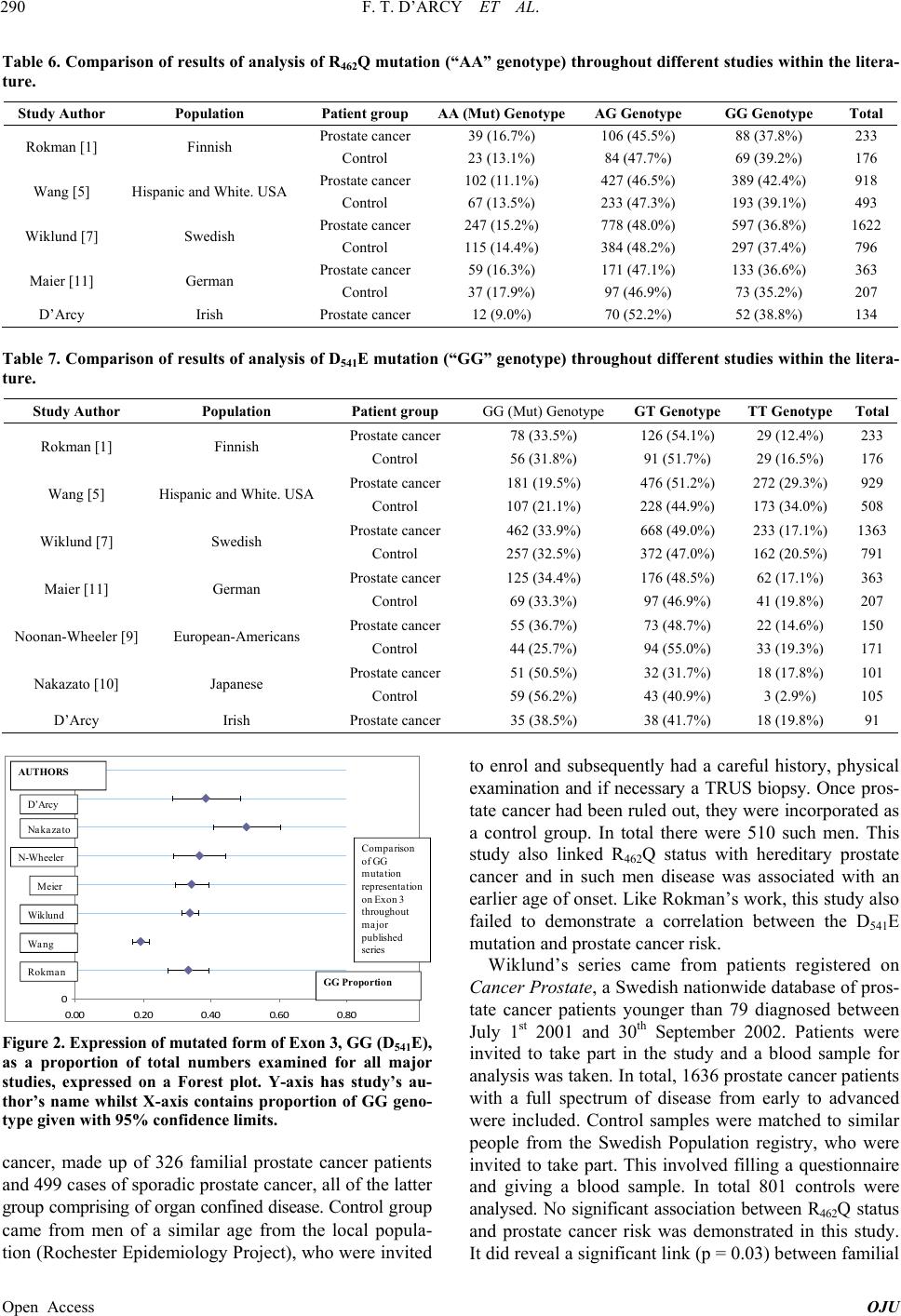 F. T. D’ARCY ET AL. 290 Table 6. Comparison of results of analysis of R462Q mutation (“AA” genotype) throughout different studies within the litera- ture. Study Author Population Patient group AA (Mut) GenotypeAG Genotype GG Genotype Total Prostate cancer 39 (16.7%) 106 (45.5%) 88 (37.8%) 233 Rokman [1] Finnish Control 23 (13.1%) 84 (47.7%) 69 (39.2%) 176 Prostate cancer 102 (11.1%) 427 (46.5%) 389 (42.4%) 918 Wang [5] Hispanic and White. USA Control 67 (13.5%) 233 (47.3%) 193 (39.1%) 493 Prostate cancer 247 (15.2%) 778 (48.0%) 597 (36.8%) 1622 Wiklund [7] Swedish Control 115 (14.4%) 384 (48.2%) 297 (37.4%) 796 Prostate cancer 59 (16.3%) 171 (47.1%) 133 (36.6%) 363 Maier [11] German Control 37 (17.9%) 97 (46.9%) 73 (35.2%) 207 D’Arcy Irish Prostate cancer 12 (9.0%) 70 (52.2%) 52 (38.8%) 134 Table 7. Comparison of results of analysis of D541E mutation (“GG” genotype) throughout different studies within the litera- ture. Study Author Population Patient group GG (Mut) Genotype GT Genotype TT Genotype Total Prostate cancer 78 (33.5%) 126 (54.1%) 29 (12.4%) 233 Rokman [1] Finnish Control 56 (31.8%) 91 (51.7%) 29 (16.5%) 176 Prostate cancer 181 (19.5%) 476 (51.2%) 272 (29.3%) 929 Wang [5] Hispanic and White. USA Control 107 (21.1%) 228 (44.9%) 173 (34.0%) 508 Prostate cancer 462 (33.9%) 668 (49.0%) 233 (17.1%) 1363 Wiklund [7] Swedish Control 257 (32.5%) 372 (47.0%) 162 (20.5%) 791 Prostate cancer 125 (34.4%) 176 (48.5%) 62 (17.1%) 363 Maier [11] German Control 69 (33.3%) 97 (46.9%) 41 (19.8%) 207 Prostate cancer 55 (36.7%) 73 (48.7%) 22 (14.6%) 150 Noonan-Wheeler [9] European-Americans Control 44 (25.7%) 94 (55.0%) 33 (19.3%) 171 Prostate cancer 51 (50.5%) 32 (31.7%) 18 (17.8%) 101 Nakazato [10] Japanese Control 59 (56.2%) 43 (40.9%) 3 (2.9%) 105 D’Arcy Irish Prostate cancer 35 (38.5%) 38 (41.7%) 18 (19.8%) 91 0 1 2 3 4 5 6 7 8 0.000.20 0.400.60 0.80 系列 1 Wan g Wiklund Meier N-Wheeler Na ka za to Rokma n Comparison of GG mutation representa tion onExon 3 throughout major published series AUT HO RS GG Proportion D’Arcy Figure 2. Expression of mutate d form of Exo n 3, GG ( D 541E), as a proportion of total numbers examined for all major studies, expressed on a Forest plot. Y-axis has study’s au- thor’s name whilst X-axis contains proportion of GG geno- type given with 95% confidence limits. cancer, made up of 326 familial prostate cancer patients and 499 cases of sporadic prostate cancer, all of the latter group comprising of organ confined disease. Control group came from men of a similar age from the local popula- tion (Rochester Epidemiology Project), who were invited to enrol and subsequently had a careful history, physical examination and if necessary a TRUS biopsy. Once pros- tate cancer had been ruled out, they were incorporated as a control group. In total there were 510 such men. This study also linked R462Q status with hereditary prostate cancer and in such men disease was associated with an earlier age of onset. Like Rokman’s work, this study also failed to demonstrate a correlation between the D541E mutation and prostate cancer risk. Wiklund’s series came from patients registered on Cancer Prostate, a Swedish nationwide database of pros- tate cancer patients younger than 79 diagnosed between July 1st 2001 and 30th September 2002. Patients were invited to take part in the study and a blood sample for analysis was taken. In total, 1636 prostate cancer patients with a full spectrum of disease from early to advanced were included. Control samples were matched to similar people from the Swedish Population registry, who were invited to take part. This involved filling a questionnaire and giving a blood sample. In total 801 controls were analysed. No significant association between R462Q status and prostate cancer risk was demonstrated in this study. It did reveal a significant link (p = 0.03) between familial Open Access OJU  F. T. D’ARCY ET AL. 291 prostate cancer risk and D541E mutation, however no link between this variant and either age at diagnosis or tu- mour aggressiveness was found. Maier’s patients were recruited from the Prostate Can- cer Genetics Project. This is a database made up of men from predominately from the South of Germany, the ma- jority of whom would have undergone radical prostatec- tomies. Patients are encouraged to enrol in this study by their urologist and there are no selection criteria. At risk families are identified by interview of the patient. There were a total of 303 of such patients. 227 sporadic cases were also sequenced as well as 207 control samples. These samples came from healthy, elderly men with no history of prostate cancer and negative DRE and/or nor- mal PSA levels. D541E or R462Q failed to demonstrate any significant association with prostate cancer in this study. Noonan-Wheeler’s study originated in Missouri, USA. It examined RNase L in men with aggressive, metastatic cancer and healthy controls. Patients were recruited from the outpatient department and were required to have a PSA over 50 ng/ml or radiological/pathological evidence of metastatic disease. The control group consisted of men older than 75 with normal PSA levels and rectal exami- nation with no background of prostate cancer. There were a total of 150 patients and 171 controls examined. It was shown that D541E was over-represented (p = 0.045) in patients with metastatic disease, and concluded that such patients were at an increased risk for sporadic, me- tastatic disease (OR = 1.68). Nakazato’s study came from Japan and examined RNase L in familial prostate cancer cases and healthy controls. It comprised 101 patients with a positive family history of cancer (29 of whom had 3 or more affected family members) and 105 controls. Prostate cancer pa- tients ranged in age from 40 to 88 years, encompassed organ confined and metastatic disease and had 76 men with a Gleason score of 7 or greater and the remaining 26 with a Gleason score of 6 or less. Control cases were recruited from the outpatients department and were of similar age. Patients with an elevated PSA or abnormal DRE were excluded from this group. Interestingly this study demonstrated a significant link between the wild- type DD variant and familial prostate cancer (p = 0.0004, OR = 7.37), possibly an observation unique to the Japa- nese population. 5. Conclusion In conclusion, no statistically significant correlation was proven in the Irish population between Gleason score, percentage gland involvement, patient age, PSA or fam- ily history with any of the studied SNPs. In particular the SNP rs3738579 failed to highlight men with significant clinical cancer. REFERENCES [1] A. Rökman, T. Ikonen, E. H. Seppälä, N. Nupponen, V. Autio, N. Mononen, J. Bailey-Wilson, J. Trent, J. Carpten, M. P. Matikainen, P. A. Koivisto, T. L. Tammela, O. P. Kallioniemi and J. Schleutker, “Germline Alterations of the RNASEL Gene, a Candidate HPC1 Gene at 1q25, in Patients and Families with Prostate Cancer,” The Ameri- can Journal of Human Genetics, Vol. 70, No. 5, 2002, pp. 1299-1304. http://dx.doi.org/10.1086/340450 [2] J. Carpten, N. Nupponen, S. Isaacs, R. Sood, C. Robbins, J. Xu, M. Faruque, T. Moses, C. Ewing, E. Gillanders, P. Hu, P. Bujnovszky, I. Makalowska, A. Baffoe-Bonnie, D. Faith, J. Smith, D. Stephan, K. Wiley, M. Brownstein, D. Gildea, B. Kelly, R. Jenkins, G. Hostetter, M. Matikainen, J. Schleutker, K. Klinger, T. Connors, Y. Xiang, Z. Wang, A. De Marzo, N. Papadopoulos, O. P. Kallioniemi, R. Burk, D. Meyers, H. Gronberg, P. Meltzer, R. Silverman, J. Bailey-Wilson, P. Walsh, W. Isaacs and J. Trent, “Germ- line Mutations in the Ribonuclease L Gene in Families Showing Linkage with HPC1,” Nature Genetics, Vol. 30, No. 2, 2002, pp. 181-184. http://dx.doi.org/10.1038/ng823 [3] A. Orr-Urtreger, A. Bar-Shira, D. Bercovich, N. Mata- rasso, U. Rozovsky, S. Rosner, S. Soloviov, G. Rennert, L. Kadouri, A. Hubert, H. Rennert and H. Matzkin, “RNASEL Mutation Screening and Association Study in Ashkenazi and Non-Ashkenazi Prostate Cancer Patients,” Cancer Epidemiology, Biomarkers & Prevention, Vol. 15, No. 3, 2006, pp. 474-479. http://dx.doi.org/10.1158/1055-9965.EPI-05-0606 [4] Y. Xiang, Z. Wang, J. Murakami, S. Plummer, E. A. Klein, J. D. Carpten, J. M. Trent, W. B. Isaacs, G. Casey and R. H. Silverman, “Effects of RNase L Mutations Associated with Prostate Cancer on Apoptosis Induced by 2’,5’-Oli- goadenylates,” Cancer Research, Vol. 63, No. 20, 2003, pp. 6795-6801. [5] L. Wang, S. K. McDonnell, D. A. Elkins, S. L. Slager, E. Christensen, A. F. Marks, J. M. Cunningham, B. J. Peter- son, S. J. Jacobsen, J. R. Cerhan, M. L. Blute, D. J. Schaid and S. N. Thibodeau, “No Association of Germ- line Alteration of MSR1 with Prostate Cancer Risk,” Na- ture Genetics, Vol. 35, 2003, pp. 128-129. http://dx.doi.org/10.1038/ng1239 [6] G. Casey, P. J. Neville, S. J. Plummer, Y. Xiang, L. M. Krumroy, E. A. Klein, W. J. Catalona, N. Nupponen, J. D. Carpten, J. M. Trent, R. H. Silverman and J. S. Witte, “RNASEL Arg462Gln Variant Is Implicated in up to 13% of Prostate Cancer Cases,” Nature Genetics, Vol. 32, No. 4, 2002, pp. 581-583. http://dx.doi.org/10.1038/ng1021 [7] F. Wiklund, B. A. Jonsson, A. J. Brookes, L. Stromqvist, J. Adolfsson, M. Emanuelsson, H. O. Adami, K. Augusts- son-Balter and H. Gronberg, “Genetic Analysis of the RNASEL Gene in Hereditary, Familial, and Sporadic Pros- tate Cancer,” Clinical Cancer Research, Vol. 10, No. 21, 2004, pp. 7150-7156. http://dx.doi.org/10.1158/1078-0432.CCR-04-0982 [8] H. Li and B. C. Tai, “RNASEL Gene Polymorphisms and the Risk of Prostate Cancer: A Meta-Analysis,” Clinical Cancer Research, Vol. 12, 2006, pp. 5713-5719. http://dx.doi.org/10.1158/1078-0432.CCR-05-2799 Open Access OJU  F. T. D’ARCY ET AL. Open Access OJU 292 [9] F. C. Noonan-Wheeler, W. Wu, K. A. Roehl, A. Klim, J. Haugen, B. K. Suarez and A. S. Kibel, “Association of Hereditary Prostate Cancer Gene Polymorphic Variants with Sporadic Aggressive Prostate Carcinoma,” Prostate, Vol. 66, No. 1, 2006, pp. 49-56. http://dx.doi.org/10.1002/pros.20320 [10] H. Nakazato, K. Suzuki, H. Matsui, N. Ohtake, S. Nakata and H. Yamanaka, “Role of Genetic Polymorphisms of the RNASEL Gene on Familial Prostate Cancer Risk in a Japanese Population,” British Journal of Cancer, Vol. 89, No. 4, 2003, pp. 691-696. http://dx.doi.org/10.1038/sj.bjc.6601075 [11] C. Maier, J. Haeusler, K. Herkommer, Z. Vesovic, J. Hoe- gel, W. Vogel and T. Paiss, “Mutation Screening and Association Study of RNASEL as a Prostate Cancer Sus- ceptibility Gene,” British Journal of Cancer, Vol. 92, 2005, pp. 1159-1164. http://dx.doi.org/10.1038/sj.bjc.6602401 [12] B. E. Madsen, E. M. Ramos, M. Boulard, K. Duda, J. Overgaard, M. Nordsmark, C. Wiuf and L. L. Hansen, “Germline Mutation in RNASEL Predicts Increased Risk of Head and Neck, Uterine Cervix and Breast Cancer,” PLoS One, Vol. 3, No. 6, 2008, Article ID: e2492. http://dx.doi.org/10.1371/journal.pone.0002492 [13] S. Nair and M. R. Pillai, “Human Papillomavirus and Disease Mechanisms: Relevance to Oral and Cervical Can- cers,” Oral Diseases, Vol. 11, 2005, pp. 350-359. http://dx.doi.org/10.1111/j.1601-0825.2005.01127.x [14] A. Leodolter, M. Naumann and P. Malfertheiner, “Pre- vention of Gastric Cancer by Helicobacter pylori Eradi- cation,” Digestive Diseases, Vol. 22, 2004, pp. 313-319. http://dx.doi.org/10.1159/000083592 [15] M. A. Epstein, B. G. Achong and Y. M. Barr, “Virus Par- ticles in Cultured Lymphoblasts from Burkitt’s Lym- phoma,” Lancet, Vol. 15, 1964, pp. 702-703. http://dx.doi.org/10.1016/S0140-6736(64)91524-7 [16] J. E. McGrory, D. J. Pritchard, K. K. Unni, D. Ilstrup and C. M. Rowland, “Malignant Lesions Arising in Chronic Osteomyelitis,” Clinical Orthopaedics and Related Re- search, Vol. 362, 1999, pp. 181-189. http://dx.doi.org/10.1097/00003086-199905000-00027 [17] L. K. Dennis and D. V. Dawson, “Meta-Analysis of Mea- sures of Sexual Activity and Prostate Cancer,” Epidemi- ology, Vol. 13, 2002, pp. 72-79. http://dx.doi.org/10.1097/00001648-200201000-00012 [18] R. B. Hayes, L. M. Pottern, H. Strickler, C. Rabkin, V. Pope, G. M. Swanson, R. S. Greenberg, J. B. Schoenberg, J. Liff, A. G. Schwartz, R. N. Hoover and J. F. Fraumeni Jr., “Sexual Behaviour, STDs and Risks for Prostate Can- cer,” British Journal of Cancer, Vol. 82, 2000, pp. 718- 725. http://dx.doi.org/10.1054/bjoc.1999.0986 [19] M. L. Taylor, A. G. Mainous 3rd and B. J. Wells, “Pros- tate Cancer and Sexually Transmitted Diseases: A Meta- Analysis,” Family Medicine Journal, Vol. 37, 2005, pp. 506-512. [20] A. V. Sarma, J. C. McLaughlin, L. P. Wallner, R. L. Dunn, K. A. Cooney, D. Schottenfeld, J. E. Montie and J. T. Wei, “Sexual Behavior, Sexually Transmitted Diseases and Prostatitis: The Risk of Prostate Cancer in Black Men,” Journal of Urology, Vol. 176, No. 3, 2006, pp. 1108-1113. http://dx.doi.org/10.1016/j.juro.2006.04.075 |