

Paper Menu >>
Journal Menu >>
 Journal of Minerals and Materials Characterization and Engineering, 2013, 1, 347-352 Published Online November 2013 (ht t p:/ / www.scirp.org/journal/jmmce ) http://dx.doi.org/10.4236/jmmce.2013.16053 Open Access JMMCE Crystallographic Study of Uranium-Thorium Bearing Minerals in Tranomaro, South-East Madagas car Frank Elliot Sahoa1, Naivo Rabesiranana1*, Raoelina Andriambololona1, Nicolas Finck2, Christian Marquardt2, Hörst Geckeis2 1Institut National des Sciences et Techniques Nucléaires (Madagascar-INSTN), Antananarivo, Madagascar 2Institute of Nuclear Waste and Disposal (INE), Karlsruhe Institute of Technology (KIT), Karlsruhe, Germany Email: *[email protected] Received September 20, 2013; revised October 21, 2013; accepted November 5, 2013 Copyright © 2013 Frank Elliot Sahoa et al. This is an open access article distributed under the Creative Commons Attribution Li- cense, which permits unrestricted use, distribution, and reproduction in any medium, provided the original work is properly cited. ABSTRACT Studies are undertaken to characterize the uranium and thorium minerals of south-east Madagascar. Seven selected uranothorianite bearing pyroxenites samples from old abandoned uranium quarries in Tranomaro, south Amboasary, Madagascar (46˚28'00"E, 24˚36'00"S) have been collected. To determine the mineral micro-structure, they were inves- tigated for qualitative identification of crystalline compounds by using X-ray powder diffraction analytical method (XRD). Results showed that the uranium and thorium compounds, as minor elements, were present in various crystal- line structures. Thorium, as thorianite, is present in a simple ThO2 cu bic crystalline system, whereas the uranium com- ponent of the Tranomaro uranothorianite samples is oxide-based and is a mixture of complex oxidation states and crys- talline systems. Generally called uraninite, its oxide compounds are present in more than eight phases. Keywords: Tranomaro; Madagascar; Uranothorianite; Crystallography; XRD 1. Introduction In 1907, a Malagasy mineralogist, J. B. Rasamoel, dis- covered euxenite-bearing tertiary lake sediments near Vinaninkarena. French interest in the source of radium led to intensive search for primary uranium minerals in the adjacent upland plateau [1]. The central island is one of the regions of interest of a Manhattan District Project. In 19 43- 1944, the Kitsamby district, located in the regio n, was then explored for potential uranium ore deposit by the project specialists [2]. But Moreau [3] has observed uranium-thorium mineralization in Tranomaro, south- east of Madagascar, where major extracting activities were undertaken by the French Commissariat à l’Energie Atomique (CEA) during the fifties and sixties, to support their effort to develo p nucl e a r pr o ject . In 1979, using new analytical technology such as gamma spectrometry system, Raoelina Andriambololona et al. [4] have further investigated the radioactivity char- acteristics of uranium and thorium minerals from Mada- gascar, in particular from the South-East region [5-7]. Rabesiranana et al. [8] reported the natural radionuclide content in soil, focusing mainly on the comparison be- tween on-site vs. off-site data, to detect significant dif- ferences, taking into account spatial variability. Using electronic microprobe analysis and X-ray fluorescence technique, Rakotondratsima and Moine et al. [9,10] studied the origin and concentration of uranothorianite bearing pyroxenite content in the region, and have demon- strated the formation of uranothorianite bearing pyrox- enites with marble at Belafa and Marosohihy and miner- alization without marble at Ambidandrakemba, two bil- lion years ago. Rakotondrazafy used XRD technique and microprobe analysis for dating and determining the foma- tion conditions of hibonite in Madagascar. He has indicated that hibonite mineral has been plainly confined in uranotho- rianite bearing skarns formed 545 million years ago [11]. Recently, in order to supply worldwide increasing need for alternative energy, and in view of future short- age of conventional fossil fuel, there is a renew interest in nuclear energy. Consequ ently, mining co mpanies are ex- ploring old and newly discovered uranium-thorium pro- vinces of the country. This is the case for the South East region of Madagascar, where the uranothorianite deposits of the Tranomaro province are re-investigated (Figure 1). 2. Rationale The uranium and thorium compounds may be mobilized *Corresponding author. 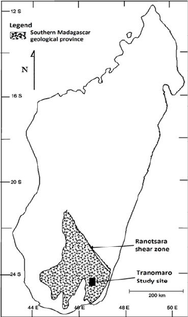 F. E. SAHOA ET AL. 348 Figure 1. Geographical location of the tranomaro study site, south-east madagascar. and transported into the environmental compartments, increasing the risk of human contamination and exposure from radioactive elements. If the mobilization is limited for an undisturbe d g eolog ical formation, human activ ities such as mining may accelerate the process. The disper- sion depends on 1) the weathering/leaching efficiency, 2) the uranium-thorium structure resistance and 3) the dis- turbance magnitude. Rock weathering is normally insti- gated by air and water from the earth’s surface gaining access to minerals previously surrounded by other min- erals. When air and water reach previously inaccessible parts of the rock matrix, chemical reaction such as oxida- tion and leaching can change the rock’s mineral compo- sition [12,13]. Uranium and thorium elements occur in the 4+ oxida- tio n st at e in p r imary igneous rocks and minerals. As s u c h , both elements are almost chemically immobile in the near surface environment at low temperature. In the oxi- dized zone of the terrestrial near-surface environment, uranium and thorium may be mobilized, but in different ways. According to Gascoyne [14], thorium is mainly transported in insoluble resistate minerals or is adsorbed on the surface of clay minerals. But uranium can be oxi- diz e d to 5 + an d 6+ states in the near surface environ me nt . The most probable oxidation reaction at normal envi- ronment is [15]: 42 22 U2HOUO4H2e The 6+ oxidation state is the most stable and forms soluble uranyl complex ions which play an important role in uranium transport during weathering [14]. Finch and Ewing [16] reported that in a highly al- tered uraninite-bearing ore body of Koongara, Australia, partially exposed to meteoric water, alteration at depth has resulted from interaction with groundwater having a reduced Eh compared to the surface. Urani nite, Pb-uranyl oxide hydrates and uranyl silicates control uranium solu- bility at depth; uranyl phosphates and uranium adsorption onto clays and FeMn-oxides control uranium solubility near the surface. So, uranium may either move in solu- tion as complex ion, or like thorium, in a sorbed or detri- tal phase. 2 2 UO Most of the studies report mobilization in terms of oxidation state. To further support the risk analysis re- lated to the dispersion of uranium and thorium from solid phases to the environment, crystallographic structure of uranium-thorium compounds in pyroxenite minerals from the Tranomaro uranium-thorium province, south- east Madagascar, is investigated. The sample characteri- zation has been conducted using X-ray powder diffrac- tion analytical technique (XRD). 3. Experimental Methodology Prior to the study, radiometric measurement within the study area had been done by the Pan African Mining Corporations-Madagascar team. Using Exploranium scintillator counters, high radioactivity spots had been localized. Sampling for the current study was subse- quently conducted in 2009, and sampling points were then selected according to counting level previously found. Only samples with count rate greater than 1500 counts per second (cps) have been selected. They were usually from abandoned mining quarries, with details shown in Table 1. Most of the samples are crude rocks, but one of them is a pure uranothorianite mineral care- fully picked from a pyroxenite mineral sample. Mineral sample is ground and sieved in order to get 10 µm to 20 µm size fine and homogeneous powder. For low diffraction background, the powder is packed into a sample holder made of silicon single crystal and smeared uniformly onto a glass slide. When packing into a sample container, care is taken to create a flat upper surface and to achieve a random distribution of the crystalline lattice orientations. The XRD analysis is carried out using a Bruker D8 Advance diffractometer equipped with a copper anode, powered at (40 kV, 40 mA), and controlled by the XRD Commander software. The sample is illuminated by the Cu-Kα radiation (λ = 1.5406 Å) and the diffracted beam Open Access JMMCE  F. E. SAHOA ET AL. Open Access JMMCE 349 Table 1. Collected mineral samples and their localization. Code Surface Counts Rate (cps) Location Longitude Latitude Mine code Name R01M37 1610 46˚30’24’’E 24˚34’43’’S M37 Ambindandrakemba R01M47 1650 46˚31’47’’E 24˚23’36’’S M47 Marosohihy R02M47 2500 46˚31’46’’E 24˚23’36’’S M47 Marosohihy R03M47 3000 46˚31’45’’E 24˚23’38’’S M47 Marosohihy R01M52 1720 46˚31’36’’E 24˚21’47’’S M52 Belafa R02M52 1725 46˚31’48’’E 24˚21’55’’S M52 Belafa Uranothorianite 2800 46˚30’27’’E 24˚4’12’’S Uranium-thorium mineral is detected with a Sol-XE energy dispersive detector. The diffractogram is collected for 2θ varying from 5˚ to 80˚ with steps of 0.01˚ and 2 seconds counting time per step, and the sample spinning at 15 rotations per minute. The diffractogram displaying the diffracted intensity as a function of the diffraction angle is processed using the Bruker Eva software. Qualitative analysis is carried out by comparison with the JCPDS database (Joint Committee for Powder Dif- fraction Studies). The more peaks match the data from the database, the higher the confidence in phase identifi- cation. For samples containing more than one single ph ase, the position of the characteristic peaks ca n ov er lap . Since each pure crystalline phase is characterized by a specific diffraction pattern, the identification of the phases present in the sample is enhanced by using the Rietweld method. Essentially, it involves fitting the ob- served diffraction pattern with a synthetic pattern which is a sum of patterns calculated for each phase in the sam- ple [17]. As such this method is known as a full-pattern fitting method [18]. The difference between the synthetic pattern and the observed pattern is minimized by an in- teractive refinement/optimization procedure. The amounts of the phases present in the sample are obtained from the final value of the refined scale factor for each phase [19]. To improve the accuracy, only compounds which are likely to be found in the studied samples are selected for identification. From previous studies [9,20], Table 2 shows major minerals and uranium-thorium compounds in py- roxenite minerals from the study site. Additionally, all uranium-thorium phase diffraction patterns are added from the JCPDS database for identification. 4. Results and Discussion Examples of the graphical output from the Bruker Eva software are shown in Figure 2. Two typical diffracto- grams are presented: Figure 2(a) shows a simple spec- trum from pure uranium-thorium minerals, whereas Fig- ure 2(b) shows a complex spectrum from the R01M37 sample, which is a natural uranium-thorium ore. A summary of the processed result is presented in Ta- ble 3. For natural ores, common major and minor mineral Table 2. Minerals expected to be found in the studied ura- nium-thorium bearing rocks. Expected elements Chemical formula Uranothorianite (Th,U)O2 Diopside pyroxenite CaMg(SiO3)2 Calcite CaCO3 Corundum CaMg(SiO3)2 Spinel FeAl2O4, MgCr2O4 Apatite Ca5(PO4)3(Cl,F,OH) Phlogopites KMg3ASi3O10 Hibonite CaAl12O19 phases such as corundum, spinel, diopside, soddyite, cal- cium silicate and calcite are usually present in the sam- ples. Interestingly, uranium-thorium compounds are pre- sent in multiple crystalline phases and systems. The uranothorianite sample contains thorianite as ThO2 in cubic system. The R01M37 sample contains 1) Th0.5U0.5O2.04 in cubic system, which can be considered as a mixture of UO2 and ThO2, and 2) Fe2UO6 in hexagonal sys- tem, which can be considered as a mixture of Fe2O3 and UO3. The R01M47 sample contains only UO3 in mono- clinic system. The R02M47 sample contains 1) UO3 in monoclinic system and 2) U2O5 in orthorhombic system. The R03M47 sample contains 1) UO3 in monoclinic system and 2) U2P2O10 as a complex system, which can be considered as a mixture of U2O5 and P2O5. The R01M52 sample contains 1) thorianite as ThO2 in cubic system, 2) uranium oxide as U3O7 in tetra- gonal system and 3) uraninite as UO2 in orthorhom- bic system. The R02M52 sample contains 1) thorianite as ThO2 in cubic system and 2) uranium oxide as UO3. The presence of both thorium and uranium oxides is expected. According to Frondel [21], uraninite is com- monly present in pegmatites. Moreau explained that pre- viously, thorium and uranium were rather concentrated in the granitic and charnocktic zones. Precambrian granite- 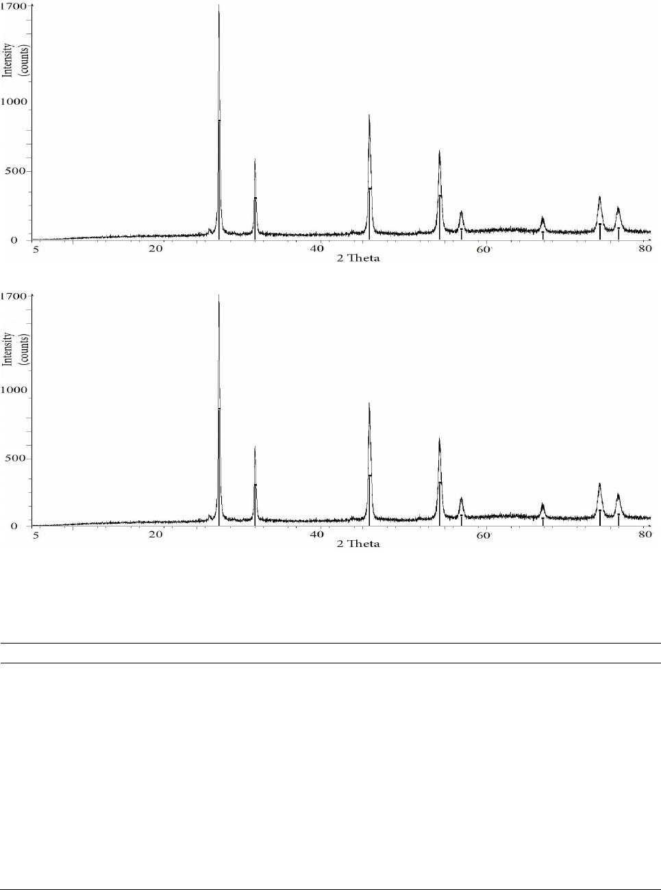 F. E. SAHOA ET AL. 350 (a) (b) Figure 2. Examples of the graphical output from the Bruker Eva software. (a) Pure XRD spectrum from uranothorianite minerals; (b) Complex XRD spectrum from R01M37 sample. Table 3. Crystalline systems of uranium-thorium minerals identified in the studied samples by XRD analysis. Sample code Identified compounds Chemical formula Crystalline systems Uranothorianite Thorianite ThO2 cubic R01M37 Thorium uranium oxide Th0.5U0.5O2.04 cubic Iron uranium o xide Fe2UO6 hexagonal R01M47 Uranium oxide UO3 monoclinic R02M47 Uranium oxide UO3 monoclinic Uranium oxide U2O5 orthorombic R03M47 Uranium oxide UO3 monoclinic Uranium oxide phosphate U2P2O10 complex R01M52 Thorianite ThO2 cubic Uranium oxide U3O7 tetragonal Uraninite UO2 orthorombic R02M52 Thorianite ThO2 cubic Uranium oxide UO3 uncertain Open Access JMMCE  F. E. SAHOA ET AL. 351 zation is followed by the formation of the main pegma- titic areas in Madagascar with thorium-uranium niobo- tantalates, uraninite and beryl. In the same time, a large uranium and thorium provinces with uranothorianite de- posits appear within the calcomagnesian series of the southern area of Madagascar [22]. Thorianite is an iso- metric ThO2 crystal (found in R01M37, R01M52, R02M52 and uranothorianite samples) in the hexoctahe- dral class _ 42 3 mm sometimes modified to {111} or {113} [21]. The uran- oth orianite diffractogram (Figure 2(a)) particularly shows the dominance of ThO2 in uranothorianite sample. This demonstrates that thorianite is the main component of the Tranomaro uranothorianite mineral, and that uranium may be contained in other co-existing phases. If thorium oxide exists only as cubic system, by con- trast, the uranium oxides are present in several and more complex crystalline systems, generally called uraninite. In fact, various definitions of uraninite are proposed by different authors. According to Rogers [23], uraninite is described as the crystalline mineral with higher specific gravity and lower water content and pitchblende for the massive mineraloid with lower specific gravity and higher water content. Following the Dana classification, uraninite is de fined as an oxide for m of uranium and tho- rium and has an isometric crystalline structure. Accord- ing to Wasserstein [24,25] who proposed a classification based on the radiogenic lead content and the redox con- ditions, α-uraninite corresponds to UO2 (found in R01M37 and R01M52 samples), β-uraninite to U3O7 (found in R01M52 sample) and τ-uraninite to U4O9. It is also known that UO2 is transformed into U3O8 and UO3 (found in R01M37, R01M47, R02M47, R03M47 and R02M52 samples) under oxidizing conditions. Ebelmen reported also that U2O5 (found in R02M47 and R03M47 samples) is an essential constituent of uraninite [26]. In the Tranomaro area, the presence of the uraninite is closely linked to that of the thorianite. This is because uranium and thorium are isometric and both present in minerals at different concentrations (88.15% of uranium in uraninite and 70% of thorium in thorianite). Some authors have proposed a solid solution model to explain the possibility of uranium-thorium exchange between the uraninite and thorianite systems, but their oxidation be- haviours differ from each another [27-29]. On one hand, this results in a simple crystal system for thorianite: it only exists in a cubic system. On the other hand, the ura- nium component of the Tranomaro uranothorianite sam- ples are oxide based and is a mixture of complex oxida- tions states and crystalline systems. The finding is in agreement with published results, showing that uraninite is a complex uranium oxide compounds [24,25,27]. 5. Conclusion Due to the presence of radioactive minerals, populations living in the Tranomaro area, south-east Madagascar may be exposed to increased environmental radiation. The study of seven mineral samples collected from the site, using X-ray diffraction, confirms the existence of ura- nium-thorium bearing minerals in the Tranomaro deposit, and moreover, gives an insight on the crystalline struc- ture and combination of the uranium-thorium minerals. The uranium and thorium are present in a complex mul- tiple mineral phases. If the thorium oxide is identified in a simple crystalline system, on the opposite, the uranium oxides, classified as uraninite, are present in more than eight phases and show multiple oxidation states and crystalline systems, including α-uraninites and β-urani- nite. As each crystalline system is more or less subject to dissolution or dispersion in the environment, further in- vestigation must be done to investigate the details of their mobilization and transport in the environment, in terms of crystallographic phases. 6. Acknowledgements This paper was completed within the framework of a cooperation between the Institut National des Sciences et Techniques Nucléaires (Madagascar-INSTN) and the Institute of Nuclear Waste and Disposal (INE). The German Academic Exchange Service (Deutscher Akade- mischer Austausch Dienst: DAAD) financed a fellowship. The Pan African Mining-Madagascar (PAM) facilitated the access to the Tranomaro sites. Technical supports fr om Madagascar-INSTN and INE are gratefully acknow- ledged. REFERENCES [1] C. Grossman, “Uranium Ores in Madagascar,” Journal of the Franklin Institute, Vol. 179, No. 5, 1915, p. 595. [2] W. Bourret, “Uranium-Bearing Pegmatites of the Antsir- abe-Kitsamby District, Madagascar,” In: J. W. Gabelman Ed., Unconventional Uranium Deposits, Ore Geology Reviews, Vol. 3, 1988, pp. 177-191. [3] M. Moreau, “Cycles Uranifères et Thorifères à Madaga- scar, ” Rapport C.E.A, n° 1685, Commissariat à l’Energie Atomique, 1960. [4] Raoelina Andriambololona, G. Rakotoson and G. Paic, “Measurement of the Escape Rate of Radon in Uranium Minerals from Madagascar,” International Journal of Ap- plied Radiation and Isotopes, Vol. 34, No. 7, 1983, pp. 1017-1018. http://dx.doi.org/10.1016/0020-708X(83)90084-4 [5] G. Paic, Raoelina Andriambololona and G. Rakotoson, “Estimation du Pouvoir d’Échappement du Rn-222 des Open Access JMMCE  F. E. SAHOA ET AL. 352 Roches Uranifères de Madagascar à l’aide d’un Détecteur Ge(Li),” Annales de l’Université de Madagascar, Série Sciences de la Nature et Mathématique, No. 16, 1979, pp. 79-91. [6] G. Paic, Raoelina Andriambololona and M. Razanajatovo, “Détermination du Spectre Énergétique Gamma de la Monazite de Fort-Dauphin à l’aide d’un Détecteur Ge(Li),” Annales de l’Université de Madagascar, Série Sciences de la Nature et Mathématique, No. 16, 1979, pp. 79-91. [7] Raoelina Andriambololona, “Etude de la Monazite Brute d’Antete et de Vohibarika Manantenina. Mesure des Teneurs en Uranium et Thorium,” Annales de l’Univesité de Madagascar, Sciences de la Nature et Mathématique, No. 17, 1980, pp. 15-32. [8] N. Rabesiranana, M. Rasolonirina, F. Terina, A. F. So- lonjara and Raoelina Andriambololona, “Top Soil Radio- activity Assessment in a High Natural Radiation Back- ground Area: The Case of Vinaninkarena, Antsirabe- Madagascar,” Applied Radiations and Isotopes, Vol. 66, No. 11, 2008, pp. 1619-1622. http://dx.doi.org/10.1016/j.apradiso.2007.12.012 [9] C. Rakotondratsima, “Les Pyrexonites à Uranothorianite du Sud-Est de Madagascar,” Ph.D Thesis, University of Claude Bernard-Lyon, 1983. [10] B. Moine, C. Rakotondratsima and M. Cuney, “Les Pyro- xénites à Urano-Thorianite du Sud-Est de Madagascar: Conditions Physico-Chimiques de la Métasomatose,” Bulletin de Minéralogie, Vol. 108, 1985, pp. 325-340. [11] A. F. M. Rakotondrazafy, “La Hibonite du Sud-Est de Madagascar,” Ph.D Thesis, University of Antananarivo, 1995. [12] J. K. Osmond and M. Ivanovich, “Uranium-Series Mobi- lization and Surface Hydrology,” In: M. Ivanovich and R. S. Harmon, Eds., Uranium Series Disequilibrium: Appli- cations to Earth, Marine and Environmental Sciences, 2nd Edition, Clarendon Press, Oxford, 1992, pp. 259-289. [13] J. N. Rosholt, “Mobilization and Weathering,” In: M. Ivanovich and R. S. Harmon, Eds., Uranium Series Dis- equilibrium: Applications to Environmental Problems, Clarendon Press, Oxford, 1982, pp. 167-180. [14] M. Gascoyne, “Geochemistry of the Actinides and Their Daughters,” In: M. Ivanovich and R. S. Harmon, Eds., Uranium Series Disequilibrium: Applications to Earth, Marine and Environmental Sciences, 2nd Edition, Clar- endon Press, Oxford, 1992, pp. 34-61. [15] D. Langmuir, “Uranium Solution-Minerals Equilibria at Low Temperatures with Application to Sedimentary Ore Deposits,” Geochimica Cosmochimica Acta, Vol. 42, No. 6, 1978, pp. 547-569. http://dx.doi.org/10.1016/0016-7037(78)90001-7 [16] R. Finch and R. Ewing, “Uraninite Alteration in an Oxy- dizing Environment and Its Relevance to the Disposal of Spent Nuclear Fuel,” Department of Geology, University of New Mexico, 1990. [17] H. M. Rietveld, “A Profile Refinement Method for Nu- clear and Magnetic Structures,” Journal of Applied Crys- tallography, Vol. 2, 1969, pp. 65-71. http://dx.doi.org/10.1107/S0021889869006558 [18] R.L. Snyder and D. L. Bish, “Quantitative Analysis,” In: D. L. Bish and J. E. Post, Eds., Modern Powder Diffrac- tion. Reviews in Mineralogy, Vol. 20, Mineralogical So- ciety of America, Washington DC, 1989, pp. 101-144. [19] L. B. Mc. Cusker, R. B. Von Dreele, D. E. Cox and P. Louër Scardi, “Rietveld Refinement Guidelines,” Journal of Applied Crystallography, Vol. 32, 1999, pp. 36-50. http://dx.doi.org/10.1107/S0021889898009856 [20] M. A. F. Rakotondrazafy, B. Moine and M. Cuney, “Mode of Formation of Hibonite (CaAl12O19) within the U-Th Skarns from the Granulites of S-E Madagascar,” Contributions to Mineralogy and Petrology, Vol. 123, No. 2, 1996, pp. 190-201. http://dx.doi.org/10.1007/s004100050150 [21] C. Frondel, “Systematic Mineralogy of Uranium and Tho- rium,” US Geological Survey Bulletin, Vol. 1064, 1958, 400 p. [22] M. Moreau, “Le Gisement des Minéralisations de Tho- rianite à Madagascar,” Annales de Géologie de Madaga- scar, Fasc. 33, 1963, pp. 175-178. [23] A. F. Rogers, “Uraninite and Pitchblende,” Proceedings of the 27th Annual Meeting of the Mineralogical Society of America, Stanford University, Chicago, 26-28 Decem- ber 1946, pp. 90-91. [24] B. Wasserstein, “Age of Uraninite by a New Method,” Nature, Vol. 174, No. 4439, 1954, pp. 1004-1005. http://dx.doi.org/10.1038/1741004b0 [25] B. Wasserstein, “Cube-Edges of Uraninite as a Criterion of Age,” Nature, Vol. 168, No. 4270 1951, p. 380. http://dx.doi.org/10.1038/168380a0 [26] J. J. Ebelman, “Note on Chemical Composition of Pitch- blende,” Journal für Praktische Chemie, Vol. 30, 1943, p. 414. [27] K. B. Alberman and J. S. Anderson, “The Oxides of Ura- nium,” Journal of the Chemical Society, 1949, pp. S303- S311. http://dx.doi.org/10.1039/jr949000s303 [28] W. Biltz and H. Muller, “Systematic-Affinity Principle; XLI, Uranium Oxides,” Zeitschrift fuer Anorganisch und Allgemeine Chemie, Vol. 163, No. 1, 1927, pp. 257-296. http://dx.doi.org/10.1002/zaac.19271630126 [29] V. M. Goldschmidt and L. Thomassen, “Die Kristall- struktur Natürlicher und Synthetischer Oxyde von Uran,” Thorium und Cerium: Norske Vidensk, Kristiana Skr Math-Naturvidensk. Kl, No. 2, 1923, pp. 1-48. Open Access JMMCE |