

Paper Menu >>
Journal Menu >>
 Open Journal of Gastroenterology, 2013, 3, 317-321 OJGas http://dx.doi.org/10.4236/ojgas.2013.37056 Published Online November 2013 (http://www.scirp.org/journal/ojgas/) Predictors of early rebleeding and mortality after acute variceal haemorrhage in patients with cirrhosis* Iliass Charif#, Kaoutar Saada, Ihsane Mellouki, Mounia El Yousfi, Dafr Allah Benajah, Mohamed El Abkari, Adil Ibrahimi, Nourdin Aqodad Department of Hepatology and Gastroenterology, Hassan II University Hospital, Faculty of Medicine and Pharmacy, Sidi Moham- med Ben Abdellah University of Fez, Fez, Morocco Email: #[email protected] Received 25 September 2013; revised 26 October 2013; accepted 14 November 2013 Copyright © 2013 Iliass Charif et al. This is an open access article distributed under the Creative Commons Attribution License, which permits unrestricted use, distribution, and reproduction in any medium, provided the original work is properly cited. ABSTRACT The upper gastrointestinal bleeding from esophageal or gastric varices is the most dangerous complication of portal hypertension. The purpose of this study was to identify the predictors of early rebleeding and mor- tality after a bleeding episode. Patients and Methods: It was a retrospective study including 215 patients admitted in our department of hepatology and gas- troenterology at the Hassan II University Hospital of Fez, from January 2001 to January 2010. Results: The mean age of our patients was 51 years. Thirty percent of patients had cirrhosis due to virus (B or C). The majority of patients (79%) had only esophageal varices. Fifty patients (23%) had a bleeding recur- rence. Twenty-five patients (11.5%) died during the first ten days, of which 52% had presented rebleeding (p = 0.01). In 30% of cases, the rebleeding was secon- dary to a fall of pressure ulcers. Univariate analysis showed that early mortality of patients was signifi- cantly associated with advanced age (p = 0.018), low prothrombin time (PT) (p = 0.022), low serum sodium (p = 0.03), low platelet count (p = 0.05), and elevated transaminases (p = 0.02). Conclusion: The survival of cirrhotic patients after a bleeding episode was influ- enced by advanced age, a low rate of PT, of serum sodium, and of the platelet count, and elevated trans- aminases. Keywords: Cirrhosis; Portal Hypertension; Gastrointestinal Bleeding; Esophageal Varices 1. INTRODUCTION The upper gastrointestinal bleeding from esophageal var- ices is one of the most dangerous complications of portal hypertension, with a consistently high morbidity and mortality [1,2]. Despite advances in the management of variceal bleeding, mortality following a bleeding epi- sode in cirrhotic patients has decreased by only 20% [3,4]. Several risk factors associated with mortality after a bleeding episode were identified. These factors are: active bleeding during the initial endoscopy, venous pressure gradient, portal vein thrombosis, alcoholic liver disease, bilirubin, albumin, hematocrit, transaminases, encephalopathy, hepatocellular carcinoma and Child Pugh score [5,6]. Many of these data were collected from retrospective studies where mortality after variceal bleeding was greater than 50%. But these studies have included a relatively small number of patients, or groups with no standard treatment especially concerning the use of antibiotics [5,7,8]. Several prognostic scores have been developed in cirrhotic patients with variceal bleed- ing. Currently, the most used are the Child-Pugh score and MELD. Recent studies have demonstrated the ability of these scores to predict early mortality in these patients [9]. Given the significant changes in the natural history of cirrhosis from the broad and standardized use of anti- biotic prophylaxis and endoscopic treatment, and given the etiological feature of cirrhosis in our population characterised by the high prevalence of viral cirrhosis, we have found that it is very interesting to investigate predictor factors of rebleeding and those involved in the short-term survival of cirrhotic patients with variceal bleeding. 2. PATIENTS AND METHODS 2.1. Study Population We present a retrospective study of patients with cirrho- sis and variceal bleeding, admitted in our department of hepatology and gastroenterology at the University Hos- *Conflicts of interest: No conflicts of interest. #Corresponding author. OPEN ACCESS 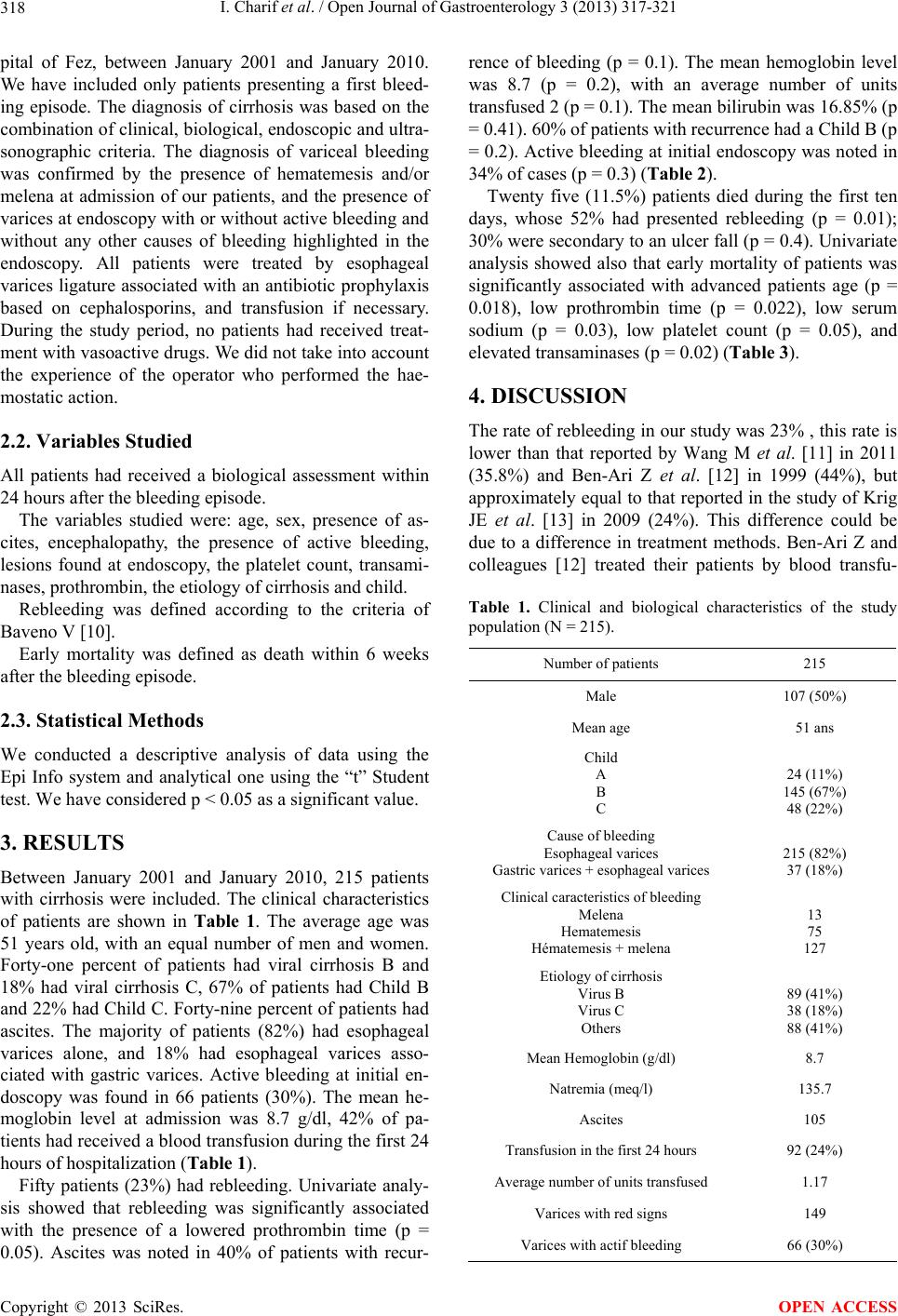 I. Charif et al. / Open Journal of Gastroenterology 3 (2013) 317-321 318 pital of Fez, between January 2001 and January 2010. We have included only patients presenting a first bleed- ing episode. The diagnosis of cirrhosis was based on the combination of clinical, biological, endoscopic and ultra- sonographic criteria. The diagnosis of variceal bleeding was confirmed by the presence of hematemesis and/or melena at admission of our patients, and the presence of varices at endoscopy with or without active bleeding and without any other causes of bleeding highlighted in the endoscopy. All patients were treated by esophageal varices ligature associated with an antibiotic prophylaxis based on cephalosporins, and transfusion if necessary. During the study period, no patients had received treat- ment with vasoactive drugs. We did not take into account the experience of the operator who performed the hae- mostatic action. 2.2. Variables Studied All patients had received a biological assessment within 24 hours after the bleeding episode. The variables studied were: age, sex, presence of as- cites, encephalopathy, the presence of active bleeding, lesions found at endoscopy, the platelet count, transami- nases, prothrombin, the etiology of cirrhosis and child. Rebleeding was defined according to the criteria of Baveno V [10]. Early mortality was defined as death within 6 weeks after the bleeding episode. 2.3. Statistical Methods We conducted a descriptive analysis of data using the Epi Info system and analytical one using the “t” Student test. We have considered p < 0.05 as a significant value. 3. RESULTS Between January 2001 and January 2010, 215 patients with cirrhosis were included. The clinical characteristics of patients are shown in Table 1. The average age was 51 years old, with an equal number of men and women. Forty-one percent of patients had viral cirrhosis B and 18% had viral cirrhosis C, 67% of patients had Child B and 22% had Child C. Forty-nine percent of patients had ascites. The majority of patients (82%) had esophageal varices alone, and 18% had esophageal varices asso- ciated with gastric varices. Active bleeding at initial en- doscopy was found in 66 patients (30%). The mean he- moglobin level at admission was 8.7 g/dl, 42% of pa- tients had received a blood transfusion during the first 24 hours of hospitalization (Table 1). Fifty patients (23%) had rebleeding. Univariate analy- sis showed that rebleeding was significantly associated with the presence of a lowered prothrombin time (p = 0.05). Ascites was noted in 40% of patients with recur- rence of bleeding (p = 0.1). The mean hemoglobin level was 8.7 (p = 0.2), with an average number of units transfused 2 (p = 0.1). The mean bilirubin was 16.85% (p = 0.41). 60% of patients with recurrence had a Child B (p = 0.2). Active bleeding at initial endoscopy was noted in 34% of cases (p = 0.3) (Table 2). Twenty five (11.5%) patients died during the first ten days, whose 52% had presented rebleeding (p = 0.01); 30% were secondary to an ulcer fall (p = 0.4). Univariate analysis showed also that early mortality of patients was significantly associated with advanced patients age (p = 0.018), low prothrombin time (p = 0.022), low serum sodium (p = 0.03), low platelet count (p = 0.05), and elevated transaminases (p = 0.02) (Table 3). 4. DISCUSSION The rate of rebleeding in our study was 23% , this rate is lower than that reported by Wang M et al. [11] in 2011 (35.8%) and Ben-Ari Z et al. [12] in 1999 (44%), but approximately equal to that reported in the study of Krig JE et al. [13] in 2009 (24%). This difference could be due to a difference in treatment methods. Ben-Ari Z and colleagues [12] treated their patients by blood transfu- Table 1. Clinical and biological characteristics of the study population (N = 215). Number of patients 215 Male 107 (50%) Mean age 51 ans Child A B C 24 (11%) 145 (67%) 48 (22%) Cause of bleeding Esophageal varices Gastric varices + esophageal varices 215 (82%) 37 (18%) Clinical caracteristics of bleeding Melena Hematemesis Hématemesis + melena 13 75 127 Etiology of cirrhosis Virus B Virus C Others 89 (41%) 38 (18%) 88 (41%) Mean Hemoglobin (g/dl) 8.7 Natremia (meq/l) 135.7 Ascites 105 Transfusion in the first 24 hours 92 (24%) Average number of units transfused 1.17 Varices with red signs 149 Varices with actif bleeding 66 (30%) Copyright © 2013 SciRes. OPEN ACCESS 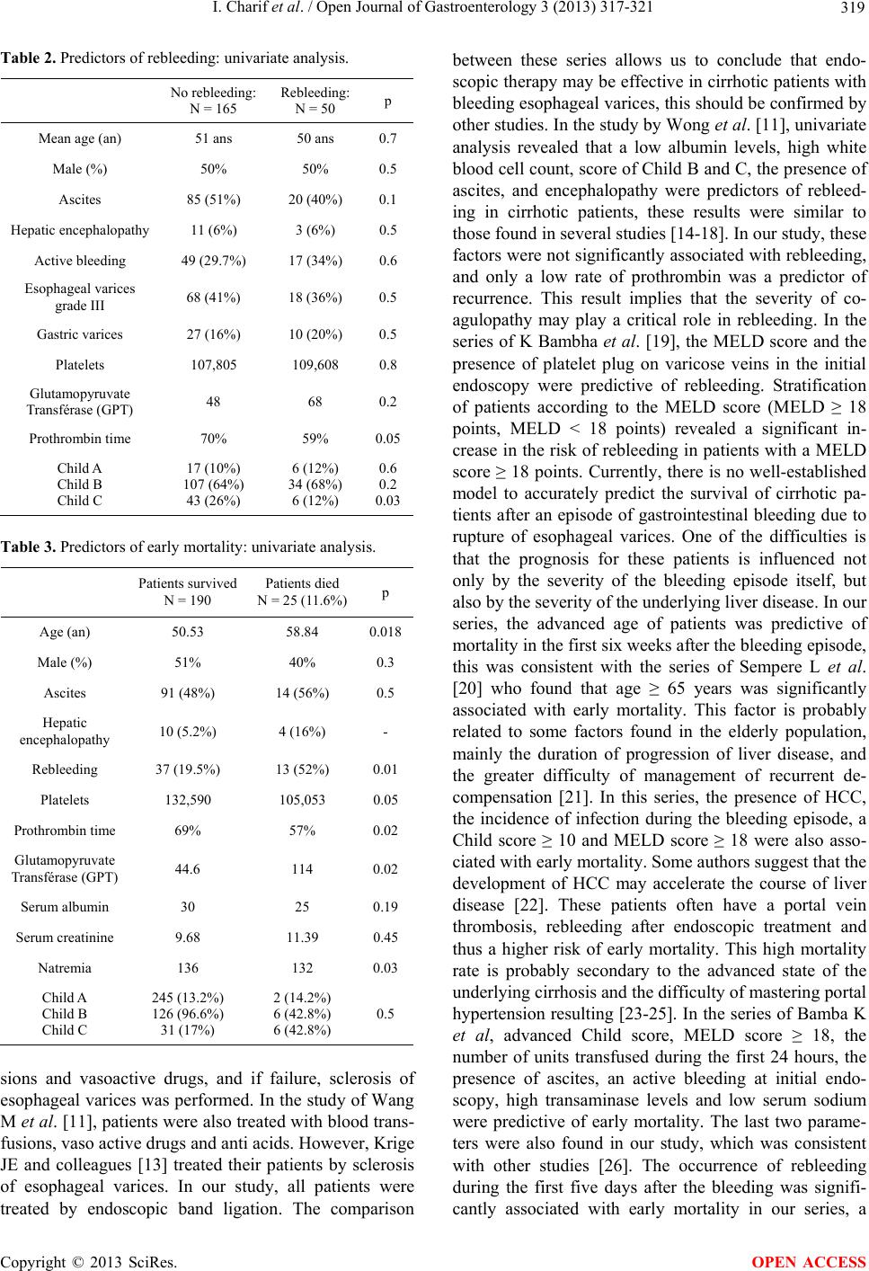 I. Charif et al. / Open Journal of Gastroenterology 3 (2013) 317-321 319 Table 2. Predictors of rebleeding: univariate analysis. No rebleeding: N = 165 Rebleeding: N = 50 p Mean age (an) 51 ans 50 ans 0.7 Male (%) 50% 50% 0.5 Ascites 85 (51%) 20 (40%) 0.1 Hepatic encephalopathy 11 (6%) 3 (6%) 0.5 Active bleeding 49 (29.7%) 17 (34%) 0.6 Esophageal varices grade III 68 (41%) 18 (36%) 0.5 Gastric varices 27 (16%) 10 (20%) 0.5 Platelets 107,805 109,608 0.8 Glutamopyruvate Transférase (GPT) 48 68 0.2 Prothrombin time 70% 59% 0.05 Child A Child B Child C 17 (10%) 107 (64%) 43 (26%) 6 (12%) 34 (68%) 6 (12%) 0.6 0.2 0.03 Table 3. Predictors of early mortality: univariate analysis. Patients survived N = 190 Patients died N = 25 (11.6%) p Age (an) 50.53 58.84 0.018 Male (%) 51% 40% 0.3 Ascites 91 (48%) 14 (56%) 0.5 Hepatic encephalopathy 10 (5.2%) 4 (16%) - Rebleeding 37 (19.5%) 13 (52%) 0.01 Platelets 132,590 105,053 0.05 Prothrombin time 69% 57% 0.02 Glutamopyruvate Transférase (GPT) 44.6 114 0.02 Serum albumin 30 25 0.19 Serum creatinine 9.68 11.39 0.45 Natremia 136 132 0.03 Child A Child B Child C 245 (13.2%) 126 (96.6%) 31 (17%) 2 (14.2%) 6 (42.8%) 6 (42.8%) 0.5 sions and vasoactive drugs, and if failure, sclerosis of esophageal varices was performed. In the study of Wang M et al. [11], patients were also treated with blood trans- fusions, vaso active drugs and anti acids. However, Krige JE and colleagues [13] treated their patients by sclerosis of esophageal varices. In our study, all patients were treated by endoscopic band ligation. The comparison between these series allows us to conclude that endo- scopic therapy may be effective in cirrhotic patients with bleeding esophageal varices, this should be confirmed by other studies. In the study by Wong et al. [11], univariate analysis revealed that a low albumin levels, high white blood cell count, score of Child B and C, the presence of ascites, and encephalopathy were predictors of rebleed- ing in cirrhotic patients, these results were similar to those found in several studies [14-18]. In our study, these factors were not significantly associated with rebleeding, and only a low rate of prothrombin was a predictor of recurrence. This result implies that the severity of co- agulopathy may play a critical role in rebleeding. In the series of K Bambha et al. [19], the MELD score and the presence of platelet plug on varicose veins in the initial endoscopy were predictive of rebleeding. Stratification of patients according to the MELD score (MELD ≥ 18 points, MELD < 18 points) revealed a significant in- crease in the risk of rebleeding in patients with a MELD score ≥ 18 points. Currently, there is no well-established model to accurately predict the survival of cirrhotic pa- tients after an episode of gastrointestinal bleeding due to rupture of esophageal varices. One of the difficulties is that the prognosis for these patients is influenced not only by the severity of the bleeding episode itself, but also by the severity of the underlying liver disease. In our series, the advanced age of patients was predictive of mortality in the first six weeks after the bleeding episode, this was consistent with the series of Sempere L et al. [20] who found that age ≥ 65 years was significantly associated with early mortality. This factor is probably related to some factors found in the elderly population, mainly the duration of progression of liver disease, and the greater difficulty of management of recurrent de- compensation [21]. In this series, the presence of HCC, the incidence of infection during the bleeding episode, a Child score ≥ 10 and MELD score ≥ 18 were also asso- ciated with early mortality. Some authors suggest that the development of HCC may accelerate the course of liver disease [22]. These patients often have a portal vein thrombosis, rebleeding after endoscopic treatment and thus a higher risk of early mortality. This high mortality rate is probably secondary to the advanced state of the underlying cirrhosis and the difficulty of mastering portal hypertension resulting [23-25]. In the series of Bamba K et al, advanced Child score, MELD score ≥ 18, the number of units transfused during the first 24 hours, the presence of ascites, an active bleeding at initial endo- scopy, high transaminase levels and low serum sodium were predictive of early mortality. The last two parame- ters were also found in our study, which was consistent with other studies [26]. The occurrence of rebleeding during the first five days after the bleeding was signifi- cantly associated with early mortality in our series, a Copyright © 2013 SciRes. OPEN ACCESS  I. Charif et al. / Open Journal of Gastroenterology 3 (2013) 317-321 320 factor also reported in the series of Kamba et al. [19]. 5. CONCLUSION The degree of liver cell failure influenced the occurrence of rebleeding after an episode of variceal bleeding in cirrhotic patients. The advanced age of patients, low prothrombin time, low rates of serum sodium and of platelets count, and high transaminase rate were predic- tors of early mortality in these patients. REFERENCES [1] Graham, D.Y. and Smith, J.L. (1981) The course of patients after variceal hemorrhage. Gastroenterology, 80, 800-809. [2] de Franchis, R. (2005) Evolving consensus in portal hypertension. Report of the Baveno IV consensus work- shop on methodology of diagnosis and therapy in portal hypertension. Journal of Hepatology, 43, 167-176. http://dx.doi.org/10.1016/j.jhep.2005.05.009 [3] Chalasani, N., Kahi, C., Francois, F., et al. (2003) Im- proved patient survival after acute variceal bleeding: A multicenter, cohort study. American Journal of Gastro- enterology, 98, 653-659. http://dx.doi.org/10.1111/j.1572-0241.2003.07294.x [4] del Olmo, J.A., Pena, A., Serra, M.A., et al. (2000) Pre- dictors of morbidity and mortality after the first episode of upper gastrointestinal bleeding in liver cirrhosis. Jour- nal of Hepatology, 32, 19-24. http://dx.doi.org/10.1016/S0168-8278(01)68827-5 [5] D’Amico, G. and De Franchis, R. (2003) Upper digestive bleeding in cirrhosis. Post-therapeutic outcome and prog- nostic indicators. Hepatology, 38, 599-612. http://dx.doi.org/10.1053/jhep.2003.50385 [6] Moitinho, E., Escorsell, A., Bandi, J.C., et al. (1999) Prognostic value of early measurements of portal pressure in acute variceal bleeding. Gastroenterology, 117, 626- 631. http://dx.doi.org/10.1016/S0016-5085(99)70455-5 [7] Le Moine, O., Adler, M., Bourgeois, N., et al. (1992) Factors related to early mortality in cirrhotic patients bleeding from varices and treated by urgent sclerotherapy. Gut, 33, 1381-1385. http://dx.doi.org/10.1136/gut.33.10.1381 [8] Siringo, S., McCormick, P.A., Mistry, P., et al. (1991) Prognostic significance of the white nipple sign in vari- ceal bleeding. Gastrointestinal Endoscopy, 37, 51-55. http://dx.doi.org/10.1016/S0016-5107(91)70621-4 [9] Wiesner, R., Edwards, E., Freeman, R., Harper, A., Kim, R., Kamath, P., et al. (2003) Model for end-stage liver disease (MELD) and allocation of donor livers. Gastro- enterology, 124, 91-96. http://dx.doi.org/10.1053/gast.2003.50016 [10] de Franchis, R. (2010) Revising consensus in portal hy- pertension: Report of the Baveno V consensus workshop on methodology of diagnosis and therapy in portal hyper- tension. Journal of Hepatology, 53, 762-768. http://dx.doi.org/10.1016/j.jhep.2010.06.004 [11] Wang, M.-T., Liu, T., Ma, X.-Q. and He, J. (2011) Prog- nostic factors associated with rebleeding in cirrhotic in- patients complicated with esophageal variceal bleeding. Chinese Medical Journal, 124, 1493-1497. [12] Ben-Ari, Z., Cardin, F., McCormick, A.P., Wannamethee, G. and Burroughs, A.K. (1999) A predictive model for failure to control bleeding during acute variceal haemor- rhage. Journal of Hepatology, 31, 443-450. http://dx.doi.org/10.1016/S0168-8278(99)80035-X [13] Krige, J.E., Kotze, U.K., Distiller, G., Shaw, J.M. and Bornman, P.C. (2009) Predictive factors for rebleeding and death in alcoholic cirrhotic patients with acute vari- ceal bleeding: A multivariate analysis. World Journal of Surgery, 33, 2127-2135. http://dx.doi.org/10.1007/s00268-009-0172-6 [14] Liu, J.S. and Liu, J. (2009) Comparison of emergency endoscopic variceal ligation plus octride or octride alone for acute esophageal variceal bleeding. Chinese Medical Journal, 122, 3003-3006. [15] Huang, L.Y., Cui, J., Wu, C.R. and Liu, Y.X. (2007) Embolization combined with endoscopic variceal ligation for the treatment of esophagogastric variceal bleeding in patients with cirrhosis. Chinese Medical Journal, 120, 36-40. [16] Heresbach, D., Bretagne, J.F., Raoul, J.L., Chaperon, J., Piette, C., Siproudhis, L., et al. (1991) Prognosis and prognostic factors of hemorrhage by rupture of varices in cirrhotic patients in the era of endoscopic sclerotherapy. Gastroentérologie Clinique et B i o l o g i q u e, 15, 838-844. [17] Bosch, J., Berzigotti, A., Garcia-Pagan, J.C. and Abraldes, J.G. (2008) The management of portal hypertension: Ra- tional basis, available treatments and future options. Jour- nal of Hepatology, 48, s68-s92. http://dx.doi.org/10.1016/j.jhep.2008.01.021 [18] Thomopoulos, K., Theocharis, G., Mimidis, K., Lam- propoulou-Karatza, Ch., Alexandridis, E. and Nikolopou- lou, V. (2006) Improved survival of patients presenting with acute variceal bleeding. Prognostic indicators of short- and long-term mortality. Digestive and Liver Dis- ease, 38, 899-904. http://dx.doi.org/10.1016/j.dld.2006.08.002 [19] Bambha, K., Kim, W.R., Pedersen, R., et al. (2008) Pre- dictors of early rebleeding and mortality after acute vari- ceal haemorrhage in patients with cirrhosis. Gut, 57, 814- 820. http://dx.doi.org/10.1136/gut.2007.137489 [20] Sempere, L., Palazón, J.M. and Sánchez-Payá, J. (2009) Assessing the short- and long-term prognosis of patients with cirrhosis and acute variceal bleeding. Revista Es- pañola de Enfermedades Digestivas, 101, 236-248. [21] Prieto, M., Clemente, G., Casafont, F., Cuende, N., Cuer- vas-Mons, V., Figueras, J., et al. (2003) Consensus docu- ment on indications for liver transplantation. 2002. Gas- troenterol Hepatol, 26, 355-375. http://dx.doi.org/10.1157/13048890 [22] D’Amico, G., Garcia-Tsao, G. and Pagliaro, L. (2006) Natural history and prognostic indicators of survival in cirrhosis: A systematic review of 118 studies. Journal of Hepatology, 44, 217-231. http://dx.doi.org/10.1016/j.jhep.2005.10.013 Copyright © 2013 SciRes. OPEN ACCESS  I. Charif et al. / Open Journal of Gastroenterology 3 (2013) 317-321 Copyright © 2013 SciRes. 321 OPEN ACCESS [23] D’Amico, G., De Franchis, R. and Cooperative Study Group (2003) Upper digestive bleeding in cirrhosis. Post- therapeutic outcome and prognostic indicators. Hepato- logy, 38, 599-612. http://dx.doi.org/10.1053/jhep.2003.50385 [24] Yeo, W., Sung, J.Y., Ward, S.C., Chung, S.C., Lee, W.Y., Li, A.K., et al. (1995) A prospective study of upper gas- trointestinal hemorrhage in patients with hepatocellular carcinoma. Digestive Diseases and Sciences, 40, 2516- 2521. http://dx.doi.org/10.1007/BF02220435 [25] Sung, J.J., Yeo, W., Suen, R., Lee, Y.T., Chung, S.C., Chan, F.K., et al. (1998) Injection sclerotherapy for vari- ceal bleeding in patients with hepatocellular carcinoma: Cyanoacrylate versus sodium tetradecyl sulphate. Gas- trointestina l E ndoscopy, 47, 235-239. http://dx.doi.org/10.1016/S0016-5107(98)70319-0 [26] D’Amico, G., Garcia-Tsao, G. and Pagliaro, L. (2006) Natural history and prognostic indicators of survival in cirrhosis: A systematic review of 118 studies. Journal of Hepatology, 44, 217-231. http://dx.doi.org/10.1016/j.jhep.2005.10.013 |