

Paper Menu >>
Journal Menu >>
 Surgical Science, 2013, 4, 509-515 Published Online November 2013 (http://www.scirp.org/journal/ss) http://dx.doi.org/10.4236/ss.2013.411099 Open Access SS Complex TAPVC-Experience with Six Patients* Baskar Ranjith Karthekeyan#, Periyasamy Thangavelu, Jebaraj Rethinasamy, Mahesh Vakamudi, Siva Muthukumar, Rajeshkumar Kodali, Kamalakannan Sambandham, Sushma Nandipati Sri Ramachandra Medical College and Research Institute, Chennai, India Email: #[email protected] Received October 30, 2013; revised November 18, 2013; accepted November 24, 2013 Copyright © 2013 Baskar Ranjith Karthekeyan et al. This is an open access article distributed under the Creative Commons Attribu- tion License, which permits unrestricted use, distribution, and reproduction in any medium, provided the original work is properly cited. ABSTRACT Total anomalous pulmonary venous connection becomes a totally different subset when associated with complex con- genital anomalies. The combination of two separate life-threatening congenital heart defects complicates the manage- ment of these patients. Six patients with total anomalous pulmonary venous connection associated with complex con- genital heart disease were studied. There were 2 girls and 4 boys. Three of them were less than 5 kg in weight, and the other 3 were more than 5 kg in weight. Four patients had severe pulmonary arterial hypertension and 2 patients had pulmonary stenosis. Three patients had supracardiac type with a right vertical vein, one had drainage to the right atrium superior vena cava junction, one patient had supra cardiac type but split flow to both the superior vena cava and one patient had cardiac type. Three patients had double outlet right ventricle. Three patients had atrioventricular canal defect and 2 patients had preoperative pulmonary vein obstruction. All patients underwent rerouting of pulmonary veins. Concomitant procedures included intraventricular tunnel repair of ventricular septal defect and infundibular resection in double outlet right ventricle. Atrioventricular canal rep air was done for Rastelli type A atrioventricular canal. Superior vena caval plasty, atrioventricular canal repair and pulmonary artery banding were done in unbalanced atrioventricular septal defect and large double outlet right ventricle. Intracardiac repair through transatrial approach was done for tetralogy of Fallot. Right ventricle-pulmonary artery conduit was done for truncus arteriosus. Single ventricle repair was done for corrected transpos ition of great arteries. There were 2 hospital deaths. Keywords: Total Anomalous; AV Canal; Complex; Congenital; Pulmonary Vein 1. Introduction Total anomalous pulmonary venous connection is char- acterized by the failed union of the pulmonary veins and incorporation of it by the developing left atrium in com- bination with a persistent embryologic connection be- tween the pulmonary and systemic venous systems. The impact of the pathology depends on the degree to which the pulmonary venous drainage is obstructed and the magnitude of the left-to-right shunt. Accurate assessment of the anatomy and detailed surgical planning become essential when total anomalous pulmonary venous con- nection is associated with other complex congenital ano- malies. This result in a combination of two separate life- threatening heart defects needs appropriate management. Most often the timing of surgery is dictated by the pres- ence or development of pulmonary venous obstruction rather than the standardized protocols that apply to the majority of patients. It is our goal that this analysis will identify some important principles in the management of these complex patients. 2. Patients and Methods Six patients with total anomalous pulmonary venous connection associated with complex congenital heart disease were diagnosed in our hospital between 2011 and 2013. Symptoms included delayed milestones, breath- lessness, cyanosis, excessive sweating, poor weight gain and recurrent lower respiratory infections. There were 2 girls and 4 boys. Three of them were less than 5 kg in weight, and the other 3 were more than 5 kg in weight. Four patients had severe pulmonary arterial hypertension and 2 patients had pulmonary stenosis. Three patients had supracardiac total anomalous pulmonary venous connection with a right vert i cal vein, one had drai nage to *Conflict of interest: none. #Corresponding author. 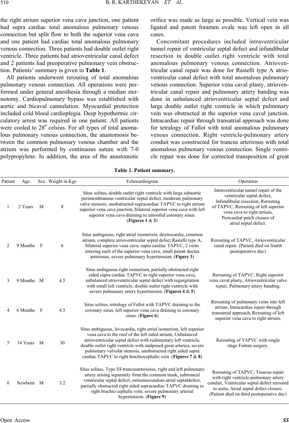 B. R. KARTHEKEYAN ET AL. 510 the right atrium superior vena cava junction, one patient had supra cardiac total anomalous pulmonary venous connection but split flow to both the superior vena cava and one patient had cardiac total anomalous pulmonary venous connection. Three patients had double outlet right ventricle. Three patients had atrioventricular canal defect and 2 patients had preoperative pulmonary vein obstruc- tion. Patients’ summary is given in Table 1. All patients underwent rerouting of total anomalous pulmonary venous connection. All operations were per- formed under general anesthesia through a median ster- notomy. Cardiopulmonary bypass was established with aortic and bicaval cannulation. Myocardial protection included cold blood cardioplegia. Deep hypothermic cir- culatory arrest was required in one patient. All patients were cooled to 280 celsius. For all types of total anoma- lous pulmonary venous connection, the anastomosis be- tween the common pulmonary venous chamber and the atrium was performed by continuous suture with 7-0 polypropylene. In addition, the area of the anastomotic orifice was made as large as possible. Vertical vein was ligated and patent foramen ovale was left open in all cases. Concomitant procedures included intraventricular tunnel repair of ventricular sep tal defect and infund ibular resection in double outlet right ventricle with total anomalous pulmonary venous connection. Atrioven- tricular canal repair was done for Rastelli type A atrio- ventricular canal defect with total anomalous pulmonary venous connection. Superior vena caval plasty, atrioven- tricular canal repair and pulmonary artery banding was done in unbalanced atrioventricular septal defect and large double outlet right ventricle in which pulmonary vein was obstructed at the superior vena caval junction. Intracardiac repair through transatrial approach was done for tetralogy of Fallot with total anomalous pulmonary venous connection. Right ventricle-pulmonary artery conduit was constructed for truncus arteriosus with total anomalous pulmonary venous connection. Single ventri- cle repair was done for corrected transposition of great Table 1. Patient summary. Patient Age Sex Weight in KgsEchocardiogram Operation 1 2 Years M 8 Situs solitus, double outlet right ventricle with large subaortic perimembranous ve ntricular septal defect, moderate pulmonary valve stenosis, unobstructed supracardiac TAPVC to right atrium superior vena cava junc tion, bilateral superior vena cava with left superior vena cava draining to unroo fed coronary sinus. (Figures 1 & 2) Intraventricular tunnel repair of the ventricular septal defect, Infundibular resection, Rerouting of TAPVC, Rerouting of left superi or vena cava to right atrium, Pericardial patch closure of atrial septal defect. 2 9 Months F 6 Situs ambiguous, ri g ht at rial isomerism, dextrocardia, comm on atrium, complete atrioventricular septal defect Rastelli type A, bilateral superior vena cava, supra cardiac TAPVC, 2 ve i ns entering each of the superior vena cava, small patent ductus arteriosus, severe pulmonary hypertension. (Figure 3) Rerouting of TAPVC, Atrioventricular canal repair. (Patient died on fourth postoperative day) 3 9 Months M 4.5 Situs ambiguous righ t i somerism, p art ia ll y obstructed right sided supra cardiac TAPVC to rig ht superior vena cava, unbalanced atrioventricular septal defect with regur g i t at i o n with small left ventricle, double outlet right ventricle with severe pulmonary artery hypertension. (Figures 4 & 5) Rerouting of TAPVC, Right superi o r vena caval plasty, Atrioventr icular valve repair, Pulmonar y artery banding. 4 6 Months F 4.5 Situs solitus, tetralogy of Fallot with TAPVC draining to the coronary sinus. left super ior vena cava draining to coronary sinus. (Figure 6) Rerouting of pulmonary veins into left atrium, Intracardiac repair through transatrial approach, Rerouting of left superior vena cava to right atrium. 5 14 Years M 30 Situs ambiguous, levo c ardia, right atrial isomerism, left superior vena cava to the roof of the left sided atrium, Unbalanced atrioventricular septal defect with rudim entary left ventricle, double outlet right ventricle with malposed g reat arteries, severe pulmonary valvu lar stenosis, unobstructed righ t si ded sup ra cardiac TAPVC to right brachiocephalic vein. (Figures 7 & 8) Rerouting of TAPVC with sin g le stage Fontan surgery. 6 Newborn M 3.2 Situs solitus, Type III-truncusarteriosus, right and left pulmonary artery arising separately f rom the common trunk, subtruncal ventricular septal defect, ostiumsecundum atrial septaldefect, partially obstructed right sided supracardiac TAPVC draining to right brachio cephalic ve i n , severe pulmonary arteri al hypertension. (Figure 9) Rerouting of TAPVC, Tr uncu s repair with right ventricle-p ulmonary artery conduit, Ventricular septal defect rerouted to aorta, Atrial septal defect closure. (Patient died on third post o pe rative day) Open Access SS 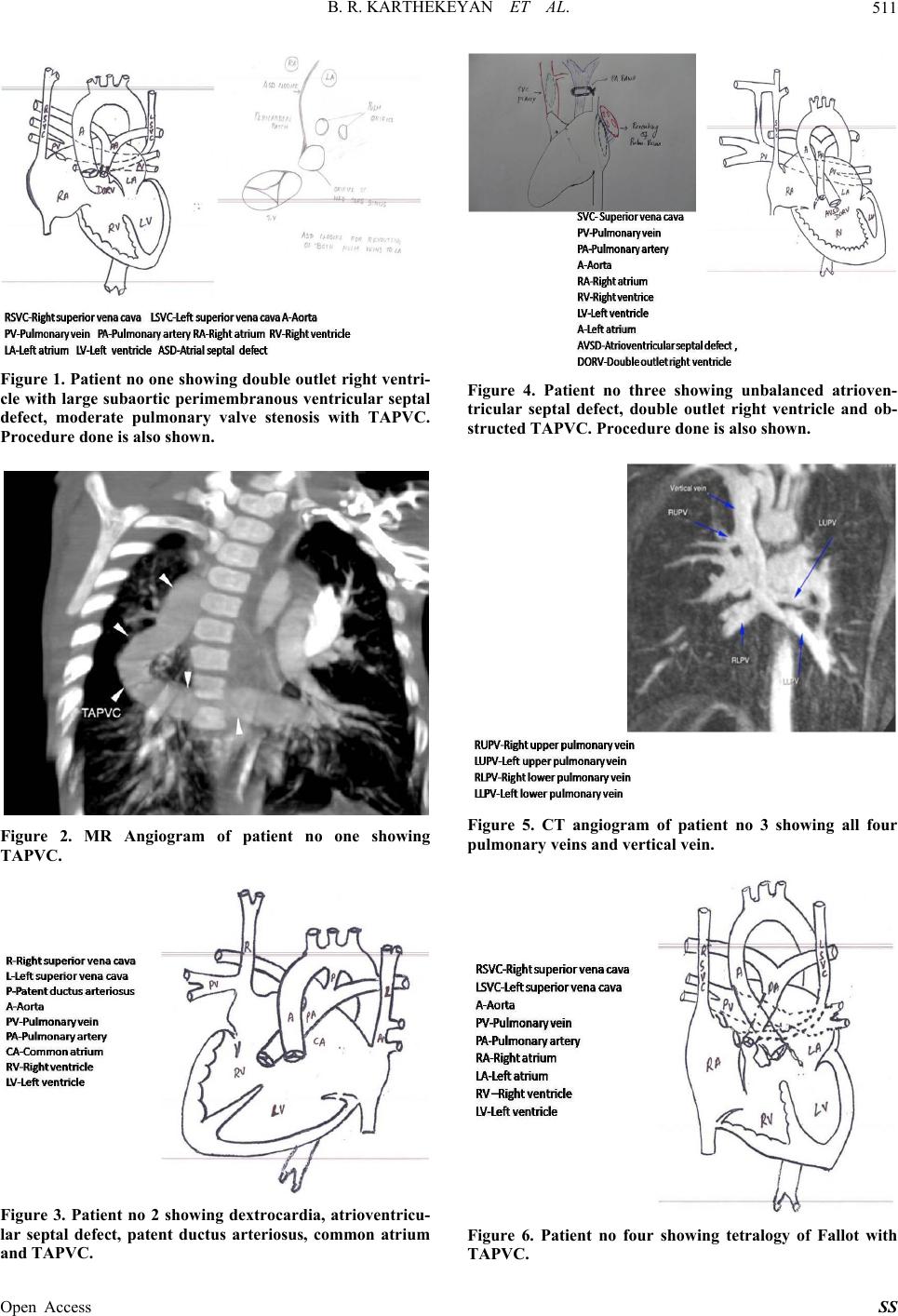 B. R. KARTHEKEYAN ET AL. 511 Figure 1. Patient no one showing double outlet right ventri- cle with large subaortic perimembranous ventricular septal defect, moderate pulmonary valve stenosis with TAPVC. Procedure done is also shown. Figure 2. MR Angiogram of patient no one showing TAPVC. Figure 3. Patient no 2 showing dextrocardia, atrioventricu- lar septal defect, patent ductus arteriosus, common atrium and TAPVC. Figure 4. Patient no three showing unbalanced atrioven- tricular septal defect, double outlet right ventricle and ob- structed TAPVC. Procedure done is also shown. Figure 5. CT angiogram of patient no 3 showing all four pulmonary veins and vertical ve in. Figure 6. Patient no four showing tetralogy of Fallot with TAPVC. Open Access SS 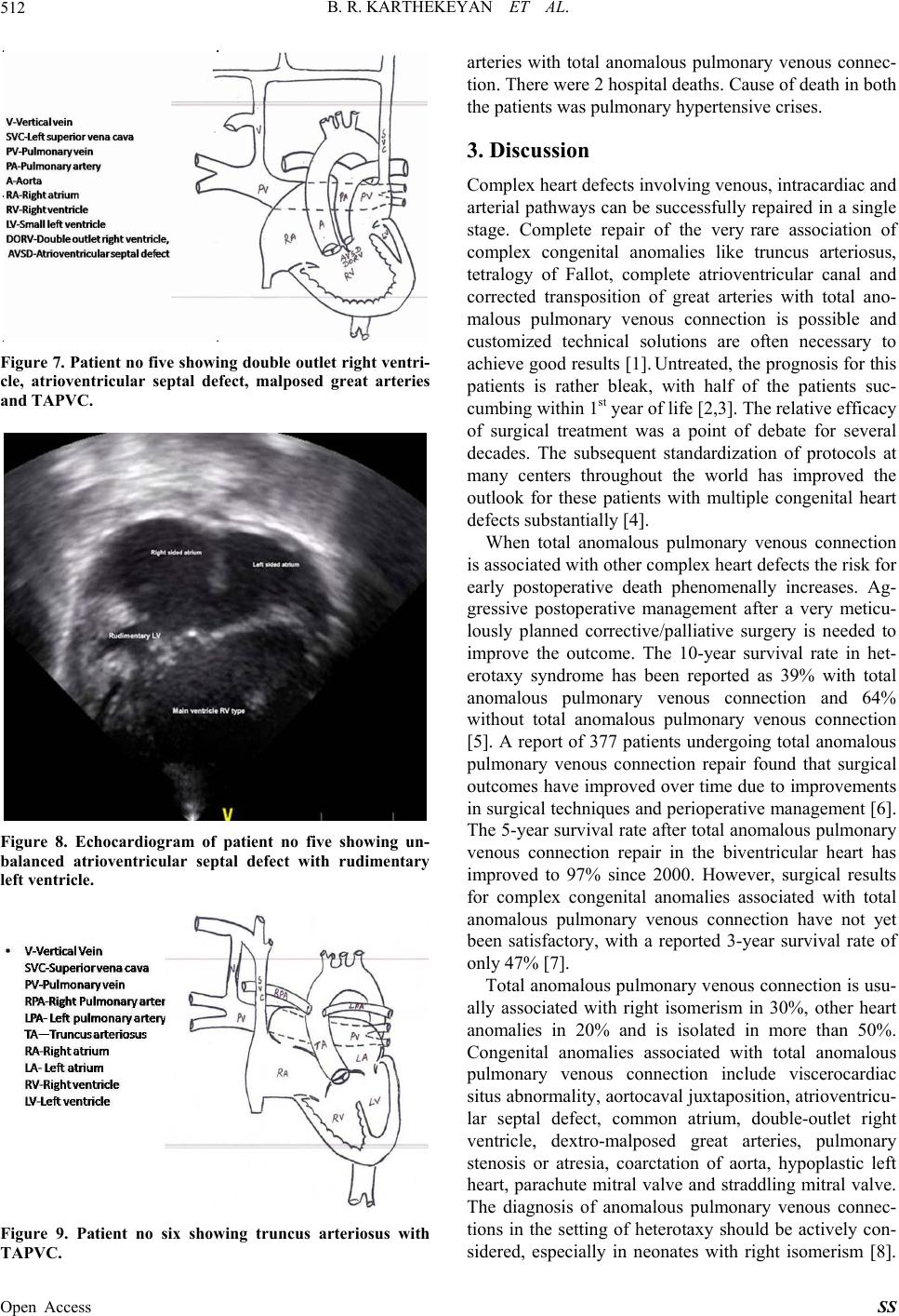 B. R. KARTHEKEYAN ET AL. 512 Figure 7. Patient no five showing double outlet right ventri- cle, atrioventricular septal defect, malposed great arteries and TAPVC. Figure 8. Echocardiogram of patient no five showing un- balanced atrioventricular septal defect with rudimentary left ventricle. Figure 9. Patient no six showing truncus arteriosus with TAPVC. arteries with total anomalous pulmonary venous connec- tion. There were 2 hospital deaths. Cause of death in both the patients was pulmonary hypertensive crises. 3. Discussion Complex heart defects involving venous, intracardiac and arterial pathways can be successfully repaired in a single stage. Complete repair of the very rare association of complex congenital anomalies like truncus arteriosus, tetralogy of Fallot, complete atrioventricular canal and corrected transposition of great arteries with total ano- malous pulmonary venous connection is possible and customized technical solutions are often necessary to achieve good results [1]. Untreated, the prognosis for this patients is rather bleak, with half of the patients suc- cumbing within 1st year of life [2,3]. The relative efficacy of surgical treatment was a point of debate for several decades. The subsequent standardization of protocols at many centers throughout the world has improved the outlook for these patients with multiple congenital heart defects substantially [4]. When total anomalous pulmonary venous connection is associated with other complex heart defects the risk for early postoperative death phenomenally increases. Ag- gressive postoperative management after a very meticu- lously planned corrective/palliative surgery is needed to improve the outcome. The 10-year survival rate in het- erotaxy syndrome has been reported as 39% with total anomalous pulmonary venous connection and 64% without total anomalous pulmonary venous connection [5]. A report of 377 patients undergoing total anomalous pulmonary venous connection repair found that surgical outcomes have improved over time due to improvements in surgical techniques and perioperative management [6]. The 5-year survival rate after total anomalous pulmonary venous connection repair in the biventricular heart has improved to 97% since 2000. However, surgical results for complex congenital anomalies associated with total anomalous pulmonary venous connection have not yet been satisfactory, with a reported 3-year survival rate of only 47% [7]. Total anomalous pulmonary venous connection is usu- ally associated with right isomerism in 30%, other heart anomalies in 20% and is isolated in more than 50%. Congenital anomalies associated with total anomalous pulmonary venous connection include viscerocardiac situs abnormality, aortocaval juxtapo sition, atrioventricu- lar septal defect, common atrium, double-outlet right ventricle, dextro-malposed great arteries, pulmonary stenosis or atresia, coarctation of aorta, hypoplastic left heart, parachute mitral valve and straddling mitral valve. The diagnosis of anomalous pulmonary venous connec- tions in the setting of heterotaxy should be actively con- sidered, especially in neonates with right isomerism [8]. Open Access SS  B. R. KARTHEKEYAN ET AL. 513 In this series of six patients total anomalous pulmonary venous connection was associated with tetralogy of Fal- lot, truncus arteriosus, unbalanced AV canal, corrected transposition of great arteries, double outlet right ventri- cle and complete AV canal with dextocardia. The two outcome factors to be considered are mortal- ity and postoperative recurrent pulmonary vein obstruc- tion. Recent reports about complex congenital heart dis- ease and total anomalous pulmonary venous connection have indicated an improvement in the outcomes over time. They showed that survival at 5 years after total anomalous pulmonary venous connection repair was 79% and the risk factors for mortality were yo unger age, pulmonary atresia, preoperative obstructed total anoma- lous pulmonary venous connection, body weight less than 3.5 kg at operation, concomitant systemic pulmo- nary shunt, postoperative pulmonary vein obstruction and infracardiac or mixed total anomalous pulmonary venous connection [9]. Body weight and age at operation was identified as a risk factor for death. Of 226 patients with repaired total anomalous pulmonary venous connection in the Society of Thoracic Surgeons database, the surgical outcome in body weight ranging from 1 to 2.5 kg at operation was worse than that in body weight ranging from 2.5 to 4 kg [10]. Increased bod y weight at operation may also reduce perioperative complications such as intracranial hemor- rhage, renal dysfunction, and coagulopathy [10]. In our series three patients weighed above 5 kgs and three pa- tients weighed less than 5 kgs. Both the child whom died was less than 5 kgs. However, waiting for an increase in body weight before operation may not be advantageous due to an increase in preoperative morbidities [11]. Age at operation tends to affect early surgical outcomes [11]. However in our series all the children were sligh tly older except the truncus arteriosus patient who was referred to us in the newborn period. Nonconfluent pulmonary arteries and pulmonary atre sia were identified as a risk factor for death in patients oper- ated for total anomalous pulmonary venous connection [12]. Because of the varied anatomic variations in the total anomalous pulmonary venous connection anatomy, especially when associated with complex congenital heart malformations, accurate preoperative diagnosis and determination of the configuration of the pulmonary vein morphology are essential for planning on surgical treat- ment of these challenging patients; else the postoperative course could be complicated [13]. Jenkins et al. reported that small pulmonary vein confluence was a strong nega- tive predictor of surv ival in patients with total anomalous pulmonary venous connection including those with two- ventricle anatomy. Generally, mixed total anomalous pulmonary venous connection has smaller pulmonary vein confluence(s) and this may contribute to its worse prognosis [14]. Most of the patients in our series had good pulmonary vein confluence. It is important to have a regulated pulmonary blood flow in total anomalous pulmonary venous connection. However patients requiring manipulation of pulmonary blood flow are difficult to manage. Concomitant sys- temic pulmonary shunt or pulmonary artery banding was identified as an added risk factor for hospital death. In the management of complex cardiac abnormalities, it is essential to have an optimal pressure-vo lume relationship in the heart (prelo ad and after load) to gain better quality of life [15]. It may be difficult to sustain an adequate circulation in patients who have had total anomalous pulmonary venous connection repair with concomitant systemic pulmonary shunt, especially in patients diag- nosed with preoperative pulmonary vein obstruction and functional univentricle physiology with right ventricular morphology. We consider that intensive management is important to adjust the pulmonary blood flow in these complex patients. Among our patients infundibular re- section was done in two patients in two patients and PA banding was done in one patient. Vertical vein if present was ligated and patent foramen ovale was left open in all patients. Atrioventricular valve regurgitation, which required surgical intervention, was a significant risk factor for postoperative recurrent pulmonary vein obstruction. Volume unloading after the atrioventricular valve repair reduces the size of the atrium and may stretch or distort the pulmonary vein(s) [9]. Atrioventricular regurgitation is associated with systemic ventricular dysfunction and subsequent pulmonary hypertension. Surgical outcomes are therefore influenced by the ability to repair atrioven- tricular valve regurgitation at total anomalous pulmonary venous connection repair [15]. We had 2 patients with atrioventricular valve regurgitation. In both the patients repair was done successfully, however one died. This patient continued to have persistent pulmonary hyperten- sion despite a satisfactory anatomic repair. This patient eventually died from a combination of hypoxemia and respiratory failure. With newer surgical techniques and better manage- ment strategies, the outcomes of patients with total anomalous pulmonary venous connection and associated congenital anomalies have improved dramatically. How- ever, when complex congenital cardiac malformations exist in association with total anomalous pulmonary ve- nous connection, it still remains a clinical challenge es- pecially if it was obstructed [9]. There is a paucity of knowledge about this subset of patients. We had 2 cases of preoperative obstructed total anomalous pulmonary venous connection of which one child died. Both the cases were obstructed at the junction with the superior vena cava. Open Access SS  B. R. KARTHEKEYAN ET AL. 514 The repair of total anomalous pulmonary venous con- nection was performed by a single surgeon, and con- formed to the technical principles of appropriate (not excessive) dissection of the pulmonary vein and minimal manipulation, taking small bites with nonabsorbable su- tures and cutting off the pectin ate muscle which is proper to the single right atrium of RAI, if necessary, to mini- mize the thickness of the anastomosis and precise anas- tomosis under short and intermittent circulatory arrest, if necessary. Surgical approaches such as primary suture- less repair reported by Yun and colleagues may be indi- cated for patients with mixed total anomalous pulmonary venous connection, especially when obstructed. However, it is not clear in the literature if primary sutureless repair will have any significant impact on the prognosis of mixed total anomalous pulmonary venous connection. The practice of cutting off the pectinate muscle is based on the theory that the thickness of the anastomosis con- tributes to the occurrence of anastomotic stenosis and recurrent pulmonary vei n o bstruction [1 6] . Univentricle or heterotaxy syndrome has been identi- fied as a risk factor for reoperation in repaired pulmonary vein stenosis. The study found that 79% of reoperations for pulmonary vein stenosis occurred within 6 months of the initial repair [17]. Another report of 20 patients iden- tified that postoperative pulmonary vein obstruction oc- curring within 6 months of the operation was a risk factor for death [18]. Residual pulmonary vein obstruction might cause subsequent pulmonary hypertension, with even worse outcomes [19]. Therefore, postoperative pul- monary vein obstruction should be repaired as soon as possible, before the pulmonary hypertension becomes persistent. Another report found that high resistance of the pulmonary vasculature, caused by postoperative pulmonary vein obstruction, was the main factor influ- encing death in 14 autopsies of patients diagnosed with univentricle and total anomalous pulmonary venous connection [20]. We think that pulmonary vein wall cannot be normal- ized immediately and that the various complications might occur due to the poor general condition of the pa- tient, even if the pulmonary vein obstruction is relieved. Recurrent pulmonary vein obstruction occurs due to sev- eral intrinsic factors and compression from the adjacent structures like the aorta, atrium, bronchus, or vertebrae. Conventional total anomalous pulmonary venous con- nection repair may be associated with later intimal hy- perplasia localized to the anastomotic site. Improvement or modification of surgical repair technique may improve the results. For example, a sutureless technique that avoids trauma to the pulmonary vein wall and minimizes the risk of distortion at the anastomosis has been reported [20]. This technique may be able to prevent postoperative pulmonary vein obstruction, although long-term surgical outcomes are unclear. More than the type of total anomalous pulmonary venous connection it is the post- operative pulmonary vein obstruction that worsens the prognosis. With our technical principles, none of the pa- tients developed anastomotic stenosis. 4. Conclusion Improvement or modification of the primary repair tech- nique is required to avoid postoperative pulmonary vein obstruction, such as using sutureless technique, espe- cially in patients of whom total anomalous pulmonary venous connection is associated with complex congenital cardiac anomalies. Prevention of postoperative pulmo- nary vein obstruction is important in this complex group of patients. Postoperative pulmonary vein obstruction should be repaired as early as possible, before the pul- monary hypertension becomes persistent. Careful post- operative management is required to control the pulmo- nary blood flow, especially with a concomitant systemic pulmonary shunt or pulmonary artery banding. Signifi- cant atrioventricular regurgitatio n should be addressed to improve postoperative morbidity and mortality. Total anomalous pulmonary venous conn ection asso ciated with complex heart disease can be successfully repaired in a single stage. We therefore consider that complete evalua- tion of the pulmonary venous anatomy, corrective sur- gery at the appropriate time, aggressive perioperative management and close follow-up after hospital discharge are essential to improve the surgical outcome when total anomalous pulmonary venous connection is associated with complex congenital cardiac anomaly. 5. Acknowledgements The authors like to acknowledge the help of Dr. Ramya, Dr. Ramkumar and Dr. Rammohan. REFERENCES [1] S. Conte, T. Jensen, J. R. Jacobsen, F. S. Joyce, P. Laur id- sen and G. Pettersson, “One-Stage Repair of Truncus Ar- teriosus, CAVC and Total Anomalous Pulmonary Venous Connection,” Annals of Thoracic Surgery, Vol. 63, No. 6, 1997, pp. 1781-1783. http://dx.doi.org/10.1016/S0003-4975(97)83863-5 [2] R. D. Mainwaring, V. M. Reddy, O. Reinhartz, R. Punn, T. Tacy and F. L. Hanley, “Surgical Results in Patients With Pulmonary Atresia-Major Aortopulmonary Collat- erals in Association with Total Anomalous Pulmonary Venous Connection,” Annals of Thoracic Surgery, Vol. 92, No. 5, 2011, pp. 1756-1760. http://dx.doi.org/10.1016/j.athoracsur.2011.06.020 [3] H. Leonard, G. Derrick, J. O’Sullivan and C. Wren, “Natural and Unnatural History of Pulmonary Atresia,” Heart, Vol. 84, No. 5, 2000, pp. 499-503. http://dx.doi.org/10.1136/heart.84.5.499 Open Access SS  B. R. KARTHEKEYAN ET AL. Open Access SS 515 [4] A. Carotti, S. B. Albanese, S. Filleppelli, et al., “Deter- minants of Outcome after Surgical Treatment of Pulmo- nary Atresia with Ventricular Septal Defect and Major Aortopulmonary Collateral Arteries,” Journal of Thoracic and Cardiovascular Surgery, Vol. 140, No. 5, 2010, pp. 1092-1103. http://dx.doi.org/10.1016/j.jtcvs.2010.07.087 [5] S. R. Foerster, K. Gauvreau, D. B. McElhinney and T. Geva, “Importance of Totally Anomalous Pulmonary Ve- nous Connection and Postoperative Pulmonary Vein Stenosis in Outcomes of Heterotaxy Syndrome,” Pediat- ric Cardiology, Vol. 29, No. 3, 2008, pp. 536-544. http://dx.doi.org/10.1007/s00246-007-9128-5 [6] T. Karamlou, R. Gurofsky, E. Al Sukhni, et al., “Factors Associated with Mortality and Reoperation in 377 Chil- dren with Total Anomalous Pulmonary Venous Connec- tion,” Circulation, Vol. 115, 2007, pp. 1591-1598. http://dx.doi.org/10.1161/CIRCULATIONAHA.106.6354 41 [7] C. L. Hanco ck Friese n, D. Zurakowski, R. R. Thiagarajan, et al., “Total Anomalous Pulmonary Venous Connection: An Analysis of Current Management Strategies in a Sin- gle Institution,” Annals of Thoracic Surgery, Vol. 79, No. 2, 2005, pp. 596-606. http://dx.doi.org/10.1016/j.athoracsur.2004.07.005 [8] C. R. Patel, J. R. Lane , M. L. Spector, P. C. Smith and S. S. Crane, “Totally Anomalous Pulmonary Venous Con- nection and Complex Congenital Heart Disease Prenatal Echocardiographic Diagnosis and Prognosis,” Journal of Ultrasound in Medicine, Vol. 24, No. 9, 2005, pp. 1191- 1198. [9] T. Nakata, Y. Fujimoto, K. Hirose and M. Osaki, “Func- tional Single Ventricle with Extracardiac Total Anoma- lous Pulmonary Venous Connection,” European Journal of Cardio-Thoracic Surgery, Vol. 36, No. 1, 2009, pp. 49- 56. http://dx.doi.org/10.1016/j.ejcts.2009.02.060 [10] C. L. Curzon, S. Milford-Beland, J. S. Li, et al., “Cardiac Surgery in Infants with Low Birth Weight Is Associated with Increased Mortality: Analysis of the Society of Tho- racic Surgeons Congenital Heart Database,” Journal of Thoracic and Cardiovascular Surgery, Vol. 135, No. 3, 2008, pp. 546-551. http://dx.doi.org/10.1016/j.jtcvs.2007.09.068 [11] V. M. Reddy, D. B. McElhinney, T. Sagrado, A. J. Parry, D. F. Teitel and F. L. Hanley, “Results of 102 Cases of Complete Repair of Congenital Heart Defects in Patients Weighting 700 to 2500 Grams,” Journal of Thoracic and Cardiovascular Surgery, Vol. 117, No. 2, 1999, pp. 324- 331. http://dx.doi.org/10.1016/S0022-5223(99)70430-7 [12] K. Takeu chi, A. Murakami, Y. Hirata, K. Kitahori, Y. Doi and S. Takamoto, “Surgical Outcome of Heterotaxy Syn- drome in a Single Institution,” Asian Cardiovascular and Thoracic Annals, Vol. 14, No. 6, 2006, pp. 489-494. http://dx.doi.org/10.1177/021849230601400610 [13] U. K. Chowdhury, B. Airan, A. Malhotra, A. K. Bisoi, A. Saxena, S. S. Kothari, M. Kalaivani and P. Venugopal, “Mixed Total Anomalous Pulmonary Venous Connection: Anatomic Variations, Surgical Approach, Techniques, and Results,” Journal of Thoracic and Cardiovascular Sur- gery, Vol. 135, No. 1, 2008, pp. 106-116. http://dx.doi.org/10.1016/j.jtcvs.2007.08.028 [14] K. J. Jenkins, S. P. Sanders, E. J. Orav, E. A. Coleman, J. E. Mayer Jr and S. D. Colan, “Individual Pulmonary Vein Size and Survival in Infants with Totally Anomalous Pulmonary Venous Connection,” Journal of the American College of Cardiology, Vol. 22, No. 1, 1993, pp. 201-206. http://dx.doi.org/10.1016/0735-1097(93)90835-O [15] Y. Nakayama, T. Hiramatsu, Y. Iwata, T. Okamura and T. Konuma, “Surgical Results for Functional Univentricular Heart with Total Anomalous Pulmonary Venous Connec- tion over a 25-Year Experience,” Annals of Thoracic Surgery, Vol. 93, No. 2, 2012, pp. 606-613. http://dx.doi.org/10.1016/j.athoracsur.2011.09.038 [16] T. J. Yun, O. O. Al-Radi, I. Adatia, C. A. Caldarone, J. G. Coles, W. G. Williams, J. Smallhorn and G. S. Van Ars- dell, “Contemporary Management of Right Atrial Isom- erism: Effect of Evolving Therapeutic Strategies,” Jour- nal of Thoracic and Cardiovascular Surgery, Vol. 131, No. 5, 2006, pp. 1108-1113. http://dx.doi.org/10.1016/j.jtcvs.2005.11.036 [17] A. M. Kelle, C. L. Backer, J. G. Gossett, S. Kaushal and C. Mavroudis, “Total Anomalous Pulmonary Venous Con- nection: Results of Surgical Repair of 100 Patients at a Single Institution,” Journal of Thoracic and Cardiovas- cular Surgery, Vol. 139, No. 6, 2010, pp. 1387-1394. http://dx.doi.org/10.1016/j.jtcvs.2010.02.024 [18] M. Ricci, M. Elliott, G. A. Cohen, et al., “Management of Pulmonary Venous Obstruction after Correction of Total Anomalous Pulmonary Venous Connection: Risk Factors for Adverse Outcome,” European Journal of Cardio- Thoracic Surgery, Vol. 24, No. 1, 2003, pp. 28-36. http://dx.doi.org/10.1016/S1010-7940(03)00180-5 [19] F. Lacour-Gayet, J. Zoghbi, A. E. Serraf, et al., “Surgical Management of Progressive Pulmonary Venous Obstruc- tion after Repair of Total Anomalous Pulmonary Venous Connection,” Journal of Thoracic and Cardiovascular Surgery, Vol. 117, No. 4, 1999, pp. 679-687. http://dx.doi.org/10.1016/S0022-5223(99)70287-4 [20] J. W. Gaynor, M. H. Collins, R. Rychik, J. P. Gaughan and T. L. Spray, “Long-Term Outcome of Infants with Single Ventricle and Total Anomalous Pulmonary Ve- nous Connection,” Journal of Thoracic and Cardiovas- cular Surgery, Vol. 117, No. 3, 1999, pp. 506-514. http://dx.doi.org/10.1016/S0022-5223(99)70330-2 |