

Paper Menu >>
Journal Menu >>
 Surgical Science, 2013, 4, 500-505 Published Online November 2013 (http://www.scirp.org/journal/ss) http://dx.doi.org/10.4236/ss.2013.411097 Open Access SS Clinical Outcome of Conservative Tr eatment of Displaced Mandibular Fracture in Adults Lipa Bodner1*, Sigal Amitay1, Ben Zion Joshua2 1Department of Oral and Maxillofacial Surgery, Soroka Medical Center, Faculty of Health Sciences, Ben-Gurion University of the Negev, Beer-Sheva, Israel 2Department of Otolaryngology Head and Neck S urgery, Soroka Medical Center, Faculty of Health Sciences, Ben-Gurion University of the Negev, Beer-Sheva, Israel Email: *[email protected] Received September 3, 2013; revised October 1, 2013; accepted October 9, 2013 Copyright © 2013 Lipa Bodner et al. This is an open access article distributed under the Creative Commons Attribution License, which permits unrestricted use, distribution, and reproduction in any medium, provided the original work is properly cited. ABSTRACT The article evaluates 12 cases of conservative treatment of displaced mandibular fractures in adults. Twelve cases of displaced mandibular fractures treated surgically, either by closed reduction (IMF) or open reduction internal fixation (ORIF) served as controls. Occlusion, maximal mouth opening, lateral jaw movements, neurological dysfunction (=sensory deficit), and bone remodeling were evaluated and scored in both groups, and results were compared. No sig- nificant differences were found between the two groups in all the evaluated parameters. It is concluded that in certain cases, with displacement of 2 - 4 mm, where a surgical approach is not feasible, reasonable spontaneous reduction and bone remodeling can occur. Meticulous follow-up is mandatory. Keywords: Mandible; Jaw Bone; Trauma; Displaced Fracture; Conservative Management 1. Introduction Fractures of the mandible are generally treated by closed or open reduction [1]. The aim of the treatment is to re- duce the displaced fracture and restore proper occlusion and facial contour. The closed reduction methods involve intermaxillary fixation (IMF) using splints, arch bars, or maxillomandibular fixation screws [2,3]. There are seve ral disadvantages with IMF, including: compromised airway, poor oral hygiene, speech difficulties, impaired nu tritional intake with weight loss, and disusing atrophy of the mas- ticatory muscles. Open reduction and internal fixation (ORIF) using wires, pins, screws, or plates are among the common methods [4]. In recent years, ORIF, by allowing immediate mobi- lization of the mandible, has won increasing acceptance as the method for treatment of mandibular fractures [5]. Conservative management as a treatment modality is well accepted in mandibular condylar fractures [6]. Con- servative treatment in cases of mandibular body fractures has been reported in children in early childhood age, [7-12] and in adults with atrophic edentulous mandible [13-15]. In some reports, closed reduction with IMF was considered as conservative treatment [16-18]. We are unaware of reports describing conservative treatment for displaced mandibular fracture in adults. The purpose of the present report is to follow the cli- nical outcome of 12 patients with displaced mandibular fracture treated conservatively in terms of restoration of mandibul a r f un c t i on. 2. Material and Methods Twenty-four cases of mandibular fracture were included in the present study. The control group (n = 12) was treated surgically by either closed reduction (IMF) or open reduction (ORIF). The experimental group (n = 12) was also advised to undergo surgery, either closed or open reduction. How- ever, due to personal or medical reasons, they were not operated on. The first patient, who was the trigger for the present study, was a 34-year-old psychiatric female patient with a displaced fracture of the mandible, who “refused” to stay at the hospital for surgery. The case was evaluated by the risk management committee of the hospital, and the advice was that as the surgery she needed was not a *Corresponding a uthor. 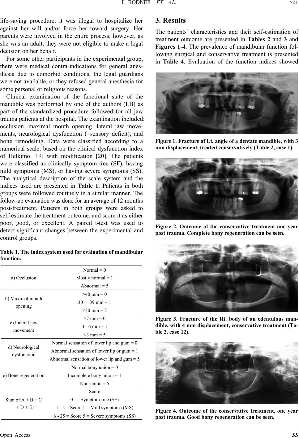 L. BODNER ET AL. 501 life-saving procedure, it was illegal to hospitalize her against her will and/or force her toward surgery. Her parents were involved in the entire process; however, as she was an adult, they were not eligible to make a legal decision on her behalf. For some other participants in the experimental group, there were medical contra-indications for general anes- thesia due to comorbid conditions, the legal guardians were not available, or they refused general anesthesia for some personal or religious reasons. Clinical examination of the functional state of the mandible was performed by one of the authors (LB) as part of the standardized procedure followed for all jaw trauma patients at the hospital. The examination included: occlusion, maximal mouth opening, lateral jaw move- ments, neurological dysfunction (=sensory deficit), and bone remodeling. Data were classified according to a numerical scale, based on the clinical dysfunction index of Helkimo [19] with modification [20]. The patients were classified as clinically symptom-free (SF), having mild symptoms (MS), or having severe symptoms (SS). The analytical description of the scale system and the indices used are presented in Table 1. Patients in both groups were followed routinely in a similar manner. The follow-up evaluation was done for an average of 12 months post-treatment. Patients in both groups were asked to self-estimate the treatment outcome, and score it as either poor, good, or excellent. A paired t-test was used to detect significant changes between the experimental and control gr oups. Table 1. The index system used for evaluation of mandibular function. Normal = 0 Mostly normal = 1 a) Occlusion Abnormal = 5 >40 mm = 0 30 - 39 mm = 1 b) Maximal mouth opening <30 mm = 5 >7 mm = 0 4 - 6 mm = 1 c) Lateral jaw movement <3 mm = 5 Normal sensation of l o w e r lip and gum = 0 Abnormal sensation of lower lip or gum = 1 d) Neurological dysfunction Abnormal sensation of lower lip and gum = 5 Normal bony union = 0 Incomplete bony union = 1 e) Bone regeneration Non-union = 5 Score 0 = Symptom free (SF) 1 - 5 = Score 1 = Mild symptoms (MS) Sum of A + B + C + D + E: 6 - 25 = Score 5 = Severe symptoms (SS) 3. Results The patients’ characteristics and their self-estimation of treatment outcome are presented in Tables 2 and 3 and Figures 1-4. The prevalence of mandibular function fol- lowing surgical and conservative treatment is presented in Table 4. Evaluation of the function indices showed Figure 1. Fracture of Lt. angle of a dentate mandible, with 3 mm displacement, treated conservatively (Table 2, case 1). Figure 2. Outcome of the conservative treatment one year post trauma. Complete bony regeneration can be seen. Figure 3. Fracture of the Rt. body of an edentulous man- dible, with 4 mm displacement, conservative treatment (Ta- ble 2, case 12). Figure 4. Outcome of the conservative treatment, one year ost trauma. Good bony regeneration can be seen. p Open Access SS 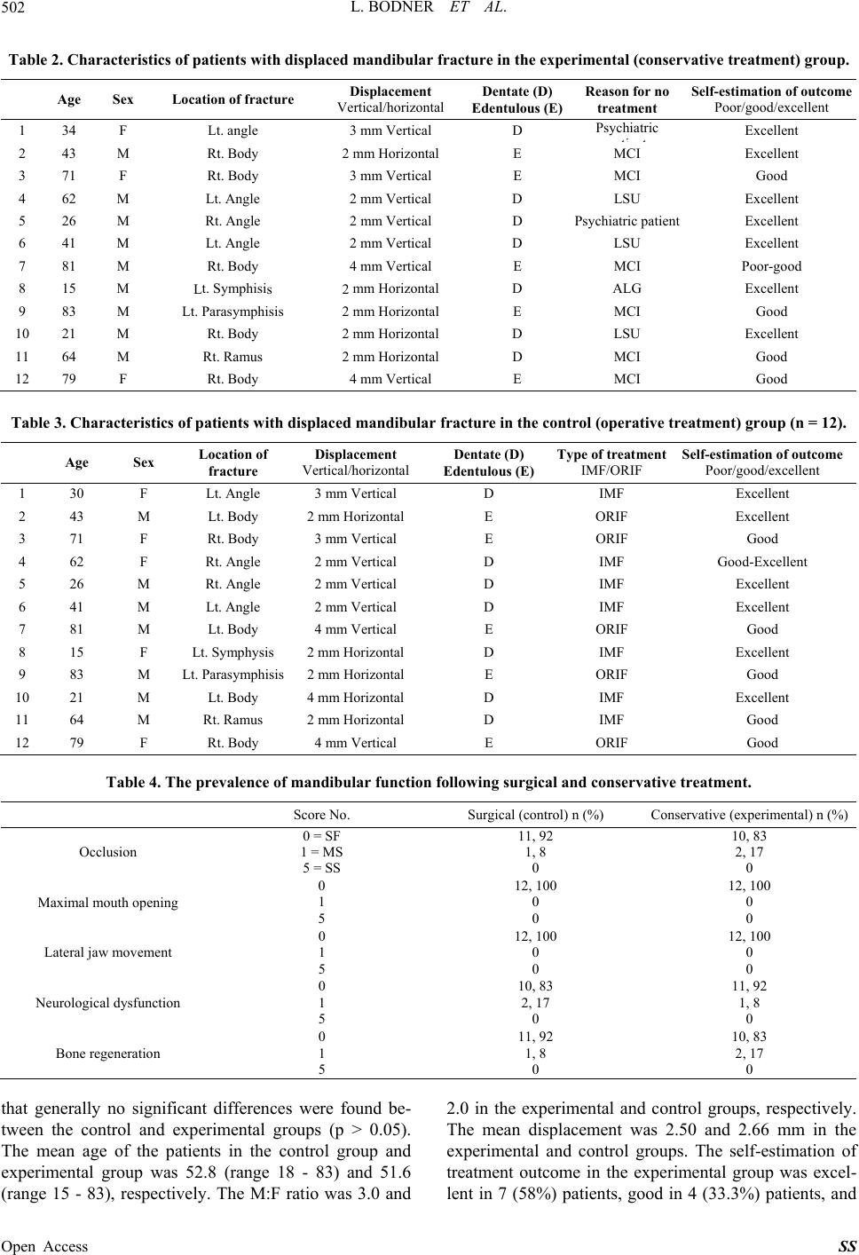 L. BODNER ET AL. Open Access SS 502 Table 2. Characteristics of patients with displaced mandibular fracture in the experimental (conservative treatment) group. Age Sex Location of fracture Displacement Vertical/horizontal Dentate (D) Edentulous (E)Reason for no treatment Self-estimation of outcome Poor/good/excellent 1 34 F Lt. angle 3 mm Vertical D Psychiatric ti t Excellent 2 43 M Rt. Body 2 mm Horizontal E MCI Excellent 3 71 F Rt. Body 3 mm Vertical E MCI Good 4 62 M Lt. Angle 2 mm Vertical D LSU Excellent 5 26 M Rt. Angle 2 mm Vertical D PsychEt Ls 2 Lt. isis 2 iatric patient xcellen 6 41 M Lt. Angle 2 mm Vertical D LSU Excellent 7 81 M Rt. Body 4 mm Vertical E MCI Poor-good 8 15 M t. Symphisi mm HorizontalD ALG Excellent 9 83 M Parasymph mm HorizontalE MCI Good 10 21 M Rt. Body 2 mm Horizontal D LSU Excellent 11 64 M Rt. Ramus 2 mm Horizontal D MCI Good 12 79 F Rt. Body 4 mm Vertical E MCI Good le 3araristics of pith displacedacture in the control (operative treatment) gn = 12). Tab. Chcteatients w mandibular frroup ( Age Sex Location of fracture Displacement Vertical/horizontal Dentate (D) Edentulous (E) Type of treatment IMF/ORIF Self-est imation of outcom e Poor/good/excellent 1 30 F Lt. Angle 3 mm Vertical D IMF Excellent 2 43 M Lt. Body 2 mm Horizontal E ORIF Excellent 3 71 F Rt. Body 3 mm Vertical E ORIF Good 4 62 F Rt. Angle 2 mm Vertical D IMF Gont Et Ls 2 Lt. Pisis 2 od-Excelle 5 26 M Rt. Angle 2 mm Vertical D IMF xcellen 6 41 M Lt. Angle 2 mm Vertical D IMF Excellent 7 81 M Lt. Body 4 mm Vertical E ORIF Good 8 15 F t. Symphysi mm HorizontalD IMF Excellent 9 83 M arasymph mm HorizontalE ORIF Good 10 21 M Lt. Body 4 mm Horizontal D IMF Excellent 11 64 M Rt. Ramus 2 mm Horizontal D IMF Good 12 79 F Rt. Body 4 mm Vertical E ORIF Good Tab. The p of mation followi surgical and vative treatmen imental) n (%) le 4revalencendibular funcngconsert. Score No. Surgical (control) n (%) Conservative (exper Occlusion 3 0 = SF 11, 92 10, 8 1 = MS 5 = SS 1, 8 0 2, 17 0 Maximening N 2, 1 al mouth op0 1 5 12, 100 0 0 12, 100 0 0 Lateral jaw m o vement 0 1 5 12, 100 0 0 12, 100 0 0 eurological dysfunction0 1 5 10, 83 17 0 11, 92 , 8 0 Bone regeneration 0 1 5 11, 92 1, 8 0 10, 83 2, 17 0 that gecant differences we found be- een the control and experimental groups (p > 0.05). (range 15 - 83), respectively. The M:F ratio was 3.0 and 2.0 in the rimental and control groively. The mean displacement was 2.50 and 2.66 mm in the lent in 7 (58%) patients, good in 4 (33.3%) patients, and nerally no signifire tw The mean age of the patients in the control group and experimental group was 52.8 (range 18 - 83) and 51.6 experimental and control groups. The self-estimation of treatment outcome in the experimental group was excel- eexpuprespects,  L. BODNER ET AL. 503 poor-good in 1 (8%) patient. The self-estimation of treatment outcome in the control group was excellent in 6 (50%) patients, good-excellent in 1 (8%) patient, and good in 5 (42%) patients. The differences were not sig- nificant (p > 0.06). The occlusion score was 92% SF and 8% MS com- pared to 83% SF and 17% MS in control and experi- mental groups, respectively. The scores for maximal mouth opening and lateral jaw movement were SF (100%) in both groups. The neurological dysfunction score was 83% SF and 17% MS compared to 92% SF an . literature on complication rates of res revealed that it ranges from 7 to 29 fixation and open reduction and in infection from ORIF, malo- cc ion is discussed with the pa ucosa at the fracture area. Only af surgery for co r treatment of clavicle fractures, where mini- m where no other treatment modality d 8% MS in control and experimental groups, res- pectively. Bone regeneration score was 92% SF and 8% MS compared to 83% SF and 17% MS in control and experimental groups, respectively. The differences be- tween the control and experimental groups were not sig- nificant (p > 0.06). 4. Discussion The study was undertaken to determine if conservative treatment of patients with displaced mandibular fracture results in normal jaw function or with increased risk of possible dysfu nct i on Reviewing the mandibular fractu per cent [21]. The complication rate has been correlated more to the severity of fracture and less to the type of treatment. No difference in complication rate was found between intermaxillary ternal fixation [22]. It was concluded that in fractures with displacement of 2 to 4 mm, there is no difference between closed reduction and open reduction. Our in- terest was to look at a group of patients with displa- cement of 2 - 4 mm, treated conservatively, and follow the rate of complications. Alpert et al. [23] described four types of complications: 1) those arising in the course of proper treatment, 2) those arising due to inappropriate treatment, 3) those due to surgical failure, and 4) those that result from no treat- ment. They also gave examples for each type of com- plication, such as wound lusion from improper treatment, injury to the marginal mandibular nerve due to technical mistakes, and malo- cclusion from no treatment. In the present series, 12 patients were treated con- servatively. The results are more than satisfactory, as they are very similar to the results of patients treated by closed (IMF) or open reduction (ORIF). It has to be emphasized that conservative treatment does not mean “no treatment”. The situat tient and/or with his family that under the specific circumstances, the conservative treatment is in fact the treatment of choice. The patients were routinely followed once a day, once a week, and later once a month, and the progress or no prog ress w as evaluated p er each visit. Th e option of chang ing treatment modality toward ORIF was always on the table. Among the dentate patients, normal occlusion is the key factor that is evaluated during each follow-up visit. Also, the need to be on a liquid/soft diet is emphasized each visit. The edentulous patients were instructed not to use their dentures for several months, in order to prevent pressure-sores of the m ter 12 - 16 weeks and radiographic evidence of bony union of the mandible, was the denture relined with a soft-liner and put back in place. Complete bony union and remodeling of the displaced fracture is an age-de- pendent process. Among the younger patients the process was faster, lasting 3 - 6 months compared to the older patients where it lasted one year or more until they were able to masticate normally with their natural teeth or dentures. Each of the patients was a unique professional challenge that was followed very carefully. Conservative or non-surgical treatment, consisting of observation and soft diet only, has been reported as a treatment option in greenstick or non-displaced mandi- bular fractures with normal occlusion [24,25]. In the report by Ellis et al., [24] 687 patients, 32% of the total sample of 2137 patients, did not undergo rrection of their mandibular fracture and were observed for 4 - 6 weeks. Ghazal et al. [25] reported on 28 cases of mandibular fractures that were managed by observation and soft diet only. This conservative appr oac h resulted in spontaneous healing of the fractures. The hypothesis is that with greenstick and non-displaced frac- ture, the periosteum is intact, and therefore may maintain sufficient stability for interfragmentary motion not to exceed the level tolerated by bone, thereby permitting ossification. However, in displaced fracture the perios- teum is probably damaged, the gap and motion between fragments are larger, which may interfere with ossifica- tion. Therefore, the common approach is closed or open reduction. In our series, the displacement was 2 - 4 mm, which is considered a mild displacement. It is hypothesized that in cases with displacement larger than 4 mm, where the risk of non-union is much greater, the conservative approach might be more risky. Similar clinical thoughts have been reported fo ally displaced or non-displaced fractures can be treated non-surgically, whereas displaced fracture has to be treated by ORIF [26]. 5. Conclusion Conservative treatment of displaced fracture of the mandible carries higher risk of complications compared to IMF or ORIF. However, in certain cases, with dis- placement of 2 - 4 mm, Open Access SS  L. BODNER ET AL. 504 is feasible, it can be a treatment option with respectable lker, Eds., Oral and Maxillofacial Trauma, 2nd Edition, W.B. Saunders, Philadelphia, 1997, pp. 473-522. [2] T. Imazawa, Yd A. Yanai, “Man- dibular Fractuomandibular Fix results. Meticulous follow-up for these patients is man- datory. REFERENCES [1] H. D. Barber, S. C. Woodbury, K. E. Silverstein and R. J. Fonseca, “Mandibular Fractures,” In: R. J. Fonseca and R. V. Wa . Komuro, M. Inoue an res Treated with Maxilla- tion Screws (MMFS Method),” Journal of Craniofacial Surgery, Vol. 2, 2006, pp. 544-549. http://dx.doi.org/10.1097/00001665-200605000-00026 [3] D. P. Colleti, A. Salama and J. J. Caccamese Jr, “Appli- cation of Intermaxillary Fixation Screws in Maxillofacial Trauma,” Journal of Oral & Maxillofacial Surgery, Vol. 65, No. 9, 2007, pp. 1746-1750. http://dx.doi.org/10.1016/j.joms.2007.04.022 027(05)80533-8 [4] T. Iizuka and C. Lindqvist, “Rigid Internal Fixation of Mandibular Fractures. An Analysis of 270 Fractures Treated Using the AO/ASIF Method,” Journal of Oral & Maxillofacial Surgery, Vol. 21, No. 2, 1992, pp. 65-69. http://dx.doi.org/10.1016/S0901-5 ures of the [5] E. Ellis 3rd, “Treatment Methods for Fract Mandibular Angle,” Journal of Oral & Maxillofacial Surgery, Vol. 28, No. 4, 1999, pp. 243-252. http://dx.doi.org/10.1016/S0901-5027(99)80152-0 [6] L. A. Assael, “Open versus Closed Reduction of Adu Mandibular Condyl Fractures: An Alternative Intel rpreta- 36-5 t tion of the Evidence,” Journal of Oral & Maxillofacial Surgery, Vol. 61, No. 11, 2003, pp. 1333-1339. http://dx.doi.org/10.1016/S0278-2391(03)007 anage- ures in S [7] C. O. Hazelrigg and J. E. Jones, “Conservative M ment of a Fractured Mandible in a Ten Month Old Child,” Journal of Oral Pathology & Medicine, Vol. 40, No. 3, 1985, pp. 112-114. [8] J. Lustmann and I. Milhem, “Mandibular Fract Infants: Review of the Literature and Report of even Cases,” Journal of Oral & Maxillofacial Surgery, Vol. 52, No. 3, 1994, pp. 240-245. http://dx.doi.org/10.1016/0278-2391(94)90291-7 [9] S. T. Crean, V. Sivarajasingam and M. J. Fardy, “Con- o. 3, 2000, pp. 229-233. .x servative Approach in the Management of Mandibular Fractures i n t h e Early D e n t ition P hase. A Case Rep o r t a n d Review of the Literature,” International Journal of Pae- diatric Dentistry, Vol. 10, N http://dx.doi.org/10.1046/j.1365-263x.2000.00196 [10] R. Scariot, I. A. de Oliveira, L. A. Passeri, N. L. Rebel- lato and P. R. Muller, “Maxillofacial Injuries in a Group of Brazilian Subjects under 18 Years of Age,” Journal of Applied Oral Science, Vol. 17, No. 3, 2009, pp. 195-198. http://dx.doi.org/10.1590/S1678-77572009000300012 [11] M. Glazer, B. Z. Joshua, Y. Woldenberg and L. Bodner, “Mandibular Fracture in Children—Analysis of 61 Cases and Review of the Literature,” International Journal of Pediatric Otorhinolaryngology, Vol. 75, No. 1, 2011, pp. 62-64. http://dx.doi.org/10.1016/j.ijporl.2010.10.008 [12] H. Kerem, A. Usluer and L. Yoleri, “Remodeling o f a Non Treated Displaced Parasymphyeal Fracture of a Child,” Journal of Craniofacial Surgery, Vol. 22, No. 4, 2011, pp. 1358-1360. http://dx.doi.org/10.1097/SCS.0b013e31821c947f [13] M. R. Cope, “Spontaneous Fracture of an Atrophic Man- dible Treated without Fixation,” British Journal of Oral Surgery, Vol. 20, No. 1, 1982, pp. 22-30. http://dx.doi.org/10.1016/0007-117X(82)90003-8 [14] K. Frame, “Conservative Management of a Patient with Fractured Atrophic Mandible,” British Dental Joa urnal, Vol. 162, 1987, pp. 27-28. http://dx.doi.org/10.1038/sj.bdj.4806014 [15] H. D. Barber, “Conservative Management of the Frac- tured Atrophic Edentulous Mandible,” Journal of Oral & Maxillofacial Surgery, Vol. 59, No. 7, 2001, pp. 789-791. http://dx.doi.org/10.1053/joms.2001.24734 [16] V. A. Petrenko, S. V. Nudelman, E. A. Vainshtein and N A. Golibkov, “Conservative Treatment . of Mandibular and Compli- . Martini, A. Takahashi, H. G. de Oliveira Neto, J. P. Fractures,” Voenno-Meditsinky Zhurnal, Vol. 4, 1988, p. 59. [17] E. Reinhart, J. Reuther, C. Michel, N. Kubler, H. Pistner, J. Bill and E. Kunkel, “Treatment Outcome cations of Surgical and Conservative Management of Mandibular Fracture,” Fortschritte der Kiefer- und Ge- sichtschirgie, Vol. 41, 1996, pp. 64-67. [18] M. Z de Carvalho Jr, R. Curcio and E. H. Shinohara, “Epide- miology of Mandibular Fractures Treated in a Brazilian Level I Trauma Public Hospital in the City of Sao Paulo, Brazil,” Brazilian Dental Journal, Vol. 17, No. 3, 2006, pp. 243-248. http://dx.doi.org/10.1590/S0103-64402006000300013 [19] M. Helkimo, “Studies on Function and Dysfunction of the Masticatory System. II. Index for Anamnestic and Clini- cal Dysfunction and Occlusal State,” Swedish Dental Journal, Vol. 67, No. 2, 1974, pp. 101-121. [20] A. E. Athanasiou, B. Melsen, D. Mavreas and F. P. Kimmel, “Stomatognatic Function of Patients Who Seek Complication Orthognatic Surgery to Correct Dentofacial Deformities,” International Journal of Adult Orthodontics and Orthog- nathic Surgery, Vol. 4, No. 4, 1989, pp. 239-254. [21] L. A. Passeri, E. Ellis 3rd and D. P. Sinn, “ of Non Rigid Fixation of Mandibular Angle Fractures,” Journal of Oral & Maxillofacial Surgery, Vol. 51, No. 4, 1993, pp. 382-384. http://dx.doi.org/10.1016/S0278-2391(10)80350-7 [22] D. B. Lois, “Complications of Mandibular Fractures: A nvited ibular Analysis of 2137 Cases,” Oral Comparison between Maxillomandibular versus Rigid Fixation,” Journal of Oral & Maxillofacial Surgery, Vol. 50, Suppl. 1, 2001, p. 94. [23] B. Alpert, M. Engelstad and G. M. Kushner, “I Review: Small versus Large Plate Fixation of Mand Fractures,” Journal of Cranio-Maxillofacial Trauma, Vol. 5, No. 3, 1999, pp. 33-39. [24] E. Ellis 3rd, K. F. Moos and A. El-Attar, “Ten Years of Mandibular Fractures: An Open Access SS  L. BODNER ET AL. Open Access SS 505 Surgery, Oral Medicine, Oral Pathology, Vol. 59, No. 2, 1985, pp. 120-129. http://dx.doi.org/10.1016/0030-4220(85)90002-7 [25] G. Ghazal, C. Jaquiery and B. Hammer, “Non-Surgical Treatment of Mandibular Fratures—Survey of 28 Pa- tients,” International Journal of Oral & Maxillofacial Surgery, Vol. 33, No. 2, 2004, pp. 141-145. http://dx.doi.org/10.1054/ijom.2003.0458 [26] O. A. Van der Meijden, T. R. Gaskill and P. J. Millet, “Treatment of Clavicle Fractures: Current C view,” Journal of Shoulder and Elbow Suoncepts Re- rgery, Vol. 21, No. 3, 2012, pp. 423-442. http://dx.doi.org/10.1016/j.jse.2011.08.053 |