

Paper Menu >>
Journal Menu >>
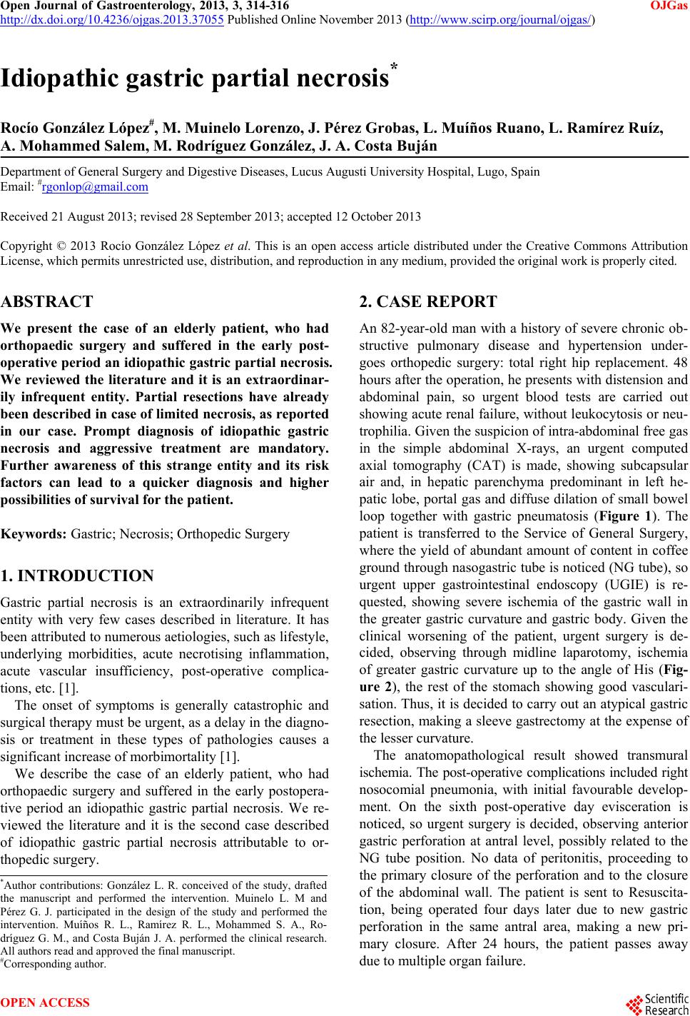 Open Journal of Gastroenterology, 2013, 3, 314-316 OJGas http://dx.doi.org/10.4236/ojgas.2013.37055 Published Online November 2013 (http://www.scirp.org/journal/ojgas/) Idiopathic gastric partial necrosis* Rocío González López#, M. Muinelo Lorenzo, J. Pérez Grobas, L. Muíños Ruano, L. Ramírez Ruíz, A. Mohammed Salem, M. Rodríguez González, J. A. Costa Buján Department of General Surgery and Digestive Diseases, Lucus Augusti University Hospital, Lugo, Spain Email: #[email protected] Received 21 August 2013; revised 28 September 2013; accepted 12 October 2013 Copyright © 2013 Rocío González López et al. This is an open access article distributed under the Creative Commons Attribution License, which permits unrestricted use, distribution, and reproduction in any medium, provided the original work is properly cited. ABSTRACT We present the case of an elderly patient, who had orthopaedic surgery and suffered in the early post- operative period an idiopathic gastric partial necrosis. We reviewed the literature and it is an extraordinar- ily infrequent entity. Partial resections have already been described in case of limited necrosis, as reported in our case. Prompt diagnosis of idiopathic gastric necrosis and aggressive treatment are mandatory. Further awareness of this strange entity and its risk factors can lead to a quicker diagnosis and higher possibilities of survival for the patient. Keywords: Gastric; Necrosis; Orthopedic Surgery 1. INTRODUCTION Gastric partial necrosis is an extraordinarily infrequent entity with very few cases described in literature. It has been attributed to numerous aetiologies, such as lifestyle, underlying morbidities, acute necrotising inflammation, acute vascular insufficiency, post-operative complica- tions, etc. [1]. The onset of symptoms is generally catastrophic and surgical therapy must be urgent, as a delay in the diagno- sis or treatment in these types of pathologies causes a significant increase of morbimortality [1]. We describe the case of an elderly patient, who had orthopaedic surgery and suffered in the early postopera- tive period an idiopathic gastric partial necrosis. We re- viewed the literature and it is the second case described of idiopathic gastric partial necrosis attributable to or- thopedic surgery. 2. CASE REPORT An 82-year-old man with a history of severe chronic ob- structive pulmonary disease and hypertension under- goes orthopedic surgery: total right hip replacement. 48 hours after the operation, he presents with distension and abdominal pain, so urgent blood tests are carried out showing acute renal failure, without leuko cytosis or neu- trophilia. Given the suspicion o f intra-abdomin al free gas in the simple abdominal X-rays, an urgent computed axial tomography (CAT) is made, showing subcapsular air and, in hepatic parenchyma predominant in left he- patic lobe, portal gas and diffuse dilation of small bowel loop together with gastric pneumatosis (Figure 1). The patient is transferred to the Service of General Surgery, where the yield of abundant amount of content in coffee ground through nasogastric tube is noticed (NG tube), so urgent upper gastrointestinal endoscopy (UGIE) is re- quested, showing severe ischemia of the gastric wall in the greater gastric curvature and gastric body. Given the clinical worsening of the patient, urgent surgery is de- cided, observing through midline laparotomy, ischemia of greater gastric curvature up to the angle of His (Fig- ure 2), the rest of the stomach showing good vasculari- sation. Thus, it is decided to carry out an atypical gastric resection, making a sleeve gastrectomy at the expense of the lesser curvature. The anatomopathological result showed transmural ischemia. The post-operative complications included right nosocomial pneumonia, with initial favourable develop- ment. On the sixth post-operative day evisceration is noticed, so urgent surgery is decided, observing anterior gastric perforation at antral level, possibly related to the NG tube position. No data of peritonitis, proceeding to the primary closure of the perforation and to the closure of the abdominal wall. The patient is sent to Resuscita- tion, being operated four days later due to new gastric perforation in the same antral area, making a new pri- mary closure. After 24 hours, the patient passes away due to multiple organ failure. *Author contributions: González L. R. conceived of the study, drafted the manuscript and performed the intervention. Muinelo L. M and Pérez G. J. participated in the design of the study and performed the intervention. Muíños R. L., Ramírez R. L., Mohammed S. A., Ro- dríguez G. M., and Costa Buján J. A. performed the clinical research. All authors read and approved the final manuscript. #Corresponding autho r . OPEN ACCESS 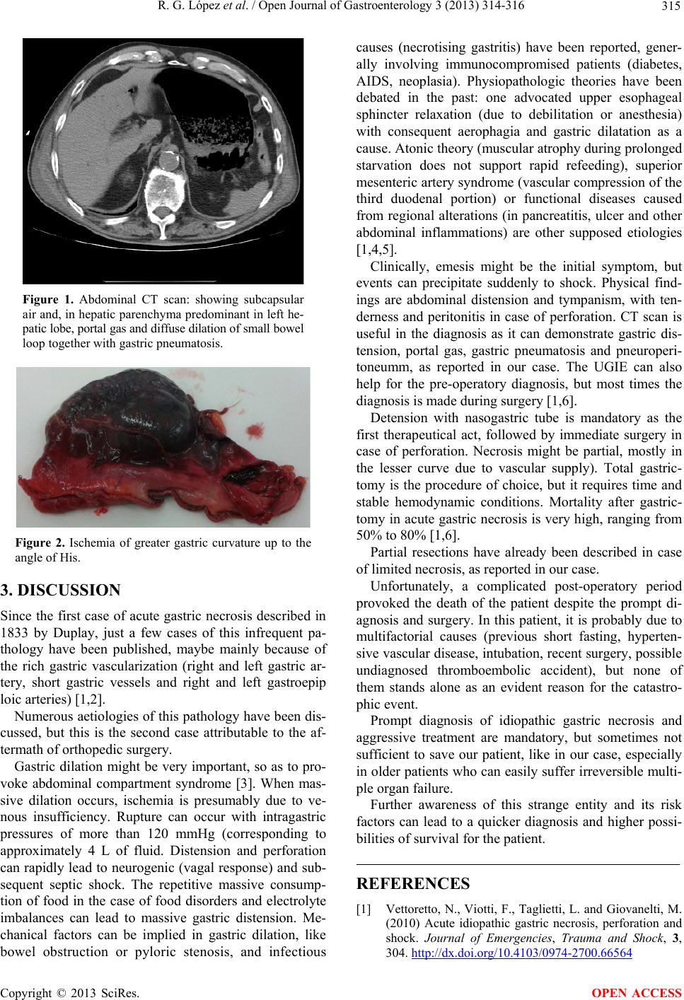 R. G. López et al. / Open Journal of Gastroenterology 3 (2013) 314-316 315 Figure 1. Abdominal CT scan: showing subcapsular air an d, in hepatic p arenchy ma predomina nt in left he- patic lobe, portal gas and diffuse dilation of small bowe l loop together with gastric pneumatosis. Figure 2. Ischemia of greater gastric curvature up to the angle of His. 3. DISCUSSION Since the first case of acute gastric necrosis described in 1833 by Duplay, just a few cases of this infrequent pa- thology have been published, maybe mainly because of the rich gastric vascularization (right and left gastric ar- tery, short gastric vessels and right and left gastroepip loic arteries) [1,2]. Numerous aetiologies of this pathology have been dis- cussed, but this is the second case attributable to the af- termath of orthopedic surgery. Gastric dilation might be very important, so as to pro- voke abdominal compartment syndrome [3]. When mas- sive dilation occurs, ischemia is presumably due to ve- nous insufficiency. Rupture can occur with intragastric pressures of more than 120 mmHg (corresponding to approximately 4 L of fluid. Distension and perforation can rapidly lead to neurogenic (vagal response) and sub- sequent septic shock. The repetitive massive consump- tion of food in the case of food disorders and electrolyte imbalances can lead to massive gastric distension. Me- chanical factors can be implied in gastric dilation, like bowel obstruction or pyloric stenosis, and infectious causes (necrotising gastritis) have been reported, gener- ally involving immunocompromised patients (diabetes, AIDS, neoplasia). Physiopathologic theories have been debated in the past: one advocated upper esophageal sphincter relaxation (due to debilitation or anesthesia) with consequent aerophagia and gastric dilatation as a cause. Atonic theory (muscular atrophy during prolonged starvation does not support rapid refeeding), superior mesenteric artery syndrome (vascular co mpression of the third duodenal portion) or functional diseases caused from regional alterations (in pancreatitis, ulcer and other abdominal inflammations) are other supposed etiologies [1,4,5]. Clinically, emesis might be the initial symptom, but events can precipitate suddenly to shock. Physical find- ings are abdominal distension and tympanism, with ten- derness and peritonitis in case of perforation. CT scan is useful in the diagnosis as it can demonstrate gastric dis- tension, portal gas, gastric pneumatosis and pneuroperi- toneumm, as reported in our case. The UGIE can also help for the pre-operatory diagnosis, but most times the diagnosis is made during surgery [1,6]. Detension with nasogastric tube is mandatory as the first therapeutical act, followed by immediate surgery in case of perforation. Necrosis might be partial, mostly in the lesser curve due to vascular supply). Total gastric- tomy is the procedure of choice, but it requires time and stable hemodynamic conditions. Mortality after gastric- tomy in acute gastric necrosis is very high, ranging from 50% to 80% [1,6]. Partial resections have already been described in case of limited necrosis, as reported in our case. Unfortunately, a complicated post-operatory period provoked the death of the patient despite the prompt di- agnosis and surgery. In this patient, it is probably due to multifactorial causes (previous short fasting, hyperten- sive vascular disease, intubation, recent surgery, possible undiagnosed thromboembolic accident), but none of them stands alone as an evident reason for the catastro- phic event. Prompt diagnosis of idiopathic gastric necrosis and aggressive treatment are mandatory, but sometimes not sufficient to save our patient, like in our case, especially in older patients who can easily suffer irreversible multi- ple organ failure. Further awareness of this strange entity and its risk factors can lead to a quicker diagnosis and higher possi- bilities of survival for the patient. REFERENCES [1] Vettoretto, N., Viotti, F., Taglietti, L. and Giovanelti, M. (2010) Acute idiopathic gastric necrosis, perforation and shock. Journal of Emergencies, Trauma and Shock, 3, 304. http://dx.doi.org/10.4103/0974-2700.66564 Copyright © 2013 SciRes. OPEN ACCESS  R. G. López et al. / Open Journal of Gastroenterology 3 (2013) 314-316 Copyright © 2013 SciRes. OPEN ACCESS 316 [2] Somervell, T.H. (1945) Physiologic gastrectomy. British Journal of Surgery, 33, 146. http://dx.doi.org/10.1002/bjs.18003313010 [3] Mahajna, A., Mitkal, S. and Krausz, M.M. (2008) Post- operative gastric dilatation causing abdominal compart- ment syndrome. World Journal of Emergency Surgery, 3, 7. http://dx.doi.org/10.1186/1749-7922-3-7 [4] Arie, E., Uri, G. and Bickel, A. (2008) Acute gastric dila- tation, necrosis and perforation complicating restrictive- type anorexia nervosa. Journal of Gastrointestinal Sur- gery, 12, 985-987. http://dx.doi.org/10.1007/s11605-007-0414-6 [5] Patocskai, E.J. and Thomas, J.M. (2002) Gastric necrosis in a patient with bulimia. European Journal of Surgery, 168, 302-304. http://dx.doi.org/10.1002/ejs.50 [6] Liu, T.H., Mehall, J.R. and Mercer, D.W. (2001) Image of the month: Acute gastric dilatation with necrosis. Ar- chives of Surgery, 136, 1437-1438. http://dx.doi.org/10.1001/archsurg.136.12.1437 |