

Paper Menu >>
Journal Menu >>
 Surgical Science, 2013, 4, 486-493 Published Online November 2013 (http://www.scirp.org/journal/ss) http://dx.doi.org/10.4236/ss.2013.411095 Open Access SS Early Gallbladder Cancer: Clinical, Morphological, Therapeutic and Evolutionary Aspects Berkane Salah*, Abid Larbi Unit of Visceral and Oncological Surgery, Bologhine Hospital, Algiers, Algeria Email: *[email protected] Received September 23, 2013; revised October 20, 2013; accepted October 28, 2013 Copyright © 2013 Berkane Salah, Abid Larbi. This is an open access article distributed under the Creative Commons Attribution License, which permits unrestricted use, distribution, and reproduction in any medium, provided the original work is properly cited. ABSTRACT Introduction: The early cancer of gallbladder is an entity which is not well recognized currently. It is a cancer which does not extend beyond the muscularis layer of the gallbladder and it is characterized in almost of cases by the absence of lymph node and visceral invasion. Patients and Method: We have conducted this retrospective study of all our cases of early gallbladder cancer treated in our surgical unit. We have studied these through clinical, morphological, thera- peutical and evolutionary aspects. Results: Of 202 gallbladder carcinoma, 33 cancers were classified as early cancer. 25 were females and 8 were males. The mean age was 56.4 years (41 - 70 years). All patients were free of g allbladder can- cer symptoms and all except one had normal CEA and CA19.9. 2 patients had synchronous tumors (one colonic cancer and one rectal cancer). For 16 patients, the diagnosis was done by ultrasonography and 17 by histological examination of the specimen removed for biliary lithiasis. 8 patients had PT1a tumor (confined only to mucosa) and 25 had PT1b tumor (tumor infiltration of the muscular layer). For 19 patients who benefited from extensive lymphadenectomy, only one (5.3%) had lymph node infiltration. 16 patients had a simple cholecystectomy and in two cases, the cholecystec- tomy was associated with bile duct resection. 17 patients had hepatectomy with extensive lymphadenectomy. 2 patients had a simultaneous right colectomy and abdominoperineal resection and another one benefited from choledocal cyst resection. 3 patients benefited from stone removal from bile duct and two had tumor removal from bile duct (ruptured tumor in the bile duct). 1 patient (3.7%) died in postoperative course (hospital mortality). In the follow-up period, 4 patients died from intercurrent causes. Two patients presented a recurrence at 14 and 36 months and died respectively at 19 and 42 months. One patient presented a bile duct cancer at 66 months. She died at 78 months after palliative treat- ment. Currently, 22 patients (66.7%) are still alive without recurrence with mean and median survival of 53 and 31 months. Conclusion: Early gallbladder cancer is an entity which must be known by the radiologist and the surgeon. Recognized on time and well treated, early gallb ladder cancer can be cured and its pro gnosis is excellent. Keywords: Early Cancer; Gallbladder Cancer; Ultrasonography; Expert Radiologist; Surgical Treatment 1. Introduction The early gallbladder cancer is an entity which is not well recognized currently. It is a tumor whose extension does not extend beyond the muscularis layer of the ga llbladder and does not have frequent lymph node infiltration and metastatic spread [1]. Moreover, it represents a proportion of all gallbladder cancer which could be easily cured. In spite of modern morphologic examination and specifi- cally an ultrasonography since twenty years, there is not a clear improvement in the diagnosis for this disease [2-4]. This form is often diagnosed after histological examina- tion of specimen removed for biliary lithiasis. Making an exact diagnosis before surgery has an importance to im- prove the prognosis of this fatal disease in its invasive form. Surgery is debated when the muscular layer is in- vaded (pT1b) between cholecystectomy and radical re- section whereas the cholecystectomy is the best treatment for the pT1a (tumor confined to mucosa). In this article, we present our experience of patients who were managed in our unit during the fifteen last years and to analyze immediate and long-term prognosis of this disease. 2. Patients and Method We analyzed all cases of our patients treated for early gallbladder cancer. We reported the clinical, morpho- *Corresponding a uthor.  B. SALAH, A. LARBI 487 logical, biological, surgical and evolutionary features of these patients. The histological specimen was reanalyzed for the cancer discovered after cholecystectomy done for biliary lithiasis. All the following features were asked to the histologist: histological form and grade (differentia- tion), parietal extension, perineural, vascular and node infiltration. In our un it, pT1a tumor (confined to mucosa) is treated by cholecystectomy and pT1b (muscular infil- tration) is treated by cholecystectomy alone for the patient above 70 years and by bisugmen tal IV-V with fatal asso- ciated disease and extensive lymphadenectomy for pa- tients under 70 years and without fatal associated disease. On the postoperative course, the patient is followed clinically, biologically (Ca 19.9 and CEA) and morpho- logically (ultrasound) each 3 months during 2 years and each 6 months during the following 2 years. After 5 years, we propose only clinically follow up. If a recurrence is diagnosed, a CT scan or MRI is done to have an exact diagnosis and if the disease is curable we propose a new surgery for resection and in the contrary only diversion is practiced. If there is no need of surgery we propose pal- liative chemotherapy. 3. Results Of 202 gallbladder cancer resected since 1996, we report 33 patients with early form (16.3%). There were 25 fe- males and 8 males, with mean age of 56.4 years (41 - 70 years). No patient had any symptoms related to cancer before the su rgery and physical examination were normal or related to lithiasis disease (pain , jaundice and palpable gallbladder in acute cholecystitis). 20 patients (60.6%) had associated biliary lithiasis. One patient had a history of choledocal cyst since 25 years. The diagnosis was done in the pr eopera tive co urse in 1 6 cases (48 .5%) by the ra di- ologist with the ultrasonography and CT Scan. 3 last pa- tients had an endoscopic ultrasonography. 17 cases were diagnosed on the histologist specimen. All of 24 car- cino-embryonary antigen (CEA) and carbohydrate anti- gen 19.9 (CA19.9) measured out were normal except for one CA19.9 which was at 5618 UI/ml for patient with ruptured tumor in the bile du ct and jaundice. One patient was treated for Hodgkin disease 10 years ago; 2 patients had their polypoid tumors known (diagnosed by ultra- sonography) respectively 1year and ten years ago. The tumoral aspect was polypoid in 22 cases (66.7%), thick- ening wall in 6 cases (18%), unapparent form in 4 cases (12%) and nodular in 1 case. 2 patients had ruptured tumor in the bile duct (pT1a and pT1b). Microscopically, all these cases were adenocarcinoma. 8 cases (24.2%) were pT1a and 25 cases (75.8%) were pT1b (Table 1: charac- teristics of 27 cases). 1 patient had a choledocal cyst di- agnosed and followed by a gastroenterologist during 16 years. 16 patients benefited from a cholecystectomy as- sociated with bile duct resection in two cases for a tumoral related cause and another had her choledocal cyst re- moved with a gallbladder. 2 patients who have had a cholecystectomy benefited from a lymphadenectomy. For 17 patients, IV-V bisugmental hepatectomy with exten- sive lymphadenectomy was done. 2 patients had respec- tively colonic and rectal resection simultaneously. 3 pa- tients benefited from stone removal from bile duct. Only one patient presented node infiltration between 19 who benefited from lymphadenectomy (5.3%). 24 patients (72.7%) had one op eration and 9 (27.8%) two op erations . The postoperative course was uneventful for 26 patients (78.8%) and complicated in 7 cases (21.2%). One patient (3%) died from myocardial infarction (Table 2: surgical treatment). Two patients benefited from systemic che- motherapy, one because she had had cystic infiltrative node and another had associated liver metastatic lesion. On the long term follow up, one patient received ex ternal beam therapy for uterine cervix cancer diagnosed 2 years after the cholecystectomy. One patient died from a colon cancer recurrence at 15 months without any evident gall- bladder cancer recurrence. 2 patients (6%) presented a recurrence at respectively 14 and 36 months. They were initially treated by cholecystectomy. One benefited from bisugmental IV-V with lymphadenectomy and chemo- therapy and died from a new recurrence at 19 m onths. The second patient had exploratory laparotomy and died at 42 months. One patient presented a bile duct cancer (upper part) at 66 months. She refused a surgery and died at 78 months. One patient died from acute diabetes complica- tions at 5 6 months a nd other di ed from gastri c hemorrhage secondary to gastric ulcus. One patient died from another disease at 67 months (portal high pressure). One was lost for follow up at 39 months. 22 patients (66.7%) are still alive with mean survival of 51 months and median sur- vival of 32 months. The global 3 and 5 year survival are respectively 53% and 34%. 11 patients (33.3%) had more than 5 year survival and the two oldest had more than 10 years. No recurrence case occurred until nowadays for PTa tumor. 4. Discussion The early cancer of gallbladder is a form which can be recognized by modern morphological examination but it is still diagnosed after surgery for biliary lithiasis. On clinical aspect, it is a silent disease (without any symp- toms). This feature c ould expl ain this absenc e of diagnosi s. Currently, it can be recognized in 2 situations: On preoperative cou rse with ultrasonogr aph y [3-5]. On the histological exam ination after cholecystectomy for biliary lithiasis [5-8]. For a clinician to make a diagnosis before a surgical step, he need s an expert ra diologist w ho should be vigilant during ultrasound examination. There are two funda- mentals lesions which are in favor of an early cancer: Open Access SS 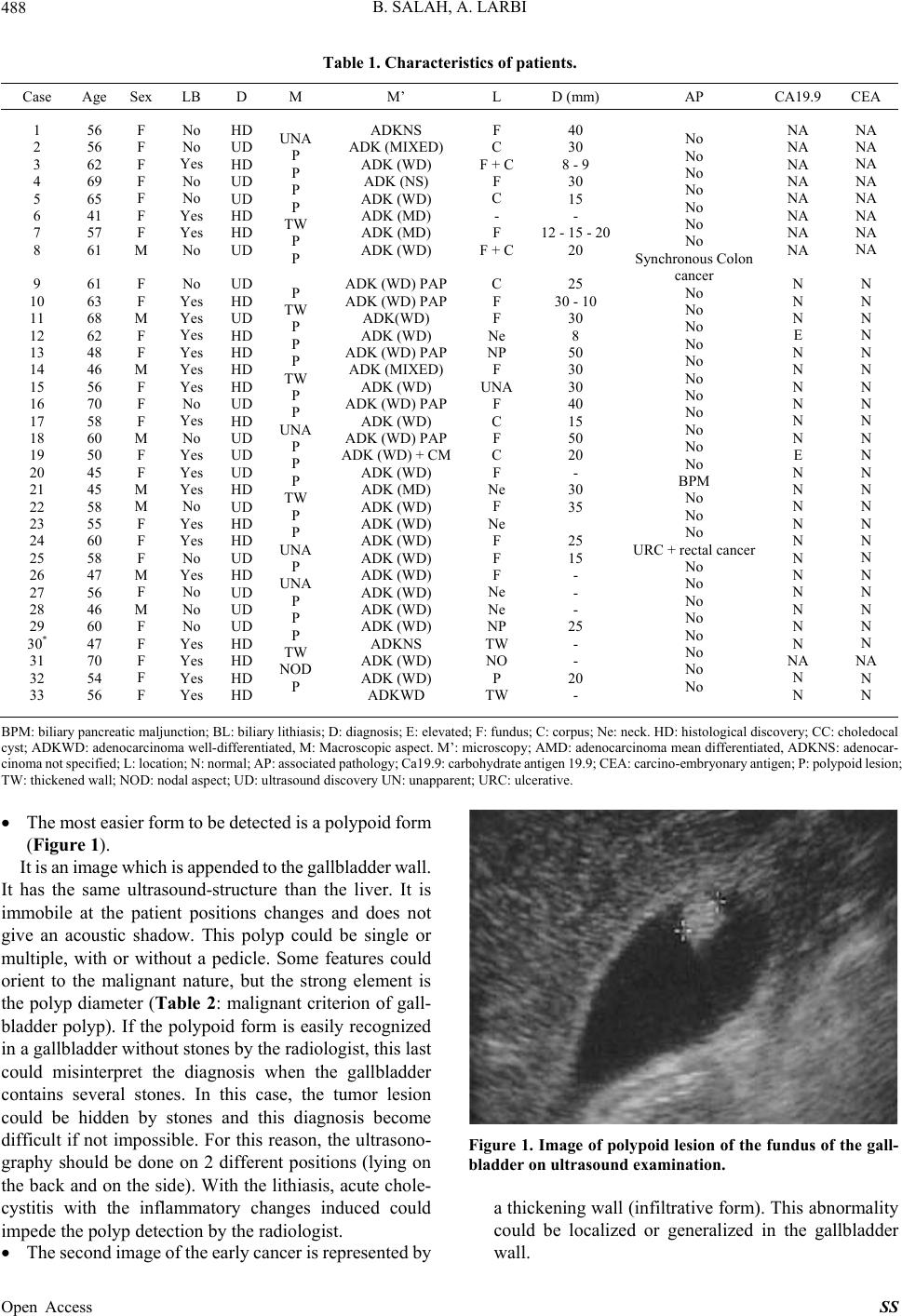 B. SALAH, A. LARBI Open Access SS 488 Table 1. Characteristics of patients. Case Age Sex LB D M M’ L D (mm) AP CA19.9CEA 1 2 3 4 5 6 7 8 9 10 11 12 13 14 15 16 17 18 19 20 21 22 23 24 25 26 27 28 29 30* 31 32 33 56 56 62 69 65 41 57 61 61 63 68 62 48 46 56 70 58 60 50 45 45 58 55 60 58 47 56 46 60 47 70 54 56 F F F F F F F M F F M F F M F F F M F F M M F F F M F M F F F F F No No Yes No No Yes Yes No No Yes Yes Yes Yes Yes Yes No Yes No Yes Yes Yes No Yes Yes No Yes No No No Yes Yes Yes Yes HD UD HD UD UD HD HD UD UD HD UD HD HD HD HD UD HD UD UD UD HD UD HD HD UD HD UD UD UD HD HD HD HD UNA P P P P TW P P P TW P P P TW P P UNA P P P TW P P UNA P UNA P P P TW NOD P ADKNS ADK (MIXED) ADK (WD) ADK (NS) ADK (WD) ADK (MD) ADK (MD) ADK (WD) ADK (WD) PAP ADK (WD) PAP ADK(WD) ADK (WD) ADK (WD) PAP ADK (MIXED) ADK (WD) ADK (WD) PAP ADK (WD) ADK (WD) PAP ADK (WD) + CM ADK (WD) ADK (MD) ADK (WD) ADK (WD) ADK (WD) ADK (WD) ADK (WD) ADK (WD) ADK (WD) ADK (WD) ADKNS ADK (WD) ADK (WD) ADKWD F C F + C F C - F F + C C F F Ne NP F UNA F C F C F Ne F Ne F F F Ne Ne NP TW NO P TW 40 30 8 - 9 30 15 - 12 - 15 - 20 20 25 30 - 10 30 8 50 30 30 40 15 50 20 - 30 35 25 15 - - - 25 - - 20 - No No No No No No No Synchronous Colon cancer No No No No No No No No No No No BPM No No No URC + rectal cancer No No No No No No No No NA NA NA NA NA NA NA NA N N N E N N N N N N E N N N N N N N N N N N NA N N NA NA NA NA NA NA NA NA N N N N N N N N N N N N N N N N N N N N N N NA N N BPM: biliary pancreatic maljunction; BL: biliary lithiasis; D: diagnosis; E: elevated; F: fundus; C: corpus; Ne: neck. HD: histological d iscovery; CC: choled ocal cyst; ADKWD: adenocarcin oma well-differentiated, M: Macr oscopic asp ect. M’: micr oscop y; AMD: adeno carcino ma mean di fferentiated, ADK NS: aden ocar- cinoma not specified; L: location; N: no rmal; AP: a ssociated path ology; Ca19 .9: carbohy drate antige n 19.9; CEA: c arcino-embry onary antigen; P: polypoid lesion; TW: thickened wall; NOD: nodal aspect; UD: ultrasound discovery UN: unapparent; URC: ulcerative. The most easie r form to be detect ed is a polypoid form (Figure 1). It is an image which is appended to the gallbladder wall. It has the same ultrasound-structure than the liver. It is immobile at the patient positions changes and does not give an acoustic shadow. This polyp could be single or multiple, with or without a pedicle. Some features could orient to the malignant nature, but the strong element is the polyp diameter (Table 2: malignant criterion of gall- bladder polyp). If the polypoid form is easily recognized in a gallbladder without stones by the radiologist, this last could misinterpret the diagnosis when the gallbladder contains several stones. In this case, the tumor lesion could be hidden by stones and this diagnosis become difficult if not impossible. For this reason, the ultrasono- graphy should be done on 2 different positions (lying on the back and on the side). With the lithiasis, acute chole- cystitis with the inflammatory changes induced could impede the polyp detection by the radiologist. Figure 1. Image of polypoid lesion of the fundus of the gall- bladder on ultrasound examination. a thickening wall (infiltrative form). This abnormality could be localized or generalized in the gallbladder wall. The second image of the early cancer is re presented by 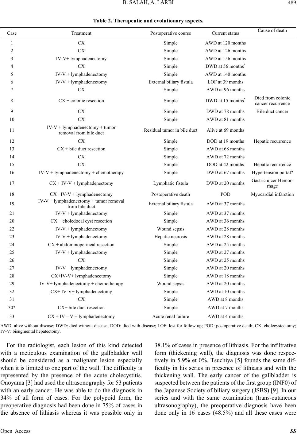 B. SALAH, A. LARBI 489 Table 2. Therapeutic and evolutionary aspec ts. Case Treatment Postoperative course Current status Cause of death 1 CX Simple AWD at 120 months 2 CX Simple AWD at 126 months 3 IV-V+ lymphadenectomy Simple AWD at 156 months 4 CX Simple DWD at 56 months* 5 IV-V + lym p h a d e nectomy Simple AWD at 140 months 6 IV-V + lymphadenectomy External biliary fistula LOF at 39 months 7 CX Simple AWD at 96 months 8 CX + colonic resection Simple DWD at 15 mon ths* Died from colonic cancer recurrence 9 CX Simple DWD at 78 months Bile duct cancer 10 CX Simple AWD at 81 months 11 IV-V + lymphadenectomy + tumor removal from bile duct Residual tumor in bile duct Alive at 69 months 12 CX Simple DOD at 19 months Hepatic recurrence 13 CX + bile duct resection Simple AWD at 68 months 14 CX Simple AWD at 72 months 15 CX Simple DOD at 42 months Hepatic recurrence 16 IV-V + lymphadenectomy + chemoth erapy Simple DWD at 67 months Hypertension portal? 17 CX + IV-V + lymphadenectomy Lymphatic fistula DWD at 20 months Gastric ulcer Hemor- rhage 18 CX+ IV-V + lymphadenectomy Postoperative death POD Myocardial infarction 19 IV-V + lym p h a d e nectomy + tumor rem o v a l from bile duct External biliary fistula AWD at 37 months 21 IV-V + lym p hadenectomy Simple AWD at 37 months 20 CX + choledocal cyst resection Simple AWD at 36 months 22 IV-V + lym p hadenectomy Wound sepsis AWD at 28 months 23 IV-V + lymphadenectomy Hepatic necrosis AWD at 28 months 24 CX + abdominoperineal resection Simple AWD at 25 months 25 IV-V + lym p hadenectomy Simple AWD at 27 months 26 CX Simple AWD at 25 months 27 IV-V lymphadenectomy Simple AWD at 20 months 28 CX+IV-V+ lymphadenectomy Simple AWD at 18 mont hs 29 IV-V+ lymphadenectomy + chemotherapy Wound sepsis AWD at 20 months 32 CX+ IV-V+ lymphadenectomy Simple AWD at 10 months 31 CX Simple AWD at 8 months 30* CX+ bile duct resection Simple AWD at 7 months 33 CX + IV – V + lymphadenectomy Acute renal failure AWD at 4 months AWD: alive without disease; DWD: died witho ut disease; DOD: died with disease; LOF: lost for follow up; POD: postoperative deat h; CX: cholecystectomy; IV-V: bisugmental he patectomy. For the radiologist, each lesion of this kind detected with a meticulous examination of the gallbladder wall should be considered as a malignant lesion especially when it is limited to one part of the wall. The difficulty is represented by the presence of the acute cholecystitis. Onoyama [3] had used the ultrasonography for 53 patients with an early cancer. He was able to do the diagnosis in 34% of all form of cases. For the polypoid form, the preoperative diagnosis had been done in 75% of cases in the absence of lithiasis whereas it was possible only in 38.1% of cases in presence of lithiasis. For the infiltrative form (thickening wall), the diagnosis was done respec- tively in 5.9% et 0%. Tsuchiya [5] founds the same dif- ficulty in his series in presence of lithiasis and with the thickening wall. The early cancer of the gallbladder is suspected b etween the patie nts of the first group (INF 0) of the Japanese Society of biliary su rgery (JSBS) [9]. In our series and with the same examination (trans-cutaneous ultrasonography), the preoperative diagnosis have been done only in 16 cases (48.5%) and all these cases were Open Access SS 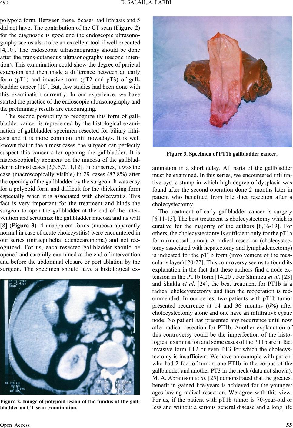 B. SALAH, A. LARBI 490 polypoid form. Between these, 5cases had lithiasis and 5 did not have. The contribution of the CT scan (Figure 2) for the diagnostic is good and the endoscopic ultrasono- graphy seems also to be an excellent tool if well executed [4,10]. The endoscopic ultrasonography should be done after the trans-cutaneous ultrasonography (second inten- tion). This examination could show the degree of parietal extension and then made a difference between an early form (pT1) and invasive form (pT2 and pT3) of gall- bladder cancer [10]. But, few studies had been done with this examination currently. In our experience, we have started the prac tice of the endo scopic ultrasonog raphy and the preliminary results are encouraging. The second possibility to recognize this form of gall- bladder cancer is represented by the histological exami- nation of gallbladder specimen resected for biliary lithi- asis and it is more common until nowadays. It is well known that in the almost cases, the surgeon can perfectly suspect this cancer after opening the gallbladder. It is macroscopically apparent on the mucosa of the gallblad- der in almost cases [2,3,6,7,11,12]. I n our se ries, it was the case (macroscopically visible) in 29 cases (87.8%) after the opening of the gallbladder by the surgeon . It was easy for a polypoid form and difficult for the thickening form especially when it is associated with cholecystitis. This fact is very important for the treatment and binds the surgeon to open the gallbladder at the end of the inter- vention and scr uti nize t he gall bl adder m ucosa and i ts wa ll [8] (Figure 3). 4 unapparent forms (mucosa apparently normal in case of acute cholecystitis) were encountered in our series (intraepithelial adenocarcinoma) and not rec- ognized. For us, each resected gallbladder should be opened and carefully ex amined at the end of intervention and before the abdominal closure or port ablation by the surgeon. The specimen should have a histological ex- Figure 2. Image of polypoid lesion of the fundus of the gall- bladder on CT scan examination. Figure 3. Specimen of PT1b gallbladder cancer. amination in a short delay. All parts of the gallbladder must be examined. In th is series, we encountered infiltra- tive cystic stump in which high degree of dysplasia was found after the second operation done 2 months later in patient who benefited from bile duct resection after a cholecystectomy. The treatment of early gallbladder cancer is surgery [6,11-15]. T he best t reatm ent is cholecystectomy which is curative for the majority of the authors [8,16-19]. For others, the cholecyst ectomy is suf ficient only for t he pT1a form (mucosal tumor). A radical resection (cholecystec- tomy associated with hepatectomy and lymphadenectomy) is indicated for the pT1b form (involvement of the mus- cularis layer) [20-22]. This controversy seems to found its explanation in the fact that these authors find a node ex- tension in the PT1b form [14,20]. For Shimizu et al. [23] and Shukla et al. [24], the best treatment for PT1b is a radical cholecystectomy and then the reoperation is rec- ommended. In our series, two patients with pT1b tumor presented recurrence at 14 and 36 months (6%) after cholecystectomy alone and on e have an infiltrative cystic node. No patient has presented any recurrence until now after radical resection for PT1b. Another explanation of this controversy could be the imperfection of the histo- logical examination and som e cases of the PT1b are in fact invasive form PT2 or even PT3 for which the cholecys- tectomy is insufficient. We have an example with patient who had 2 foci of tumor, one PT1b in the corpus of the gallbladder and another PT3 in the neck (data not shown). M. A. Abramson et al. [25] demonstrated that the greatest benefit in gained life-years is achieved for the youngest ages having radical resection. We agree with this view. For us, if the patient with pT1b tumor is 70-year-old or less and without a serious general disease and a long life Open Access SS 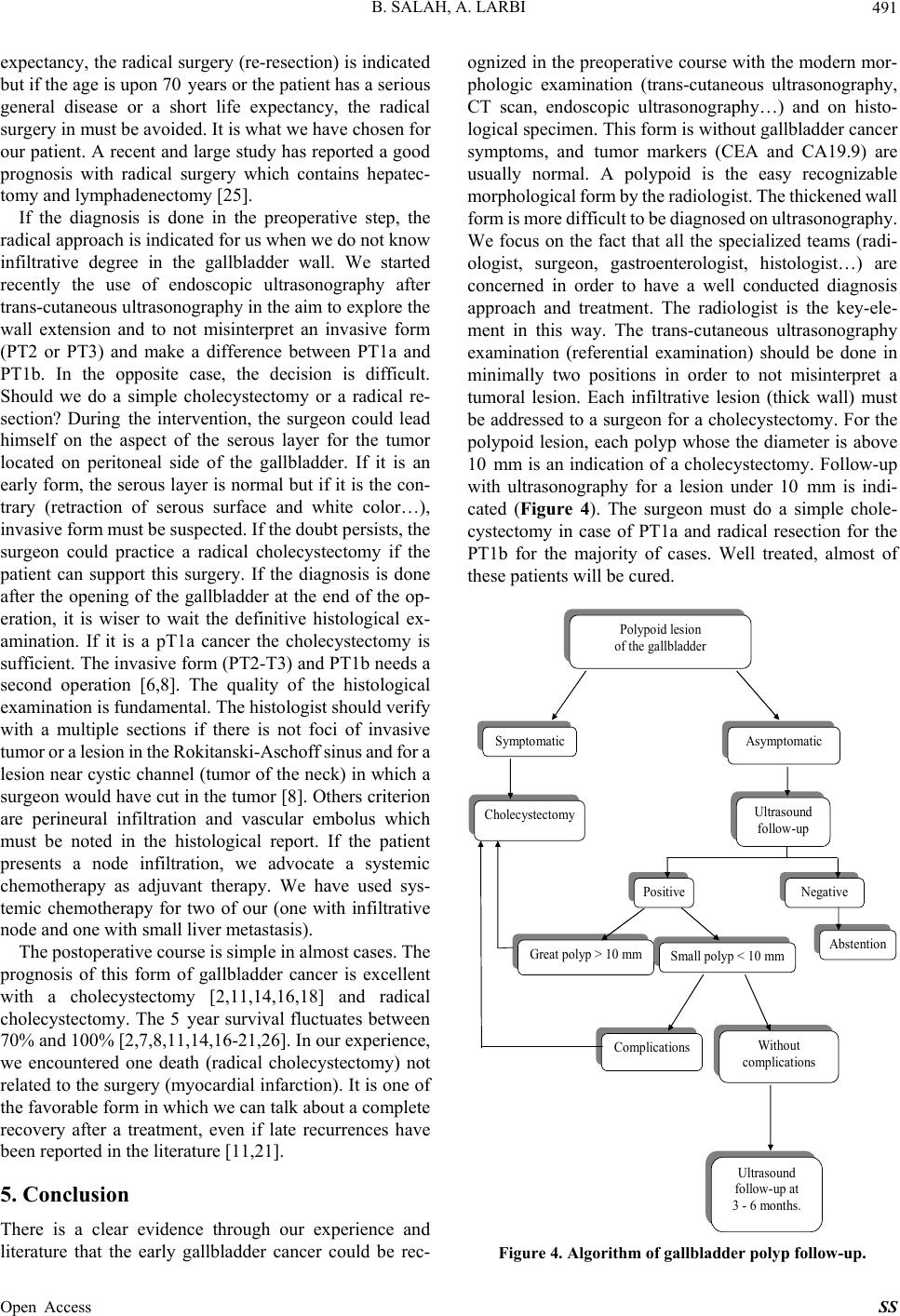 B. SALAH, A. LARBI 491 expectancy, the radical surgery (re-resection) is indicated but if the age is upon 70 years or the patient has a serious general disease or a short life expectancy, the radical surgery in must be avoided. It is what we have chosen for our patient. A recent and large study has reported a good prognosis with radical surgery which contains hepatec- tomy and lymphadenectomy [25]. If the diagnosis is done in the preoperative step, the radical approach is indicated for us when we do not know infiltrative degree in the gallbladder wall. We started recently the use of endoscopic ultrasonography after trans-cutan eous ultr asonogr aphy in the aim to exp lore th e wall extension and to not misinterpret an invasive form (PT2 or PT3) and make a difference between PT1a and PT1b. In the opposite case, the decision is difficult. Should we do a simple cholecystectomy or a radical re- section? During the intervention, the surgeon could lead himself on the aspect of the serous layer for the tumor located on peritoneal side of the gallbladder. If it is an early form, the serous layer is normal but if it is the con- trary (retraction of serous surface and white color…), invasi ve f orm m ust be susp ec t ed. I f t he dou b t pers is ts , t he surgeon could practice a radical cholecystectomy if the patient can support this surgery. If the diagnosis is done after the opening of the gallbladder at the end of the op- eration, it is wiser to wait the definitive histological ex- amination. If it is a pT1a cancer the cholecystectomy is sufficient. The invasive form (PT2-T3) and PT1b needs a second operation [6,8]. The quality of the histological examination is fundamental. The histologist should verify with a multiple sections if there is not foci of invasive tumor or a lesi on in the Rokitanski-As choff sinus and f or a lesion near cystic channel (tumor of the neck) in which a surgeon would h ave cut in the tumor [8]. Others criterion are perineural infiltration and vascular embolus which must be noted in the histological report. If the patient presents a node infiltration, we advocate a systemic chemotherapy as adjuvant therapy. We have used sys- temic chemotherapy for two of our (one with infiltrative node and one with small liver metastasis). The postoperative course is sim ple in almost cases. T he prognosis of this form of gallbladder cancer is excellent with a cholecystectomy [2,11,14,16,18] and radical cholecystectomy. The 5 year survival fluctuates between 70% and 100% [2,7,8,11, 14,16-21,26]. In our experience, we encountered one death (radical cholecystectomy) not related to the surgery (myocardial infarction). It is one of the favorable form in which we can talk about a complete recovery after a treatment, even if late recurrences have been reported in the literature [11,21]. 5. Conclusion There is a clear evidence through our experience and literature that the early gallbladder cancer could be rec- ognized in the preop erative course with the modern mor- phologic examination (trans-cutaneous ultrasonography, CT scan, endoscopic ultrasonography…) and on histo- logical specimen. This form is without gallbladder cancer symptoms, and tumor markers (CEA and CA19.9) are usually normal. A polypoid is the easy recognizable morpholo gical form by the ra diologist. The t hickened wall form is more difficult to be diagnosed on ultras onography. We focus on the fact that all the specialized teams (radi- ologist, surgeon, gastroenterologist, histologist…) are concerned in order to have a well conducted diagnosis approach and treatment. The radiologist is the key-ele- ment in this way. The trans-cutaneous ultrasonography examination (referential examination) should be done in minimally two positions in order to not misinterpret a tumoral lesion. Each infiltrative lesion (thick wall) must be addressed to a surgeon for a cholecystectomy. For the polypoid lesion, each polyp whose the diameter is above 10 mm is an indication of a cholecystectomy. Follow-up with ultrasonography for a lesion under 10 mm is indi- cated (Figure 4). The surgeon must do a simple chole- cystectomy in case of PT1a and radical resection for the PT1b for the majority of cases. Well treated, almost of these patients will be cured. Polypoid lesion of the gallbladder Symptomatic Asymptomatic Cholecystecto my Ultrasound follow-up Negative Positive Abstention Great polyp > 10 mm Small polyp < 10 mm Complications Without complications Ultrasound follow-up at 3 - 6 mo nths. Figure 4. Algorithm of gallbladder polyp follow-up. Open Access SS 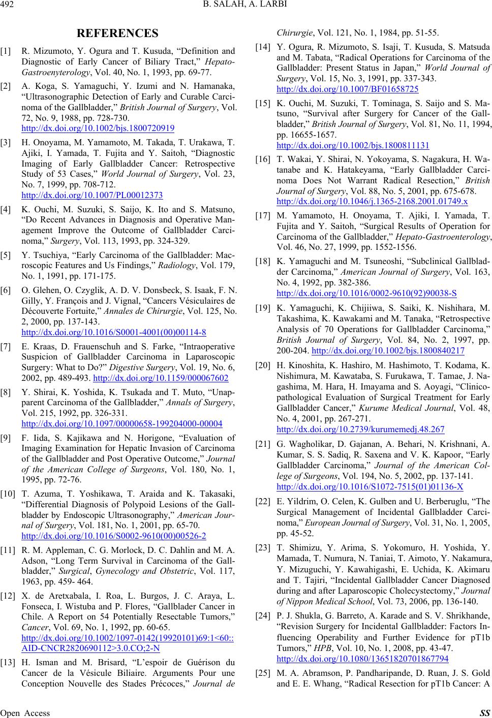 B. SALAH, A. LARBI 492 REFERENCES [1] R. Mizumoto, Y. Ogura and T. Kusuda, “Definition and Diagnostic of Early Cancer of Biliary Tract,” Hepato- Gastroenyterology, Vol. 40, No. 1, 1993, pp. 69-77. [2] A. Koga, S. Yamaguchi, Y. Izumi and N. Hamanaka, “Ultrasonographic Detection of Early and Curable Carci- noma of the Gallbladder,” British Journal of Surgery, Vol. 72, No. 9, 1988, pp. 728-730. http://dx.doi.org/10.1002/bjs.1800720919 [3] H. Onoyama, M. Yamamoto, M. Takada, T. Urakawa, T. Ajiki, I. Yamada, T. Fujita and Y. Saitoh, “Diagnostic Imaging of Early Gallbladder Cancer: Retrospective Study of 53 Cases,” World Journal of Surgery, Vol. 23, No. 7, 1999, pp. 708-712. http://dx.doi.org/10.1007/PL00012373 [4] K. Ouchi, M. Suzuki, S. Saijo, K. Ito and S. Matsuno, “Do Recent Advances in Diagnosis and Operative Man- agement Improve the Outcome of Gallbladder Carci- noma,” Surgery, Vol. 113, 1993, pp. 324-329. [5] Y. Tsuchiya, “Early Carcinoma of the Gallbladder: Mac- roscopic Features and Us Findings,” Radiology, Vol. 179, No. 1, 1991, pp. 171-175. [6] O. Glehen, O. Czyglik, A. D. V. Donsbeck, S. Isaak, F. N. Gilly, Y. François and J. Vignal, “Cancers Vésiculaires de Découverte Fortuite,” Annales de Chirurgie, Vol. 125, No. 2, 2000, pp. 137-143. http://dx.doi.org/10.1016/S0001-4001(00)00114-8 [7] E. Kraas, D. Frauenschuh and S. Farke, “Intraoperative Suspicion of Gallbladder Carcinoma in Laparoscopic Surgery: What to Do?” Digestive Surgery, Vol. 19, No. 6, 2002, pp. 489-493. http://dx.doi.org/10.1159/000067602 [8] Y. Shirai, K. Yoshida, K. Tsukada and T. Muto, “Unap- parent Carcinoma of the Gallbladder,” Annals of Surgery, Vol. 215, 1992, pp. 326-331. http://dx.doi.org/10.1097/00000658-199204000-00004 [9] F. Iida, S. Kajikawa and N. Horigone, “Evaluation of Imaging Examination for Hepatic Invasion of Carcinoma of the Gallbladder and Post Operative Outcome,” Journal of the American College of Surgeons, Vol. 180, No. 1, 1995, pp. 72-76. [10] T. Azuma, T. Yoshikawa, T. Araida and K. Takasaki, “Differential Diagnosis of Polypoid Lesions of the Gall- bladder by Endoscopic Ultrasonography,” American Jour- nal of Surgery, Vol. 181, No. 1, 2001, pp. 65-70. http://dx.doi.org/10.1016/S0002-9610(00)00526-2 [11] R. M. Appleman, C. G. Morlock, D. C. Dahlin and M. A. Adson, “Long Term Survival in Carcinoma of the Gall- bladder,” Surgical, Gynecology and Obstetric, Vol. 117, 1963, pp. 459- 464. [12] X. de Aretxabala, I. Roa, L. Burgos, J. C. Araya, L. Fonseca, I. Wistuba and P. Flores, “Gallblader Cancer in Chile. A Report on 54 Potentially Resectable Tumors,” Cancer, Vol. 69, No. 1, 1992, pp. 60-65. http://dx.doi.org/10.1002/1097-0142(19920101)69:1<60:: AID-CNCR2820690112>3.0.CO;2-N [13] H. Isman and M. Brisard, “L’espoir de Guérison du Cancer de la Vésicule Biliaire. Arguments Pour une Conception Nouvelle des Stades Précoces,” Journal de Chirurgie, Vol. 121, No. 1, 1984, pp. 51-55. [14] Y. Ogura, R. Mizumoto, S. Isaji, T. Kusuda, S. Matsuda and M. Tabata, “Radical Operations for Carcinoma of the Gallbladder: Present Status in Japan,” World Journal of Surgery, Vol. 15, No. 3, 1991, pp. 337-343. http://dx.doi.org/10.1007/BF01658725 [15] K. Ouchi, M. Suzuki, T. Tominaga, S. Saijo and S. Ma- tsuno, “Survival after Surgery for Cancer of the Gall- bladder,” Briti sh Journal of Surgery, Vol. 81, No. 11, 1994, pp. 16655-1657. http://dx.doi.org/10.1002/bjs.1800811131 [16] T. Wakai, Y. Shirai, N. Yokoyama, S. Nagakura, H. Wa- tanabe and K. Hatakeyama, “Early Gallbladder Carci- noma Does Not Warrant Radical Resection,” British Journal of Surgery, Vol. 88, No. 5, 2001, pp. 675-678. http://dx.doi.org/10.1046/j.1365-2168.2001.01749.x [17] M. Yamamoto, H. Onoyama, T. Ajiki, I. Yamada, T. Fujita and Y. Saitoh, “Surgical Results of Operation for Carcinoma of the Gallbladder,” Hepato-Gastroenterology, Vol. 46, No. 27, 1999, pp. 1552-1556. [18] K. Yamaguchi and M. Tsuneoshi, “Subclinical Gallblad- der Carcinoma,” American Journal of Surgery, Vol. 163, No. 4, 1992, pp. 382-386. http://dx.doi.org/10.1016/0002-9610(92)90038-S [19] K. Yamaguchi, K. Chijiiwa, S. Saiki, K. Nishihara, M. Takashima, K. Kawakami and M. Tanaka, “Retrospective Analysis of 70 Operations for Gallbladder Carcinoma,” British Journal of Surgery, Vol. 84, No. 2, 1997, pp. 200-204. http://dx.doi.org/10.1002/bjs.1800840217 [20] H. Kinoshita, K. Hashiro, M. Hashimoto, T. Kodama, K. Nishimura, M. Kawataba, S. Furukawa, T. Tamae, J. Na- gashima, M. Hara, H. Imayama and S. Aoyagi, “Clinico- pathological Evaluation of Surgical Treatment for Early Gallbladder Cancer,” Kurume Medical Journal, Vol. 48, No. 4, 2001, pp. 267-271. http://dx.doi.org/10.2739/kurumemedj.48.267 [21] G. Wagholikar, D. Gajanan, A. Behari, N. Krishnani, A. Kumar, S. S. Sadiq, R. Saxena and V. K. Kapoor, “Early Gallbladder Carcinoma,” Journal of the American Col- lege of Surgeons, Vol. 194, No. 5, 2002, pp. 137-141. http://dx.doi.org/10.1016/S1072-7515(01)01136-X [22] E. Yildrim, O. Celen, K. Gulben and U. Berberuglu, “The Surgical Management of Incidental Gallbladder Carci- noma,” European Journal of Surgery, Vol. 31, N o . 1, 2005, pp. 45-52. [23] T. Shimizu, Y. Arima, S. Yokomuro, H. Yoshida, Y. Mamada, T. Numura , N. Tania i, T. Aimoto, Y. Nakamura, Y. Mizuguchi, Y. Kawahigashi, E. Uchida, K. Akimaru and T. Tajiri, “Incidental Gallbladder Cancer Diagnosed during and after Laparoscopic Cholecystectomy,” Journal of Nippon Medical School, Vol. 73, 2006, pp. 136-140. [24] P. J. Shukla, G. Barreto, A. Karade and S. V. Shrikhande, “Revision Surgery for Incidental Gallbladder: Factors In- fluencing Operability and Further Evidence for pT1b Tumors,” HPB, Vol. 10, No. 1, 2008, pp. 43-47. http://dx.doi.org/10.1080/13651820701867794 [25] M. A. Abramson, P. Pandharipande, D. Ruan, J. S. Gold and E. E. Whang, “Radical Resection for pT1b Cancer: A Open Access SS  B. SALAH, A. LARBI Open Access SS 493 Decision Analysis,” HPB, Vol. 11, No. 8, 2009, pp. 656- 663. http://dx.doi.org/10.1111/j.1477-2574.2009.00108.x [26] D. M. Hari, J. H. Howard, A. M. Leung, C. G. Chui, M. S. Sim and A. J. Bilchik, “A 21-Year Analysis of Stage I Gallbladder Carcinoma: Is Cholecystectomy Alone Ade- quate?” HPB, Vol. 15, No. 1, 2013, pp. 40-48. http://dx.doi.org/10.1111/j.1477-2574.2012.00559.x |