

Paper Menu >>
Journal Menu >>
 Advances in Bioscience and Biotechnology, 2013, 4, 41-47 ABB http://dx.doi.org/10.4236/abb.2013.411A2006 Published Online November 2013 (http://www.scirp.org/journal/abb/) Melatonin modulates the expression of Bcl-2 family proteins in liver after thermal injury in rats Ganka Bekyarova1*, Maria Tzaneva2, Minka Hristova1 1Division of Pathophysiology, Medical University of Varna, Varna, Bulgaria 2Department of General and Clinical Pathology, Medical University of Varna, Varna, Bulgaria Email: *[email protected] Received 26 September 2013; revised 28 October 2013; accepted 9 November 2013 Copyright © 2013 Ganka Bekyarova et al. This is an open access article distributed under the Creative Commons Attribution License, which permits unrestricted use, distribution, and reproduction in any medium, provided the original work is properly cited. ABSTRACT Melatonin, the principal secretory product of the pineal gland, functions as a potent antioxidant and free radical scavenger. Additionally, the antiapoptotic effect of melatonin has been observed both in vivo and in vitro. The aim of this study was to investigate the protective effects of melatonin against burn-in- duced injury in rat liver and whether these changes were associated with oxidative stress and changes in the expression of apoptosis related genes Bcl-2 and Bax. Melatonin (10 mg/kg, i.p.) was applied immedi- ately after 30% of total body surface area (TBSA) burns of male Wistar rats. Malondialdehyde (MDA) as marker of oxidative stress and tumor necrosis fac- tor (TNF-α) as inflammatory marker were assayed by biochemical methods. The hepatic apoptosis related genes Bcl-2 and Bax using light immunоchistochem- istry were investigated, too. Hepatic TNF-α and MDA levels were increased significantly following severe burn. Thermal trauma increased the Bax expression without any changes of anti-apoptotic Bcl-2 protein in sinusoidal endothelial cells (SECs) of burn-treated animals compared with the control group animals as well as elevated ratio Bax/Bcl-2 suggesting the sus- ceptibility of these cells to apoptosis. Melatonin sig- nificantly decreased the MDA and TNF-α levels in the liver tissue. It decreased also expression of Bax, in- creased expression of Bcl-2 and reduced Bax/Bcl-2 ratio. In conclusion, experimental data show that me- latonin modulates the expression of Bcl-2 family pro- teins by increasing anti-apoptotic Bcl-2, inhibits apop- tosis and restricts the burn-induced damage. Keywords: Melatonin; Liver; Bcl-2; Bax; Tumor Necrosis Factor; Lipid Peroxidation; Thermal Skin Injury 1. INTRODUCTION Severe burn is still a serious clinical problem in emer- gency medicine causing damages to organs distant from burn wound. Clinical studies have shown that liver dys- function and damage are essential for survival in cri- tically ill and severely burned patients [1,2]. Various cel- lular and molecular interactions such as neutrophil and macrophage activation, mitochondrial dysfunction oxy- gen radical, cytokine overproduction, depletion of glu- tathione, may be involved in the processes [3-6]. These complex mechanisms cause liver dysfunction, alteration of hepatic parenchyma and eventually cell death (apopto- sis). The increased apoptosis in liver and other organs is a result of “systemic apoptotic response” after burns [7]. Pathophysiology of burn-induced liver injury includes many mechanisms and is not entirely defined yet. The possible mechanisms for increased apoptosis are a de- creased perfusion and elevation of pro-inflammatory cytokines early post burn. Oxidative stress is recognized as an important mechanism of apoptosis of hepatocytes in liver injury [8,9]. The increased ROS production re- sults in an imbalance between anti-apoptotic Bcl-2 and pro-apoptotic Bax proteins which contributes to the sus- ceptibility of the cell to apoptosis. Bax and Bcl-2 pro- teins are among the most commonly used markers for apoptosis [10]. Literature data about the role of free ra- dicals in liver apoptosis after burns are scarce. Melatonin (N-acetyl-5-methoxytryptamine) is an in- doleamine endogenously produced in the pineal gland and extrapineal tissues, liver including [11-13]. Mela- tonin possesses a wide variety of biologic effects such as sedative, anxiolytic, antinociceptive. Melatonin plays a direct role in mitochondrial homeostasis by scavenging reactive oxygen species (ROS) and reactive nitrogen species (RNS), improving mitochondrial respiration, increasing the level of ATP synthesis, and reducing the *Corresponding author. OPEN ACCESS  G. Bekyarova et al. / Advances in Bioscience and Biotechnology 4 (2013) 41-47 42 harmful decrease in the mitochondrial membrane poten- tial that can trigger the apoptotic cascade [14,15]. Mela- tonin is widely used to prevent oxidative stress induced by ischemia/reperfusion (I/R) in the liver [9,16,17] as well as other organs such as gastric mucosa [10]. The aim of this experimental study was to investigate the protective effects of melatonin against burn-induced injury and whether these changes were associated with oxidative stress and changes in the expression of apop- tosis-related proteins Bcl-2 and Bax in rat liver. 2. MATERIAL AND METHODS 2.1. Animals The experimental procedure was approved by the Home Office for Care and Use of Laboratory Animals and per- formed with a strong consideration for ethics of animal experimentation. Age-matched male rats weighing be- tween 220 and 250 g fasted for 12 h were allowed free access to water before injury. Animals were housed in a 20˚C and offered rat chow and water ad libitum. They were kept in dark: light cycles (DL = 12:12 h) in indi- vidual wire-bottomed cages. Thus, lights were turned off at 8:00 p.m. and turned on at 8:00 a.m. for achieving satisfactory photoperiod. 2.2. Thermal Injury and Melatonin Treatment After light ether inhalation, general anesthesia was per- formed using thiopental (30 mg/kg i.p.). In order to ac- complish 30% of third degree burn hot boiling water (90˚C) was applied on the back of the animals during a period of 10 sec. For those rats which were subjected to burn injury 4 mL of physiological saline was applied intraperitoneally (i.p) for immediate resuscitation fol- lowing burn injury. No animals died within the first 24 h of post-burn period. Twenty-four male Wistar rats were divided into three equal (n = 8) groups: 1. control—non- burned, non-treated group (C); 2. burned, nontreated (B), 3. burned rats, melatonin-treated (B+Mel). All the ani- mals were given buprenorphine (0.3 mg/kg i.p. b.w.) twice daily for pain control post burn. They were re- anesthetized with thiopental and sacrificed 24 h after burns as liver was sampled. 2.3. Biochemical Analysis Livers was gently separated from the underlying tissue and homogenized in 1:5 w/v 50 mM phosphate buffer (pH 7.4) containing 0.1 mM EDTA at 4000 rpm for 10 min. The homogenate was centrifuged at 800 × g rpm for 15 min to discard the sediment and supernatant was fro- zen until analysis. All the manipulations were performed at 4˚C - 8˚C. Analysis was carried out immediately after thawing of the samples. For apoptosis and immunohis- tochemical analysis tissue samples were fixed in 10% neutral buffered formalin and embedded in paraffin. Membrane lipid peroxidation was assayed by MDA measured by its thiobarbituric acid (TBA) reactivity of liver homogenate using the method of Porter et al. (1976) [18]. Results were expressed as nmol MDA/g tissue. They were determined using the extinction co-efficient of MDA-TBA complex at 532 nm = 1.56 × 10⁻⁵ cm−1·M−1 solution. Hepatic tumor necrosis factor (TNF)-α level was quan- tified using enzyme-linked immunosorbent assay (ELISA) kit (Diaclone Gen-Probe, France) specific for the previ- ously mentioned rat cytokines according to the manu- facturer’s instructions and guidelines. Hepatic TNF-α level was expressed as pg/mL. 2.4. Immunohistochemical Examination for Bcl-2 and Bax in Liver Immunohistochemical staining was performed with rab- bit anti-rat Bcl-2 or Bax on paraffin sections according to the manufacturer’s instructions (Santa Cruz). Morphometric method was used to assess quantita- tively enzyme contents of Bax and Bcl-2. Enzyme con- tent was determined as: strong, score 3; moderate, score 2; weak, score1, and lacking, score 0 on the basis of the occurrence of immunodeposits (Tzaneva, 2001) [19]. Bax and Bcl-2 contents of the liver cells were defined as the enzyme content of each cell was multiplied by their scoring factors and divided by total number of cells. Morphometric investigation was performed on 50 cells from each sample. 2.5. Histopathological Examinations Tissue specimens were fixed in 10% buffered formalin (pH 7.2), dehydrated in ascending series of ethyl alcohol (70% - 100%), cleared in methyl benzoate and embedded in paraffin wax. Tissue sections of 5 μm were stained with hematoxylin and eosin (H&E) and examined using light microscope (Olympus BH-2, Tokyo, Japan). Apop- totic SECs were examined by light microscopy at a mag- nification of 400× (high power field). Approximately 500 SECs were examined in each high power field. The apoptotic index was defined as the number of stained cells per high power field. Statistics Our data were log-transformed to satisfy the assumptions required to perform parametric tests and, therefore, pre- sented as geometric mean and 95% confidence intervals of the mean. Orthogonal contrasts in ANOVA were used to statistically analyze the difference between any two specified groups. Copyright © 2013 SciRes. OPEN ACCESS 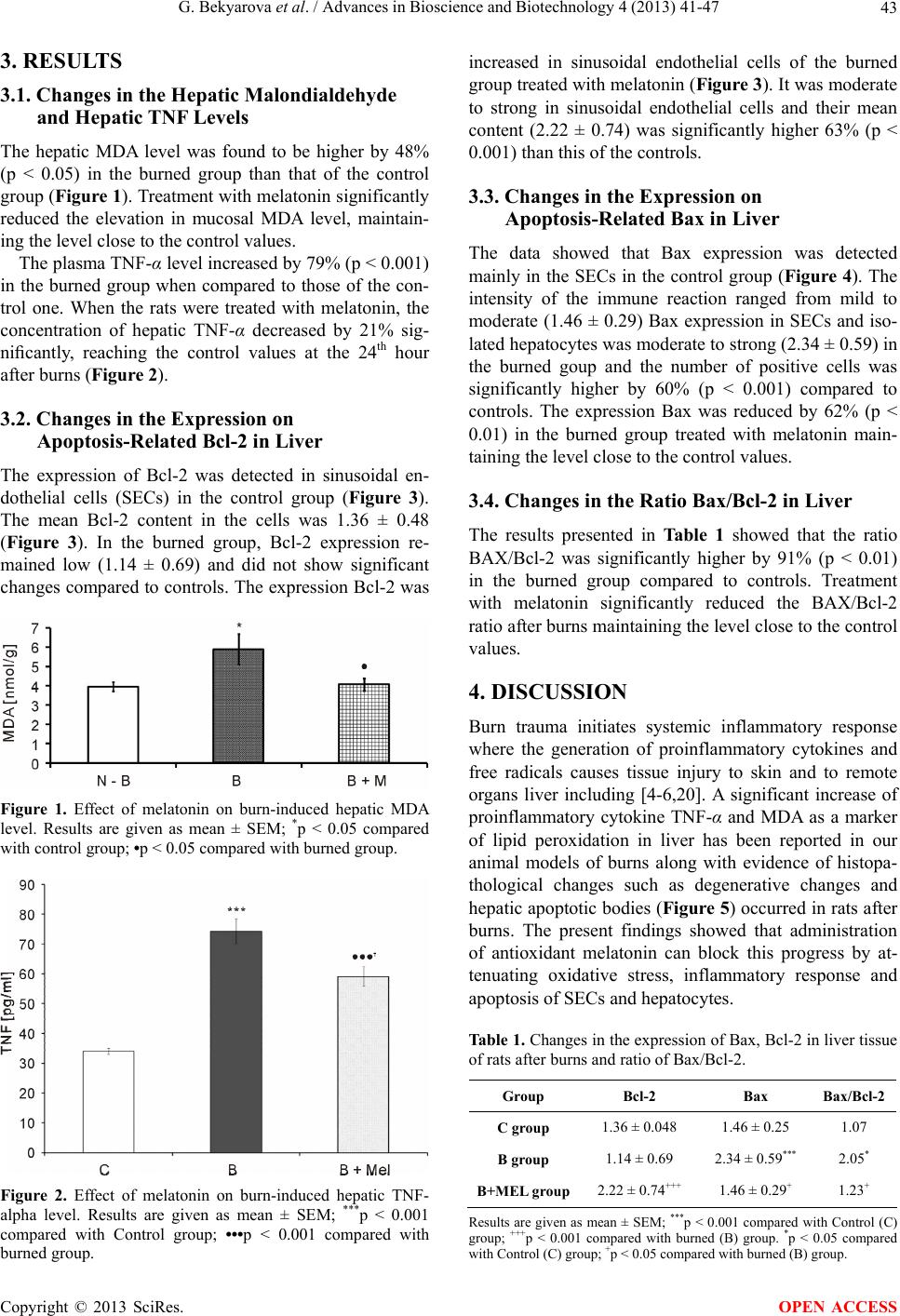 G. Bekyarova et al. / Advances in Bioscience and Biotechnology 4 (2013) 41-47 43 3. RESULTS 3.1. Changes in the Hepatic Malondialdehyde and Hepatic TNF Levels The hepatic MDA level was found to be higher by 48% (p < 0.05) in the burned group than that of the control group (Figure 1). Treatment with melatonin significantly reduced the elevation in mucosal MDA level, maintain- ing the level close to the control values. The plasma TNF-α level increased by 79% (p < 0.001) in the burned group when compared to those of the con- trol one. When the rats were treated with melatonin, the concentration of hepatic TNF-α decreased by 21% sig- nificantly, reaching the control values at the 24th hour after burns (Figure 2). 3.2. Changes in the Expression on Apoptosis-Related Bcl-2 in Liver The expression of Bcl-2 was detected in sinusoidal en- dothelial cells (SECs) in the control group (Figure 3). The mean Bcl-2 content in the cells was 1.36 ± 0.48 (Figure 3). In the burned group, Bcl-2 expression re- mained low (1.14 ± 0.69) and did not show significant changes compared to controls. The expression Bcl-2 was Figure 1. Effect of melatonin on burn-induced hepatic MDA level. Results are given as mean ± SEM; *p < 0.05 compared with control group; •p < 0.05 compared with burned group. Figure 2. Effect of melatonin on burn-induced hepatic TNF- alpha level. Results are given as mean ± SEM; ***p < 0.001 compared with Control group; •••p < 0.001 compared with burned group. increased in sinusoidal endothelial cells of the burned group treated with melatonin (Figure 3). It was moderate to strong in sinusoidal endothelial cells and their mean content (2.22 ± 0.74) was significantly higher 63% (p < 0.001) than this of the controls. 3.3. Changes in the Expression on Apoptosis-Related Bax in Liver The data showed that Вax expression was detected mainly in the SECs in the control group (Figure 4). The intensity of the immune reaction ranged from mild to moderate (1.46 ± 0.29) Bax expression in SECs and iso- lated hepatocytes was moderate to strong (2.34 ± 0.59) in the burned goup and the number of positive cells was significantly higher by 60% (p < 0.001) compared to controls. The expression Bax was reduced by 62% (p < 0.01) in the burned group treated with melatonin main- taining the level close to the control values. 3.4. Changes in the Ratio Bax/Bcl-2 in Liver The results presented in Table 1 showed that the ratio BAX/Bcl-2 was significantly higher by 91% (p < 0.01) in the burned group compared to controls. Treatment with melatonin significantly reduced the BAX/Bcl-2 ratio after burns maintaining the level close to the control values. 4. DISCUSSION Burn trauma initiates systemic inflammatory response where the generation of proinflammatory cytokines and free radicals causes tissue injury to skin and to remote organs liver including [4-6,20]. A significant increase of proinflammatory cytokine TNF-α and MDA as a marker of lipid peroxidation in liver has been reported in our animal models of burns along with evidence of histopa- thological changes such as degenerative changes and hepatic apoptotic bodies (Figure 5) occurred in rats after burns. The present findings showed that administration of antioxidant melatonin can block this progress by at- tenuating oxidative stress, inflammatory response and apoptosis of SECs and hepatocytes. Table 1. Changes in the expression of Bax, Bcl-2 in liver tissue of rats after burns and ratio of Bax/Bcl-2. Group Bcl-2 Bax Bax/Bcl-2 C group 1.36 ± 0.048 1.46 ± 0.25 1.07 B group 1.14 ± 0.69 2.34 ± 0.59*** 2.05* B+MEL group2.22 ± 0.74+++ 1.46 ± 0.29+ 1.23+ Results are given as mean ± SEM; ***p < 0.001 compared with Control (C) group; +++p < 0.001 compared with burned (B) group. *p < 0.05 compared ith Control (C) group; +p < 0.05 compared with burned (B) group. w Copyright © 2013 SciRes. OPEN ACCESS 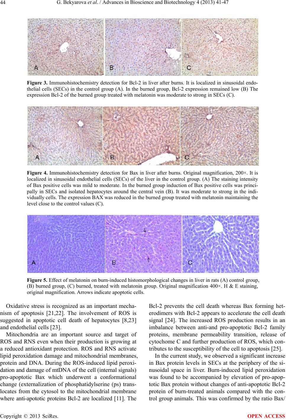 G. Bekyarova et al. / Advances in Bioscience and Biotechnology 4 (2013) 41-47 Copyright © 2013 SciRes. 44 Figure 3. Immunohistochemistry detection for Bcl-2 in liver after burns. It is localized in sinusoidal endo- thelial cells (SECs) in the control group (A). In the burned group, Bcl-2 expression remained low (B) The expression Bcl-2 of the burned group treated with melatonin was moderate to strong in SECs (C). Figure 4. Immunohistochemistry detection for Bax in liver after burns. Original magnification, 200×. It is localized in sinusoidal endothelial cells (SECs) of the liver in the control group. (A) The staining intensity of Bax positive cells was mild to moderate. In the burned group induction of Bax positive cells was princi- pally in SECs and isolated hepatocytes around the central vein (B). It was moderate to strong in the indi- vidually cells. The expression BAX was reduced in the burned group treated with melatonin maintaining the level close to the control values (C). Figure 5. Effect of melatonin on burn-induced histomorphological changes in liver in rats (A) control group, (B) burned group, (C) burned, treated with melatonin group. Original magnification 400×. H & E staining, original magnification. Arrows indicate apoptotic cells. Oxidative stress is recognized as an important mecha- nism of apoptosis [21,22]. The involvement of ROS is suggested in apoptotic cell death of hepatocytes [8,23] and endothelial cells [23]. Mitochondria are an important source and target of ROS and RNS even when their production is growing at a reduced antioxidant protection. ROS and RNS activate lipid peroxidation damage and mitochondrial membranes, protein and DNA. During the ROS-induced lipid peroxi- dation and damage of mtDNA of the cell (internal signals) pro-apoptotic Bax which underwent a conformational change (externalization of phosphatidylserine (ps) trans- locates from the cytosol to the mitochondrial membrane where anti-apototic proteins Bcl-2 are localized [11]. The Bcl-2 prevents the cell death whereas Bax forming het- erodimers with Bcl-2 appears to accelerate the cell death signal [24]. The increased ROS production results in an imbalance between anti-and pro-apoptotic Bcl-2 family proteins, membrane permeability transition, release of cytochome C and further production of ROS, which con- tributes to the susceptibility of the cell to apoptosis [25]. In the current study, we observed a significant increase in Bax protein levels in SECs at the periphery of the si- nusoidal space in liver. Burn-induced lipid peroxidation was found to be accompanied by elevation of pro-apop- totic Bax protein without changes of anti-apoptotic Bcl-2 protein of burn-treated animals compared with the con- trol group animals. This was confirmed by the ratio Bax/ OPEN ACCESS  G. Bekyarova et al. / Advances in Bioscience and Biotechnology 4 (2013) 41-47 45 Bcl-2 which was higher in the burned group compared with the control group and the large number of Bax pos- sitive stained SECs and hepatocytes together with few Bcl-2 possitive stained SEC and hepatocytes (Table 1) indicating the susceptibility of these cells to apoptosis. Our results were consistent with privious reports about elevation of Bax protein levels and reduction of the anti-apoptotic protein Bcl-2 and its translocation to mi- tochondria causing apoptosis in the gut after experiment- tal thermal trauma [26]. Similar findings for apoptotic proteins Вax and Bcl-2 in SECs and hepatocytes are demonstrated in cold I/R model [27]. The expression of proapototic BAX protein can also be increased and under the action of external death signals [28,29]. The increased Bax proapoptotic protein expression may associate with translocation of proapop- totic Bid protein to mitochondria by the Fas/TNF-R1 pathway [30]. There is strong evidence that burn induces Fas/FasL [31] representing one possible pathway for burn induction of hepatic apoptosis and dysfunction. Bid-mediated mitochondria pathway seems to be critical in certain types of cells, such as hepatocytes [32]. Thus, Bid connects the death receptor pathway and the mito- chondria pathway and is responsible for cytochrome C release and the downstream caspase activities after Fas/ TNF-R1 activation. Overexpression of pro-inflamema- tory cytokines and decreased level of Bcl-2 protein were reported in liver after burns [26]. Jeschke et al. (2001) investigating molecular mechanisms of burn-induced apoptosis and dysfunction found out that burn injury leads to endoplasmic reticulum (ER) stress, mitochondrial da- mage with release of cytochome C and apoptotic cell death. Other authors suggest that Bid can induce release of cytochrome C by mechanism independent of Bax and increasing permeability of the mitochondrial membrane [16]. Treatment of the animals with melatonin (2 × 10 mg/kg) significantly prevented those changes resulting in a sig- nificantly lower ratio Bax/Bcl-2 compared with burned untreated rats. Melatonin is able to shift the balance to- wards a cell-protective state. Indeed many studies dem- onstrate that antiapoptotic effect involves the down re- gulation of BAX or up regulation of Bcl-2 [11]. Mela- tonin increased expression of Bcl-2, decreased expres- sion of Bax in SECs and hepatocytes, indicating de- creased susceptibility of these cells to apoptosis and he- patic damage in burned rats. Therefore, anti-apoptotic effects of melatonin are related with decrease of ROS production in mitochondria and activate of anti-apoptotic and redox-sensitive system Bcl-2/Bax. The present study provides morphological and mo- lecular biological evidence for the protective role of the melatonin in ameliorating oxidative stress, hepatic apop- tosis, and inflammation in experimental thermal trauma. Melatonin may protect liver from oxidative stress by scavenging the free radicals in mitochondria inhibiting lipid peroxidation and production of proinflammatory cytokine TNF-α. Thus, melatonin modulates the expres- sion of Bcl-2 family proteins by increasing anti-apoptotic Bcl-2 in SECs and by inhibiting apoptosis restricts the damage of the liver. REFERENCES [1] Lausevic, Z., Lausevic, M., Trbojevic-Stankovic, B., Krs- tic, S. and Stojimirovic, B. (2008) Predicting multiple organ failure in patients with severe trauma. Canadian Journal of Surgery, 51, 97-102. [2] Jeschke, M.G. (2009) The hepatic response to thermal injury: Is the liver important for postburn outcomes. Mo- lecular Medicine, 15, 337-351. http://dx.doi.org/10.2119/molmed.2009.00005 [3] Nishiura, T., Nishimura, T., de Serres, S., Godfrey, V., Bradham, C.A., Nakagawa, T., Brenner, D.A. and Meyer, A.A. (2000) Gene expression and cytokine and enzyme activation in the liver after a burn injury. Journal of Burn Care & Rehabilitation, 21, 135-141. http://dx.doi.org/10.1097/00004630-200021020-00009 [4] Chen, X.L., Xia, Z.F., Yu, Y.X., Wei, D., Wang, C.R. and Ben, D.F. (2005) p38 mitogen-activated protein ki- nase inhibition attenuates burn-induced liver injury in rats. Burns, 31, 320-330. http://dx.doi.org/10.1016/j.burns.2004.10.015 [5] Gauglitz, G.G., Song, J., Herndon, D.N., Finnerty, C.C., Boehning, D., Barral, J.M. and Jeschke, M.G. (2008) Characterization of the inflammatory response during acute and post-acute phases after severe burn. Shock, 30, 503-507. http://dx.doi.org/10.1097/SHK.0b013e31816e3373 [6] Agay, D., Andriollo-Sanchez, M., Claeyssen, R., Touvard, L., Denis, J., Roussel, A.M. and Chancerelle, Y. (2008) Interleukin-6, TNF-alpha and interleukin-1 beta levels in blood and tissue in severely burned rats. European Cyto- kine Network, 19, 1-7. [7] Gravante, G., Delogu, D. and Sconocchia, G. (2007) “Sys- temic apoptotic response” after thermal burns. Apoptosis, 12, 259-270. http://dx.doi.org/10.1007/s10495-006-0621-8 [8] Kurose, I., Higuchi, H., Miura, S., Saito, H., Watanabe, N., Hokari, R., Hirokawa, M., Takaishi, M., Zeki, S., Nakamura, T., Ebinuma, H., Kato, S. and Ishii, H. (1997) Oxidative stress-mediated apoptosis of hepatocytes exposed to acute ethanol intoxication.Hepatology, 25, 368-378. http://dx.doi.org/10.1002/hep.510250219 [9] Guha, M., Maity, P., Choubey, V., Mitra, K., Reiter, R.J. and Bandyopadhyay, U. (2007) Melatonin inhibits free radical-mediated mitochondrial-dependent hepatocyte apop- tosis andliver damage induced during malarial infection. Journal of Pineal Research, 43, 72-81. http://dx .do i.org/1 0.1111 /j.1600-079X.2007.00488.x [10] Maity, P., Bindu, S., Dey, S., Goyal, M., Alam, A., Pal, C., Reiter, R. and Bandyopadhyay, U. (2009) Melatonin Copyright © 2013 SciRes. OPEN ACCESS  G. Bekyarova et al. / Advances in Bioscience and Biotechnology 4 (2013) **-** Copyright © 2013 SciRes. 46 OPEN ACCESS reduces indomethacin-induced gastric mucosal cell apo- ptosis by preventing mitochondrial oxidative stress and the activation of mitochondrial pathway of apoptosis. Journal of Pineal Research, 46, 314-323. http://dx .do i.org/1 0.1111 /j.1600-079X.2009.00663.x [11] Reiter, R.J., Tan, D.X., Manchester, L.C. and Qi, W. (2001) Biochemical reactivity of melatonin with reactive oxygen and nitrogen species: A review of the evidence. Cell Biochemistry and Biophysics, 34, 237-256. http://dx.doi.org/10.1385/CBB:34:2:237 [12] Reiter, R.J. (2000) Melatonin. Lowering the high price of free radicals. News in Physiological Sciences, 15, 246- 250. [13] Rodriguez, C., Mayo, J.C., Sainz, R.M., Antolнn, I., Her- rera, F., Martнn, V. and Reiter, R.J. (2004) Regulation of antioxidant enzymes: A significant role for melatonin. Journal of Pineal Research, 36, 1-9. http://dx.doi.org/10.1046/j.1600-079X.2003.00092.x [14] López, A., García, J.A., Escames, G., Venegas, C., Ortiz, F., López, L.C. and Acuña-Castroviejo, D. (2009) Mela- tonin protects the mitochondria from oxidative damage reducing oxygen consumption, membrane potential, and superoxide anion production. Journal of Pineal Research, 46, 188-198. http://dx .do i.org/1 0.1111 /j.1600-079X.2008.00647.x [15] Acuña Castroviejo, D., López, L.C., Escames, G., López, A., García, J.A. and Reiter, R.J. (2011) Melatonin-mi- tochondria interplay in health and disease. Current Topics in Medicinal Chemistry, 11, 221-240. [16] Kim, S.H. and Lee, S.M. (2011) Cytoprotective effects of melatonin against necrosis and apoptosis induced by ischemia/reperfusion injury in rat liver. Journal of Pineal Research, 44, 165-171. http://dx .do i.org/1 0.1111 /j.1600-079X.2007.00504.x [17] Tuñón, M.J., San Miguel, B., Crespo, I., Jorquera, F., Santamaría, E., Alvarez, M., Prieto, J. and González- Gallego, J. (2011) Melatonin attenuates apoptotic liver damage in fulminant hepatic failure induced by the rabbit hemorrhagic disease virus. Journal of Pineal Research, 50, 38-48. http://dx .do i.org/1 0.1111 /j.1600-079X.2010.00807.x [18] Porter, N.A., Nixon, J.R. and Isaac, R. (1976) Cyclic pero- xidase and thiobarbituric assay. Biochimica et Biophysica Acta, 441, 596-599. [19] Tzaneva, M. (2001) Electron microscopic immunohis- tochemical investigation of chromogranin A in endocrine cells in human oxyntic gastric mucosa. Acta Histochemi- ca, 103, 179-194. http://dx.doi.org/10.1078/0065-1281-00585 [20] Jeschke, M.G., Einspanier, R., Klein, D. and Jauch, K.W. (2002) Insulin attenuates the systemic inflammatory res- ponse to thermal trauma. Molecular Medicine, 8, 443- 450. [21] Sarafian, T.A. and Bredesen, D.E. (1994) Is apoptosis mediated by reactive oxygen species? Free Radical Research, 21, 1-8. http://dx.doi.org/10.3109/10715769409056549 [22] Singh, R. and Czaja, M.J. (2007) Regulation of hepato- cyte apoptosis by oxidative stress. Journal of Gastroen- terology and Hepatology, 22, S45-S48. http://dx .do i.org/1 0.1111 /j.1440-1746.2006.04646.x [23] Rauen, U., Polzar, B., Stephan, H., Mannherz, H.G. and de Groot, H. (1999) Cold-induced apoptosis in cultured hepatocytes and liver endothelial cells: Mediation by reactive oxygen species. FASEB Journal, 13, 155-168. [24] Radogna, F., Diederich, M. and Ghibelli, L. (2010) Mela- tonin: A pleiotropic molecule regulating inflammation. Biochemical Pharmacology, 80, 1844-1852. http://dx.doi.org/10.1016/j.bcp.2010.07.041 [25] Lucken-Ardjomande, S. and Martinou, J.C. (2005) Regu- lation of Bcl-2 proteins and of the permeability of the outer mitochondrial membrane. Comptes Rendus Biolo- gies, 328, 616-631. http://dx.doi.org/10.1016/j.crvi.2005.05.002 [26] Klein, D., Schubert, T., Horch, R.E., Jauch, K.W. and Jeschke, M.G. (2004) Insulin treatment improves hepatic morphology and function through modulation of hepatic signals after severe trauma. Annals of Surgery, 240, 340- 349. http://dx.doi.org/10.1097/01.sla.0000133353.57674.cd [27] Yue, L.H., Zhao, Y.L., Chen, J. and Lu, D.R. (2010) Effect of fusion protein TAT and heme oxygenase-1 on liver sinusoidal endothelial cells apoptosis during pre- servation injury. Chinese Medical Journal, 123, 68-73. [28] Ding, W.X. and Yin, X.M. (2004) Dissection of the multiple mechanisms of TNF-alpha-induced apoptosis in liver injury. Journal of Cellular and Molecular Medicine, 8, 445-454. http://dx .do i.org/1 0.1111 /j.1582-4934.2004.tb00469.x [29] Shuh, M., Bohorquez, H., Loss Jr., G.E. and Cohen, A.J. (2013) Tumor necrosis factor-α: Life and death of he- patocytes during liver ischemia/reperfusion injury. Ochs- ner Journal, 13, 119-130. [30] Yin, XM. (2000) Bid, a critical mediator for apoptosis induced by the activation of Fas/TNF-R1 death receptors in hepatocytes. Journal of Molecular Medicine, 78, 203- 211. http://dx.doi.org/10.1007/s001090000099 [31] Jeschke, M.G., Micak, R.P., Finnerty, C.C. and Herndon, D.N. (2007) Changes in liver function and size after a severe thermal injury. Shock, 28, 172-177. http://dx.doi.org/10.1097/shk.0b013e318047b9e2 [32] Chen, X., Ding, W.X., Ni, H.M., Gao, W., Shi, Y.H., Gam- botto, A.A., Fan, J., Beg, A.A. and Yin, X.M. (2007) Bid-independent mitochondrial activation in tumor necro- sis factor alpha-induced apoptosis and liver injury. Mole- cular and Cellular Biology, 27, 541-553. http://dx.doi.org/10.1128/MCB.01166-06  G. Bekyarova et al. / Advances in Bioscience and Biotechnology 4 (2013) 41-47 47 ABBREVIATIONS SECs—Sinusoidal Endothelial Cells, ROS—Reactive Oxygen Species, RNS—Reactive Nitrogen Species, MDA—Malondialdehyde, TNF-α—Tumor Necrosis Factor, Bcl-2—Anti-Apoptotic Protein, Bax—Pro-Apoptotic Protein, I/R—Ischemia/Reperfusion, TBSA—Total Body Surface Area. Copyright © 2013 SciRes. OPEN ACCESS |