

Paper Menu >>
Journal Menu >>
 J. Biomedical Science and Engineering, 2013, 6, 1077-1084 JBiSE http://dx.doi.org/10.4236/jbise.2013.611135 Published Online November 2013 (http://www.scirp.org/journal/jbise/) Physio-pathology of induced endotoxaemia in bovine and its treatment regimen Irtiza Nabi1, Digvijay Singh1, Naresh Kumar Sood2 1Department of Veterinary Physiology and Biochemistry, College of Veterinary Science, Guru Angad Dev Veterinary and Animal Sciences University, Ludhiana, India 2Department of Veterinary Pathology, College of Veterinary Science, Guru An gad Dev Veteri nary and Animal Sciences University, Ludhiana, India Email: [email protected] Received 30 September 2013; revised 2 November 2013; accepted 18 November 2013 Copyright © 2013 Irtiza Nabi et al. This is an open access article distributed under the Creative Commons Attribution License, which permits unrestricted use, distribution, and reproduction in any medium, provided the original work is properly cited. ABSTRACT Endotoxic shock was induced in five apparently healthy male buffalo calves by i.v infusion of Escherichia coli endotoxin at 5microgram/kilogram (μg/Kg) body weight/hour (BW/hr) for 3 hours. Endotoxin infusion caused clinical signs of restlessness, respiratory dis- tress, snoring, diarrhoea, profuse salivation along with the significant hypoproteinemia, hypoalbuminemia and hypokalemia in all the animals. The animals were ob- served up to day 4 or death, whichever was earlier. The treatment with one time intravenous infusion of hypertonic saline solution @ 4milliliter/Kilogram body weight (ml/Kg·BW), dextran-40 @ 10 ml/Kg·BW, flu- nixin meglumine @ 1.1 milligram/Kg·BW (mg/Kg·BW) and blood @ 20 ml/Kg·BW to these animals alleviated the clinical signs and significantly raised the circulat- ing glucose level at 4.5 and 5.5 hrs. The treatment led to survival of three of the five endotoxemic buffalo cal- ves. The significant hypoproteinemia, hypoalbumine- mia, hypokalemia and hypoglobulinemia continued even after treatment. Gross and histopathologic find- ings of congestion, haemorrhage, necrosis in vital or- gans viz., lungs, liver, kidneys, brain and intestines were suggestive of endotoxin induced hypoxia and multi-organ failure. Additionally, emphysema and fi- brinous thrombi in microvasculature of lungs were salient histopathological findings indicating terminal respiratory failure in the remaining two dead endo- toxemic buffalo calves. From clinical signs, plasma chemistry and pathological lesions, it was concluded that endotoxemia led to a disruption of critical life processes, but a timely and effective treatment could counter these deleterious effects and save precious lives. Keywords: Blood; Buffalo Calves; Dextran-40; Endotoxemia; Flunixin Meglumine; Hypertonic Saline; Physiology; Pathology 1. INTRODUCTION Endotoxic shock is an acute circulatory failure occurring in the presence of severe infection and represents an im- balance between the body’s oxygen demand and supply. It is principally of the distributive type, although cardio- genic and hypovolemic components may also be invol ved [1]. Endotoxin is an integral component of the outer me m- brane of gram-negative bacteria and the effects of expo- sure of host cells to it include the un controlled release of cytokines and eicosanoids, kinins and other short, me- dium and long-term reactants that upset the balance be- tween pro-inflammatory and anti-inflammatory pathways, causing hypotension, disseminated intravascular coagula- tion, abortion and death [2]. Endotoxemia is a life-thr ea te n- ing inflammatory condition, which can lead to shock, mul- tiple organ failure, suppression of immune system and in- terferes with wound-healing processes [3]. Despite many limitations, animal models remain es- sential for the development of new therapeutic regimens of endotoxic shock, which can’t be replicated by in-vitro studies [4]. Therefore, the present investigation was planned with the objectives to study the major physio-pathological changes during induced endotoxemia in buffalo calves and the effects of treatment regimen of hypertonic saline solution, Dextran-40, Flunixin meglumine and whole blo od infusion on these profiles. The studies could also help in finding suitable therapeutic regimens applicable in other species of animals and man. 2. MATERIAL AND METHODS Five apparently healthy 6-month to one-year-old male OPEN ACCESS  I. Nabi et al. / J. Biomedical Science and Engineering 6 (2013) 1077-1084 1078 buffalo calves with body weight range of 70 - 140 Kg were used in the present investigation. These calves were kept under the good managemental conditions as are practiced at the university dairy farm. All the animals were de-wormed and vaccinated against haemorrhaegic septicemia well before the start of the experiment. The Endotoxin1 was reconstituted by dissolving it in normal saline solution (0.9%NaCl) to make a stock solu- tion of 1 mg/ml. Endotoxin concentration of 5 µg/ml was prepared by dissolving 1 ml of stock solution in 199 ml of normal saline to make a total volume 200 ml. The en- dotoxin was then infused intravenously through jugular vein @ 5 µg/kg BW/hr for 3 hours, followed immedi- ately by a rapid infusion of hypertonic saline solution @ 4 milliliter/Kilogram body weight (ml/Kg·BW), dextran- 40 @ 10 ml/Kg·BW, flunixin meglumine @ 1.1 milli- gram/Kg·BW (mg/Kg·BW) and blood @ 20 ml/Kg·BW Body as one time treatment. The HSS had been prepared by dissolving 72 gm of sodium chloride in one liter of double distilled water (7.2% Nacl aqueous solution) and autoclaved at a pressure of 15 lbs for 20 minutes, 16 - 18 hrs prior to infusion. The animals were casted in right lateral recumbency on operation table. Before endotoxin infusion, an area over jugular furrow was shaved and disinfected. The lo ca l anesthetic Lignocaine (2%) @ 90 - 120 ml was injected subcutaneously and intra-muscularly before catheteriza- tion of the jugular vein and the carotid artery. The blood samples from jugular vein of the experimental buffalo calves were collected in heparin immediately before and after 1.5, 2.5, 3.5, 4.5, 5.5, 6.5 hrs of the start of endo- toxin infusion, follow ed by samples at 24 hr of last sam- ple of day 1, upto 4th day. The following physiological constituents like the plasma total protein, acute phase proteins like albumin, fibrinogen, globulins, alkaline phos- phatase, creatinine, glucose, sodium, potassium, calcium and phosphorus were estimated by dry stat clinical chem- istry analyser2. Plasma fibrinogen was estimated using a portable refractometer by comparing the protein in non- heated plasma and that in the respective sample heated to 56˚ to 58˚ Celsius for 3 minutes [5]. The data so gener- ated was pooled and analysed with CRD Anova [6]. All the values obtained were compared with the normal pre- infusion values within the group. The study design and animal experimental protocols were approved by the In- stitutional Animal Ethics Committee (IAEC) of the Uni- versity. 3. RESULTS AND DISCUSSION 3.1. Clinical Signs Clinical signs observed due to endotoxemia consisted of restlessness, respiratory distress, forceful abdominal res- piration, diarrhoea and profuse salivation. Three endoto xe- mic buffalo calves out of five animals survived beyond the observation period, whereas, the remaining two buf- falo calves d ied duri ng the observ ation pe r iod. 3.2. Clinical Chemistry The results of the physiological p rofiles estimated during present investigation are presented in Table 1 and Fig- ures 1-3. The normal mean pre-infusion total protein was found to be 6.62 ± 0.17 g/dl, which is close to 6.82 ± 0.24 g/dl reported by Sobti et al. (1981) [7], 6.40 ± 0.19 g/dl [8], but lower than 7.54 ± 0.25 g/dl [9]. In the present study, however, a significant (P < 0.05) hypoproteinemia was observed at 3.5 hr after the endotoxin infusion (Figure 1). [10,11] also observed hypoproteinemia on E. coli endotoxin infusion in bovine calv e s. The hypoproteinemia as observed in present investiga- tion was perhaps due to increased protein catabolism coupled with decreased ability of anoxic liver to metabo- lize amino acid to synthesize protein, besides sequestra- tion of plasma proteins into tissues [8]. A significant (P < 0.05) hypoproteinemia continued even after treatment with HSS, Dextran-40, flunixin me- glumine and blood at 4.5, 5.5 and 6.5 hrs., which may be attributed to the rapid plasma volume expansion and the redirected splanchnic perfusion following HSS infusion [12]. In fact, the HSS acutely increases the plasma osmo- larity and draws intra cellular and interstitial water in to t he vascular space. There was plasma volume expansion of 3ml for every 1ml of hypertonic saline infused [13]. The pre-infusion plasma albumin was found to be 2.78 ± 0.10 g/dl, which is similar to 2.70 ± 0.12g/dl [14] but lower than 3.29 ± 0.13 g/dl and 3.20 ± 0.19 g/dl [15,16]. A significant (P < 0.05) hypo-albumine mia was observed throughout th e endotox in infusion (Figure 1). Hypoalb u- minemia persisted even after treatment at 4.5, 5.5 and 6.5 hrs. in a previous study [8]. A significant (P < 0.05) hy- poalbuminemia in endotoxin infused buffalo calves was also observed [8]. The fall in albumin can be attributed to loss of blood and plasma in tissues, besides diarrhoea, the common manifestations in endotoxic shock. The hy- po-albuminemia, in turn, might contribute to hypoprote- inemia [15]. The pre-infusion mean plasma fibrinogen recorded was 0.24 ± 0.04 g/dl, which is lower than 0.30 to 0.8 gm/dl [5] and 0.35 to 0.60 g/dl [16]. In the pr esent study, only non- significant post-infusion alterations in fibrinogen level were observ ed throughout th e experiment (Figure 1). The normal or pre-infusion mean plasm a globulins were found to be 3.54 ± 0.22 g/dl, which is close to 3.90 ± 0.39 g/dl [14], but higher than 3.24 ± 0.24 g/dl [15], 2.34 ± 0.25 g/dl [17] and 2.79 ± 0.13 g/dl [18]. In the present 1Escherichia coli endotoxin Lyophilized (Phenol extracted) 0111:B4 lipopolysacharide, SIGMA Chemicals USA. 2Dr y stat clinical dr y chemist r y anal y ser, Johnson & Johnson, USA. Copyright © 2013 SciRes. OPEN ACCESS 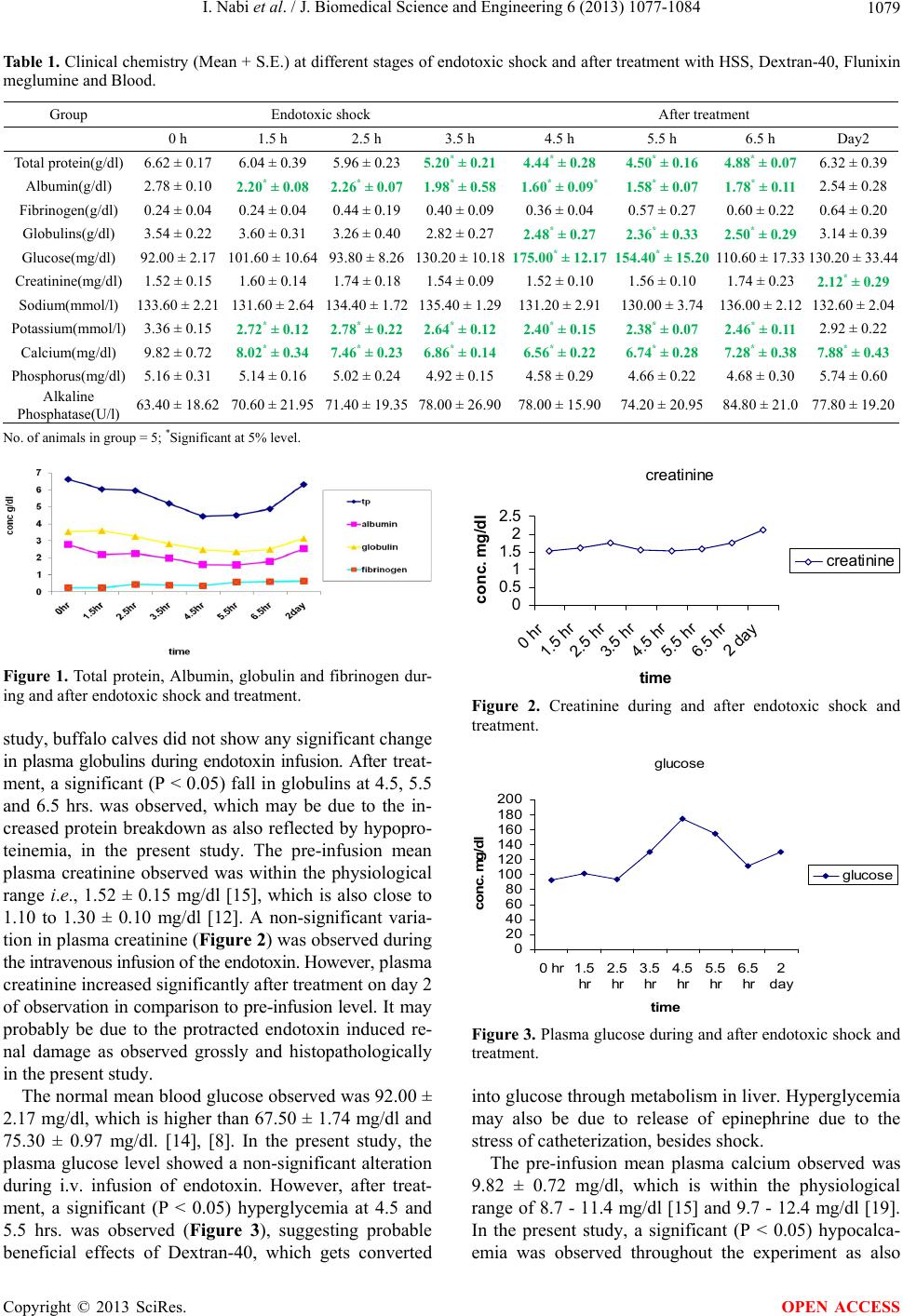 I. Nabi et al. / J. Biomedical Science and Engineering 6 (2013) 1077-1084 Copyright © 2013 SciRes. 1079 Table 1. Clinical chemistry (Mean + S.E.) at different stages of endotoxic shock and after treatment with HSS, Dextran-40, Flunixin meglumine and Blood. Group Endotoxic shock After treatment 0 h 1.5 h 2.5 h 3.5 h 4.5 h 5.5 h 6.5 h Day2 Total protein(g/dl) 6.62 ± 0.17 6.04 ± 0.39 5.96 ± 0.235.20* ± 0.214.44* ± 0.284.50* ± 0.16 4.88* ± 0.07 6.32 ± 0.39 Albumin(g/dl ) 2.78 ± 0.10 2.20* ± 0.08 2.26* ± 0.071.98* ± 0.581.60* ± 0.09*1.58* ± 0.07 1.78* ± 0.11 2.54 ± 0.28 Fibrinogen(g/dl) 0.24 ± 0.04 0.24 ± 0.04 0.44 ± 0.190.40 ± 0.09 0.36 ± 0.04 0.57 ± 0.27 0. 60 ± 0.22 0.64 ± 0.20 Globulins(g/dl) 3.54 ± 0.22 3.60 ± 0.31 3.26 ± 0.402.82 ± 0.27 2.48* ± 0.272.36* ± 0.33 2.50* ± 0.29 3.14 ± 0.39 Glucose(mg/dl) 92.00 ± 2.17 101.60 ± 10.64 93.80 ± 8.26130.20 ± 10.18175.00* ± 12.17154.40* ± 15.20 110.60 ± 17.33 130.20 ± 33.44 Creatinine(mg/dl) 1.52 ± 0.15 1.60 ± 0.14 1.74 ± 0 .181.54 ± 0.09 1.52 ± 0.10 1.56 ± 0.10 1.7 4 ± 0 .23 2.12* ± 0.29 Sodium(mmol/l) 133.60 ± 2.21 131.60 ± 2 .64 134.40 ± 1.72135.40 ± 1.29131.20 ± 2.91130.00 ± 3.74 136.00 ± 2.12 132.60 ± 2.04 Potassium(mmol/l) 3.36 ± 0.15 2.72* ± 0.12 2.78* ± 0.222.64* ± 0.122.40* ± 0.152.38* ± 0.07 2.46* ± 0.11 2.92 ± 0.22 Calcium(mg/dl) 9.82 ± 0.72 8.02* ± 0.34 7.46* ± 0.236.86* ± 0.146.56* ± 0.226.74* ± 0.28 7.28* ± 0.38 7.88* ± 0.43 Phosphorus(mg/dl) 5.16 ± 0.31 5.14 ± 0.16 5.02 ± 0.244.92 ± 0.15 4.58 ± 0.29 4.66 ± 0. 22 4.68 ± 0.30 5.74 ± 0.60 Alkaline Phosphatase(U/l) 63.40 ± 18.62 70.60 ± 21 .95 71.40 ± 19.3578.00 ± 26.9078.00 ± 15.9074.20 ± 20.95 84.80 ± 21.0 77.80 ± 19.20 No. of animals in group = 5; *Significant at 5% level. creatinine 0 0.5 1 1.5 2 2.5 0 hr 1.5 hr 2.5 hr 3.5 hr 4.5 hr 5.5 hr 6.5 hr 2 day time conc. mg/dl creatinine Figure 1. Total protein, Albumin, globulin and fibrinogen dur- ing and after endotoxic shock and treatment. Figure 2. Creatinine during and after endotoxic shock and treatment. study, buffalo calves did not show any significant change in plasma globulins during endotoxin infusion. After tr eat- ment, a significant (P < 0.05) fall in globulins at 4.5, 5.5 and 6.5 hrs. was observed, which may be due to the in- creased protein breakdown as also reflected by hypopro- teinemia, in the present study. The pre-infusion mean plasma creatinine observed was within the physiological range i.e., 1.52 ± 0.15 mg/dl [15], which is also close to 1.10 to 1.30 ± 0.10 mg/dl [12]. A non-significant varia- tion in plasma creatinine (Figure 2) was ob served during the intravenous infusion of the endotoxin. However, plasma creatinine increased significantly after treatment on day 2 of observation in comparison to pre-infusion level. It ma y probably be due to the protracted endotoxin induced re- nal damage as observed grossly and histopathologically in the present study. glucose 0 20 40 60 80 100 120 140 160 180 200 0 hr1.5 hr 2.5 hr 3.5 hr 4.5 hr 5.5 hr 6.5 hr 2 day time conc. m g/dl glucose Figure 3. Plasma glucose during and after endotoxic shock and treatment. The normal mean b lood gluco se observed w as 92.00 ± 2.17 mg/dl, which is higher th an 67.50 ± 1.74 mg/dl and 75.30 ± 0.97 mg/dl. [14], [8]. In the present study, the plasma glucose level showed a non-significant alteration during i.v. infusion of endotoxin. However, after treat- ment, a significant (P < 0.05) hyperglycemia at 4.5 and 5.5 hrs. was observed (Figure 3), suggesting probable beneficial effects of Dextran-40, which gets converted into glucose through metabolism in liver. Hyperglycemia may also be due to release of epinephrine due to the stress of ca theterization, besides sh o ck. The pre-infusion mean plasma calcium observed was 9.82 ± 0.72 mg/dl, which is within the physiological range of 8.7 - 11.4 mg/dl [15] and 9.7 - 12.4 mg/dl [19]. In the present study, a significant (P < 0.05) hypocalca- emia was observed throughout the experiment as also OPEN ACCESS  I. Nabi et al. / J. Biomedical Science and Engineering 6 (2013) 1077-1084 1080 reported previously [20]. The pre-infusion mean plasma phosphorus observed was 5.16 ± 0.31 mg/dl., which is close to physiological value of 5.6 - 6.5 mg/dl [19] and the plasma phosphorus did not show any significant va r ia - tion during and after the intravenous infusion of endo- toxin and the treatment. The pre-infusion mean plasma sodium observed was 133.6 ± 2.21 mmol/l, which is close to 136.6 ± 5.51 mmol/l [16 ], 134.48 ± 4.07 mmol/l [9] an d 133.40 ± 3.19 mmol/l [20]. However, a generalized non-significant in- crease in sodium was noticed throughout the period of experiment as also recorded in endotoxemic cows [21] and buffalo calves [14], previously. In the present inves- tigation, the mean sodium level at the end of the obser- vation period was almost equal to pre-infusion level. Ab- sence of any significant increase in plasma sodium even after infusion of HSS seems to be advantageous as it makes the infusion of HSS safe [22]. The pre-infusion mean plasma potassium ranged be- tween 3.36 ± 0.15 mmol/l, which is close to 3.60 ± 0.2 mmol/l [21] but higher than 2.28 ± 0.18 mmol/l [9]. A significant (P < 0.05) hypokalemia was observed at 1.5, 2.5 and 3.5 hrs. i.e., throughout the endotoxin infusion and even after treatment at 4.5, 5.5 and 6.5 hrs. This fall in potassium level may be regarded as an attempt by the body to sequester potassium as a part of the mechanism, whereby endotoxins promote the release of endogenous pyrogens from leucocytes. Decrease in potassium level could also be attributed to release of histamine during en- dotoxic shock, which increases the capillary permeability, besides regulating the secretion of adrenaline and nor- adrenaline together [23]. Hypokalemia after treatment can rather be attributed to rapid volume expansion fol- lowing HSS infusion [12]. Decrease in plasma levels of sodium and potassium may furthermore be attributed to the greatly diminished active transport of sodium and po- tassium across the cell membrane [23]. The pre-infusion mean plasma alkaline phosphatase was 63.40 ± 18.62 U/l, which is lower than 173 ± 40 U/l as reported by [12]. A non-significant variation in plasma alkaline phosphatase was recorded throughout the period of observation. 4. PATHOLOGICAL LESIONS Two out of five endotoxemic buffalo calves that died during observ ation period were su bjected to necrop sy ex- amination in order to study the gross and histopathologi- cal changes. 4.1. Gross Lesions Gross lesions varied from mild to clear cut haemorrhages along with congestion and emphysema in lungs (Figure 4) in both the endotoxemic buffalo calves. In addition, congestion, odema and dilation of intestines (Figure 5), haemorrhages on its mucosal surface as well as in medi- astinal lymph nodes and in the gall bladder mucosa were noticed. Severe congestion leading to reddish discolora- tion of cortex of the kidneys (Figure 6) was conspicuous. The gross lesions were suggestive of toxemia, enteropa- thy and pulmonary insult accompanying endotoxic shock. Figure 4. Lung-haemorrhages and emphysema. Figure 5. Intestines-congestion, edema and dilatation with di- arrheic contents. Figure 6. Kidney-marked congestion in cortex. Copyright © 2013 SciRes. OPEN ACCESS 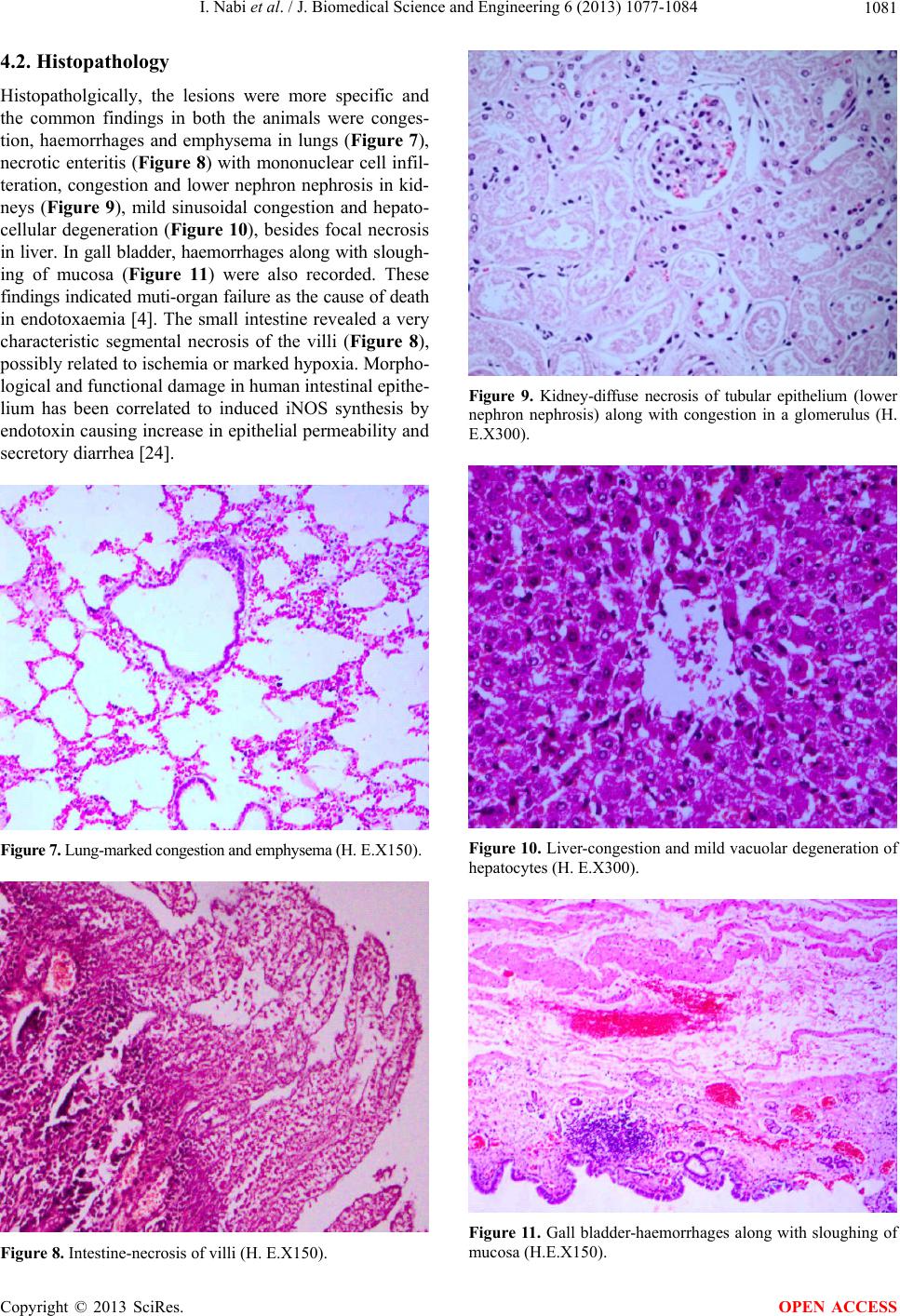 I. Nabi et al. / J. Biomedical Science and Engineering 6 (2013) 1077-1084 1081 4.2. Histopathology Histopatholgically, the lesions were more specific and the common findings in both the animals were conges- tion, haemorrhages and emphysema in lungs (Figure 7), necrotic enteritis (Figure 8) with mononuclear cell infil- teration, congestion and lower nephron nephrosis in kid- neys (Figure 9), mild sinusoidal congestion and hepato- cellular degeneration (Figure 10), besides focal necrosis in liver. In gall bladder, haemorrhages along with s lough - ing of mucosa (Figure 11) were also recorded. These findings indicated muti-organ failure as the cause of death in endotoxaemia [4]. The small intestine revealed a very characteristic segmental necrosis of the villi (Figure 8), possibly related to ischemia or marked hypoxia. Morpho- logical and functiona l damage in human intestinal epithe- lium has been correlated to induced iNOS synthesis by endotoxin causing in crease in epithelial permeability and secretory diarrhea [24]. Figure 7. L u n g - m a r k e d c o n g e s t i o n a n d e m p h y s e m a ( H . E.X150). Figure 8. Intestine-necrosis of villi (H. E.X150). Figure 9. Kidney-diffuse necrosis of tubular epithelium (lower nephron nephrosis) along with congestion in a glomerulus (H. E.X300). Figure 10. Liver-congestion and mild vacuolar degeneration of hepatocytes (H. E.X300). Figure 11. Gall bladder-haemorrhages along with sloughing of mucosa (H.E.X150). Copyright © 2013 SciRes. OPEN ACCESS 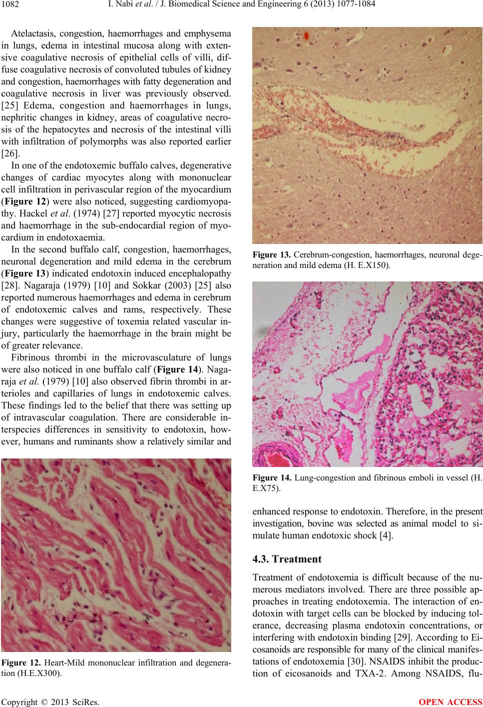 I. Nabi et al. / J. Biomedical Science and Engineering 6 (2013) 1077-1084 1082 Atelactasis, congestion, haemorrhages and emphysema in lungs, edema in intestinal mucosa along with exten- sive coagulative necrosis of epithelial cells of villi, dif- fuse coagulative necrosis of convoluted tubules of kidney and congestion, haemorrhages with fatty degeneration and coagulative necrosis in liver was previously observed. [25] Edema, congestion and haemorrhages in lungs, nephritic changes in kidney, areas of coagulative necro- sis of the hepatocytes and necrosis of the intestinal villi with infiltration of polymorphs was also reported earlier [26]. In one of the endotoxemic buffalo calves, degenerative changes of cardiac myocytes along with mononuclear cell infiltration in perivascular region of the myocardium (Figure 12) were also noticed, suggesting cardiomyopa- thy. Hackel et al. (1974) [27] reported myocytic necrosis and haemorrhage in the sub-endocardial region of myo- cardium in endotoxaemia. In the second buffalo calf, congestion, haemorrhages, neuronal degeneration and mild edema in the cerebrum (Figure 13) indicated endotoxin induced encephalopathy [28]. Nagaraja (1979) [10] and Sokkar (2003) [25] also reported numerous haemorrhages and edema in cerebrum of endotoxemic calves and rams, respectively. These changes were suggestive of toxemia related vascular in- jury, particularly the haemorrhage in the brain might be of greater relevance. Fibrinous thrombi in the microvasculature of lungs were also noticed in one buffalo calf (Figure 14). Naga- raja et al. (1979) [10] also observed fibrin thrombi in ar- terioles and capillaries of lungs in endotoxemic calves. These findings led to the belief that there was setting up of intravascular coagulation. There are considerable in- terspecies differences in sensitivity to endotoxin, how- ever, humans and ruminants show a relatively similar and Figure 12. Heart-Mild mononuclear infiltration and degenera- tion (H.E.X300). Figure 13. Cerebrum-congestion, haemorrhages, neuronal dege- neration and mild edema (H. E.X150). Figure 14. Lung-congestion and fibrinous emboli in vessel (H. E.X75). enhanced response to endotoxin. Therefore, in the present investigation, bovine was selected as animal model to si- mulate human endotoxic shock [4]. 4.3. Treatment Treatment of endotoxemia is difficult because of the nu- merous mediators involved. There are three possible ap- proaches in treating endotoxemia. The interaction of en- dotoxin with target cells can be blocked by inducing tol- erance, decreasing plasma endotoxin concentrations, or interfering with endotoxin binding [29]. According to Ei- cosanoids are responsible for many of the clinical m an if e s- tations of endotoxemia [30]. NSAIDS inhibit the produc- tion of eicosanoids and TXA-2. Among NSAIDS, flu- Copyright © 2013 SciRes. OPEN ACCESS 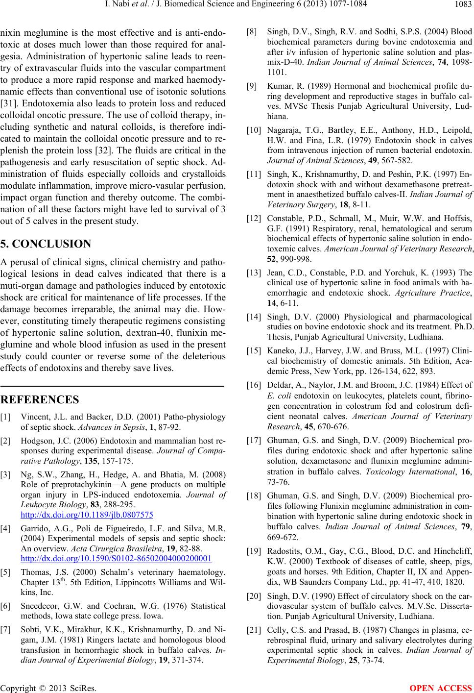 I. Nabi et al. / J. Biomedical Science and Engineering 6 (2013) 1077-1084 1083 nixin meglumine is the most effective and is anti-endo- toxic at doses much lower than those required for anal- gesia. Administration of hypertonic saline leads to reen- try of extravascular fluid s into the vascular compartment to produce a more rapid response and marked haemody- namic effects than conventional use of isotonic solutions [31]. Endotoxemia also leads to protein loss and reduced colloidal oncotic pressure. The use of colloid therapy, in- cluding synthetic and natural colloids, is therefore indi- cated to mainta in the colloidal o ncotic pr ess ure and to r e- plenish the protein loss [32]. The fluids are critical in the pathogenesis and early resuscitation of septic shock. Ad- ministration of fluids especially colloids and crystalloids modulate inflammation, improve micro - v asu l ar p erf u si on , impact organ function and thereby outcome. The combi- nation of all these factors might have led to survival of 3 out of 5 calves in the present st udy . 5. CONCLUSION A perusal of clinical signs, clinical chemistry and patho- logical lesions in dead calves indicated that there is a muti-organ damage and pathologies induced by entotoxic shock are critical for maintenance of life processes. If the damage becomes irreparable, the animal may die. How- ever, constituting timely therapeu tic regimens co ns is ti n g of hypertonic saline solution, dextran-40, flunixin me- glumine and whole blood infusion as used in the present study could counter or reverse some of the deleterious effects of endotoxins and th ereby save lives. REFERENCES [1] Vincent, J.L. and Backer, D.D. (2001) Patho-physiology of septic shock. Advances in Sepsis, 1, 87-92. [2] Hodgson, J.C. (2006) Endotoxin and mammalian host re- sponses during experimental disease. Journal of Compa- rative Pathology, 135, 157-175. [3] Ng, S.W., Zhang, H., Hedge, A. and Bhatia, M. (2008) Role of preprotachykinin—A gene products on multiple organ injury in LPS-induced endotoxemia. Journal of Leukocyte Biology, 83, 288-295. http://dx.doi.org/10.1189/jlb.0807575 [4] Garrido, A.G., Poli de Figueiredo, L.F. and Silva, M.R. (2004) Experimental models of sepsis and septic shock: An overview. Acta Cirurgica Brasileira, 19, 82-88. http://dx.doi.org/10.1590/S0102-86502004000200001 [5] Thomas, J.S. (2000) Schalm’s veterinary haematology. Chapter 13th. 5th Edition, Lippincotts Williams and Wil- kins, Inc. [6] Snecdecor, G.W. and Cochran, W.G. (1976) Statistical methods, Iowa state college press. Iowa. [7] Sobti, V.K., Mirakhur, K.K., Krishnamurthy, D. and Ni- gam, J.M. (1981) Ringers lactate and homologous blood transfusion in hemorrhagic shock in buffalo calves. In- dian Journal of Experimental Biology, 19, 371-374. [8] Singh, D.V., Singh, R.V. and Sodhi, S.P.S. (2004) Blood biochemical parameters during bovine endotoxemia and after i/v infusion of hypertonic saline solution and plas- mix-D-40. Indian Journal of Animal Sciences, 74, 1098- 1101. [9] Kumar, R. (1989) Hormonal and biochemical profile du- ring development and reproductive stages in buffalo cal- ves. MVSc Thesis Punjab Agricultural University, Lud- hiana. [10] Nagaraja, T.G., Bartley, E.E., Anthony, H.D., Leipold, H.W. and Fina, L.R. (1979) Endotoxin shock in calves from intravenous injection of rumen bacterial endotoxin. Journal of Animal Sciences, 49, 567-582. [11] Singh, K., Krishnamurthy , D. and Peshin, P.K. (1997) En - dotoxin shock with and without dexamethasone pretreat- ment in anaesthetized buffalo calves-II. Indian Journal of Veterinary Surgery, 18, 8-11. [12] Constable, P.D., Schmall, M., Muir, W.W. and Hoffsis, G.F. (1991) Respiratory, renal, hematological and serum biochemical effects of hypertonic saline solution in endo- toxemic calves. American Journal of Veterinary Research, 52, 990-998. [13] Jean, C.D., Constable, P.D. and Yorchuk, K. (1993) The clinical use of hypertonic saline in food animals with ha- emorrhagic and endotoxic shock. Agriculture Practice, 14, 6-11. [14] Singh, D.V. (2000) Physiological and pharmacological studies on bovine endotoxic shock and its treatment. Ph.D. Thesis, Punjab Agricultural University, Ludhiana. [15] Kaneko, J.J., Harvey, J.W. and Bruss, M.L. (1997) Clini- cal biochemistry of domestic animals. 5th Edition, Aca- demic Press, New York, pp. 126-134, 622, 893. [16] Deldar, A., Naylor, J.M. and Broom, J.C. (1984) Effect of E. coli endotoxin on leukocytes, platelets count, fibrino- gen concentration in colostrum fed and colostrum defi- cient neonatal calves. American Journal of Veterinary Research, 45, 670-676. [17] Ghuman, G.S. and Singh, D.V. (2009) Biochemical pro- files during endotoxic shock and after hypertonic saline solution, dexametasone and flunixin meglumine admini- stration in buffalo calves. Toxicology International, 16, 73-76. [18] Ghuman, G.S. and Singh, D.V. (2009) Biochemical pro- files following Flunixin me glumine administration in com- bination with hypertonic saline during endotoxic shock in buffalo calves. Indian Journal of Animal Sciences, 79, 669-672. [19] Radostits, O.M., Gay, C.G., Blood, D.C. and Hinchcliff, K.W. (2000) Textbook of diseases of cattle, sheep, pigs, goats and horses. 9th Edition, Chapter II, IX and Appen- dix, WB Saunders Company Ltd., pp. 41-47, 410, 1820. [20] Singh, D.V. (1990) Effect of circulatory shock on the car- diovascular system of buffalo calves. M.V.Sc. Disserta- tion. Punjab Agricultural University, Ludhiana. [21] Celly, C.S. and Prasad, B. (1987) Changes in plasma, ce- rebrospinal fluid, urinary and salivary electrolytes during experimental septic shock in calves. Indian Journal of Experimental Biology, 25, 73-74. Copyright © 2013 SciRes. OPEN ACCESS  I. Nabi et al. / J. Biomedical Science and Engineering 6 (2013) 1077-1084 Copyright © 2013 SciRes. 1084 OPEN ACCESS [22] Singh, D.V., Singh, R.V. and Sodhi, S.P.S. (2007) Some biochemical profiles during bovine endotoxic (lipopoly- saccharide) shock and after treatment with hypertonic sa- line solution. Indian Journal of Animal Sciences, 77, 438- 441. [23] Singh, D.V., Sodhi, S.P.S. and Singh, R. (1994) Effect of shock in macro and microelements status in Cow and Buffalo Calves. Indian Veterinary Medical Journal, 18, 142-148. [24] Vincent, J.L., Zhang, H., Szabo, C. and Preiser, J.C. (2000) Effects of nitric oxide in septic shock. American Journal of Respiratory and Critical Care Medicine, 6, 1781-1785. http://dx.doi.org/10.1164/ajrccm.161.6.9812004 [25] Singh, D.V., Sodhi, S.P.S., Brar, A.P.S. and Grewal, J.S. (1996) Certain pathological profiles of endotoxic shock in buffalo calves. Buffalo Journal, 19, 283-288. [26] Sokkar, S.M., Darwiesh, G. and Madbooly, A. (2003) Study of the pathological effects of Escherichia coli en- dotoxin in rams. Journal of Veterinary Medicine, 50, 226- 230. http://dx.doi.org/10.1046/j.1439-0450.2003.00651.x [27] Hackel, D.B., Ratliff, N.B. and Mikat, E. (1974) The heart in shock: Brief review. Circulation Research, 35, 805-811. http://dx.doi.org/10.1161/01.RES.35.6.805 [28] Papadopoulos, M.C., Davies, D.C., Moss, R.F., Tighe, D. and Bennett, E.D. (2000) Pathophysiology of septic en- cephalopathy: A review. Critical Care Medicine, 28, 3011- 3024. http://dx.doi.org/10.1097/00003246-200008000-00057 [29] Hardie, E.M. and Krusse-Elliott, K. (2008) Endotoxic shock Part II: A review of treatment. Journal of Internal Medicine, 4, 306-314. [30] Moses, V.S. and Bertone, A.L. (2002) Non-steriodal anti- inflammatory drugs. Veterinary Clinics of North America: Equine Practice, 18, 21-37. http://dx.doi.org/10.1016/S0749-0739(01)00002-5 [31] Zafar, M.A., Hussain, M.H., Muhammad, G. and Saqib, M. (2004) Potential use of hypertonic saline solution (7-7.5% Nacl) resuscitation in hypovolemic and endoto- xic shock. International Journal of Agriculture and Biol- ogy, 6, 926-930. [32] Kelmer, G. (2009) Update on treatment of endotoxemia. Veterinary Clinics of North America: Equine Practice, 25, 259-270. http://dx.doi.org/10.1016/j.cveq.2009.04.012 |