

Paper Menu >>
Journal Menu >>
 Open Journal of Urology, 2013, 3, 276-280 Published Online November 2013 (http://www.scirp.org/journal/oju) http://dx.doi.org/10.4236/oju.2013.37052 Open Access OJU Laparoscopic Management for Non-Palpable Testis Abdel-Aziz Ali Emara1,2 1Department of Urology, Al-Azhar University, Cairo, Egypt 2Department of Urology, Ghodran Hospital, Baljurashi, KSA Email: abdelazi[email protected] Received October 10, 2013; revised November 5, 2013; accepted November 12, 2013 Copyright © 2013 Abdel-Aziz Ali Emara. This is an open access article distributed under the Creative Commons Attribution License, which permits unrestricted use, distribution, and reproduction in any medium, provided the original work is properly cited. ABSTRACT Purpose: To present our experience with laparoscopic management of the non-palpable undescended testis. Patients and Methods: Between Nov. 2010 and Oct. 2012, 47 non-palpable testes in 41 patients were evaluated prospectively by laparoscopy. The age of the patients at the time of surgery varied from 1 to 9 years with a mean age of 2.85 years. Testicular viability and location were evaluated by physical examination and Doppler ultrasonography after 1 and 3 months. Results: Out of 49 testicular units, 47 (95.9%) were successfully treated by laparoscopic orchiopexy. 45 tes- ticular units (91.8%) were treated by one-stage laparoscopic orchiopexy, 2 (4.1%) were treated by two-stage laparo- scopic orchiopexy and 2 (4.1%) diagnosed as vanishing testis with detection of blind end spermatic vessels and vas def- erens during laparoscopy. Physical examination and Doppler study demonstrated that 46 of 47 testes (97.9%) were vi- able and 45 of 47 (95.7 %) were located in the low er scrotum and 2 of 47 (4.3%) in the upp er scrotum at the end of fol- low-up. Conclusion: The laparoscopy is a reliable technique for diagnosis and treatment of the non-palpable in- tra-abdominal testis with high success and survival rates of the testes. Keywords: Laparoscopy; Orchiopexy; Undesc ended Testis 1. Introduction An undescended testis is one of the most common clini- cal disorders of childhood, occurring in approximately 3% of full-term newborns, 21% of premature newborns, and 0.8% - 1.8% of 1-year-old boys [ 1]. In the boys w ith undescended testis, the testis is palpable in the groin in 80% [2] and non-palpable in 20% and 20% - 50% of those with non-palpable testis, the testis is absent [3]. A non-palpable testis is defined as, a testis not palpated by any examination technique, including pre-operatively un- der anaesthesia [4]. Non-palpable testes have an in- creased risk of malignant transformation, infertility and epididymal malformation [5]. Many diagnostic methods have been described of whi c h the most promising method is the use of diagnostic lapa- roscopy. Non-palpable undescended testis management basically includes four surgical techniques: 1) Open or- chidopexy in stages; 2) Fowler-Stephens orchidopexy in which spermatic vessels are sectioned and the deferential artery is preserved in 1 or 2 stages by open or laparo- scopic approach; 3) Testicular autotransplant with sper- matic vessel section and microsurgical anastomosis to the epigastric ve ssels and 4) laparosco pic orchid o pexy. Of the above mentioned techniques, laparoscopic or- chidopexy is gaining in popularity, and has an 88% - 100% cure rate [4]. Its advantages are greater dissection of spermatic vessels and vas defer ens and the creation of a medial neohiatus that consequently shortens the dis- tance between the abdominal cavity and the scrotum during testicular descent. This technique was originally described for intra-abdominal undescended testis man- agement but it has also been employed in high palpable undescended testis with good results [6]. We aimed to present our experience with laparoscopic management of the non-palpable, intra-abdominal, undescended testis. 2. Patients and Methods Study design, patient selection and assessment: This prospective study was conducted between Nov. 2010 and Oct. 2012, Patients were consecutively enrolled from patients referred to the urology and pediatric clinics at Ghodran Hospital, Baljurashi, Kingdom of Saudi Arabia (KSA) for treatment of undescended testis. All patients underwent thorough history-taking, physical examination and abdominal ultrasonography. A testis was considered non-palpable if it was not palpable clinically or detected  A. A. EMARA 277 by abdominal ultrasonography. The intra-abdominal van- ishing testis syndrome (unilateral anorchia) was diag- nosed if the spermatic vessels and vas deferens were noted to end blindly during laparoscopy. Accordingly, surgical exploration was not performed in these cases. If an intraabdominal testis was identified laparoscopically, orchiopexy was done. The study protocol was approved by the Ethics Committee of Ghodran Hospital, KSA and parents of all patients enrolled in this study provided written informed consent. Laparoscopic technique: Diagnostic laparoscopy was performed in 41 patients with non-palpable testis. A clear diet was started on the day before surgery and a fasting of 4 - 8 hours according to their age was applied before laparoscopy. A single dose of cefazolin sodium 40 mg/kg was given by IM route for prophylaxis. Before laparo- scopy, physical examination was repeated under general anaesthesia in order to palpate the testes. A urethral catheter was inserted into the bladder. A patient was placed supine in the frog-leg position and in the 30 de- grees Trendelenburg position. A U-shaped incision of 1 cm length was made just below the umbilicus. The ante- rior wall of the abdomen was pulled upwards and then a 10 mm trocar was inserted into the abdominal cavity by open technique. CO2 was insufflated to achieve pneu- moperitoneum at a pressure of 10 mm Hg. A zero degree 10 mm laparoscope was inserted and the abdominal or- gans were inspected. During the diagnostic laparoscopy, intraperitoneal examination for a unilateral non-palpable undescended testis began with examination of the normal contralateral internal inguinal ring, vas deferens and spermatic vessels. Followed by determination and evalu- ation of the median (urachus), medial (obliterated um- bilical artery) and lateral (inferior epigastric artery) um- bilical ligaments, external iliac vessels, any potential intra-abdominal testis, the vas deferens, spermatic ves- sels and the patency of internal ring of the affected side were determined. Anatomic orientation of the localiza- tion and volume, mobility of the testis, the distance be- tween the testes and the inguinal canal, paratesticular anomalies, lengths of vas deferens and spermatic vessels and presence of inguinal hernia were assessed to proceed with either orchiectomy or orchiopexy and either one or two stage orchiopexy. Blind-ended vas deferens and sper- matic vessels were considered as vanishing testis. Mobilization of the testis was performed by incisin g of the peritoneum over the superior border of the internal ring. The gubernaculum was identified and mobilized circumferentially to provide traction by grasping its tes- ticular end. Dissection was continued distally along the gubernaculum until the scrotum began to invaginate. The gubernaculum was transected using electrocautery as far as possible. Dissection was continued cranially toward the renal hilum as far as possible to gain enough length on the spermatic cord to allow tension free orchiopexy. The peritoneum over the vas may also be incised to gain additional length. If the spermatic vessels remained too short, we performed a first stage Fowler Stephens pro- cedure by placing 2 endoscopy clips as far proximal as possible on the cord vessels, the vessels were transected between clips. If adequate leng th was obtained, the tip of the endoscopy dissector was placed medial to the inferior epigastric vessels and lateral to the medial umbilical ligaments on the anterior abdominal wall. Then the dis- sector tip was directed toward the ipsilateral hemiscro- tum. A 5 mm trocar cannula was passed over the dissec- tor through the scrotal incision into the abdominal cavity and the free end of gubernaculum was grasped and the testis was brought into approximation with the end of the trocar. The testis, grasping forceps and trocar were with- drawn through the scrotum. The cord structures were inspected to verify that were not twisted. Two 3-zero polydioxanone sutures were used to complete the or- chiopexy distally as possible in created sub-dartos pouch. Finally, the pneumoperitoneum was deflated and the fas- cia and skin were closed. The patients underwent the hospital care and medica- tions (cefazolin sodium 40 mg/kg) and discharged on ou t- patient medications (amoxicillin 45 mg/kg). They were invited to visit the outpatient clinic for follow-up. Follow up: Patien ts wer e follo wed af ter on e week , one month and 3 months from laparoscopic orchiopexy op- eration. Detailed medical history and physical examina- tion were done at each follow-up visit. Scrotal Doppler ultrasound was performed at 1, 3-month visits. The time of the procedure, intra-operative complica- tions, hospital stay, recovery from operation, defined as the time at which the child returned to his normal pre- operative activities, and testicular location and viability were estimated. 3. Results Forty one patients with non-palp able testis were enrolled in this study. The mean age of studied patients was 2.85 ± 0.85 years (range, one year to 9 years). The non-pal- pable testis was bilateral in 16 (32.7%), right un ilateral in 23 (46.9%) and left unilateral in 10 (20.4%) (Table 1). Out of 49 testicular units, diagno stic laparoscop y showed that 45 were located within 3 cm of the internal inguinal ring, 2 were more than 3 cm from the internal inguinal ring and 2 diagnosed as vanishing testis with detection of blind end spermatic vessels and vas deferens, one at right side and other at left. None of the patients associated with oblique inguinal hernia. Of 49 testicular units, 45 (91.8%) were treated by one-stage laparoscopic orchio- pexy in which the non-palpable testis was located within 3 cm from the internal inguinal ring and two-stage lap ar o- scopic orchiopexy was performed in 2 (4.1%) in which the non-palpable testis was located more than 3 cm from the internal inguin al ring side (Table 2). Open Access OJU 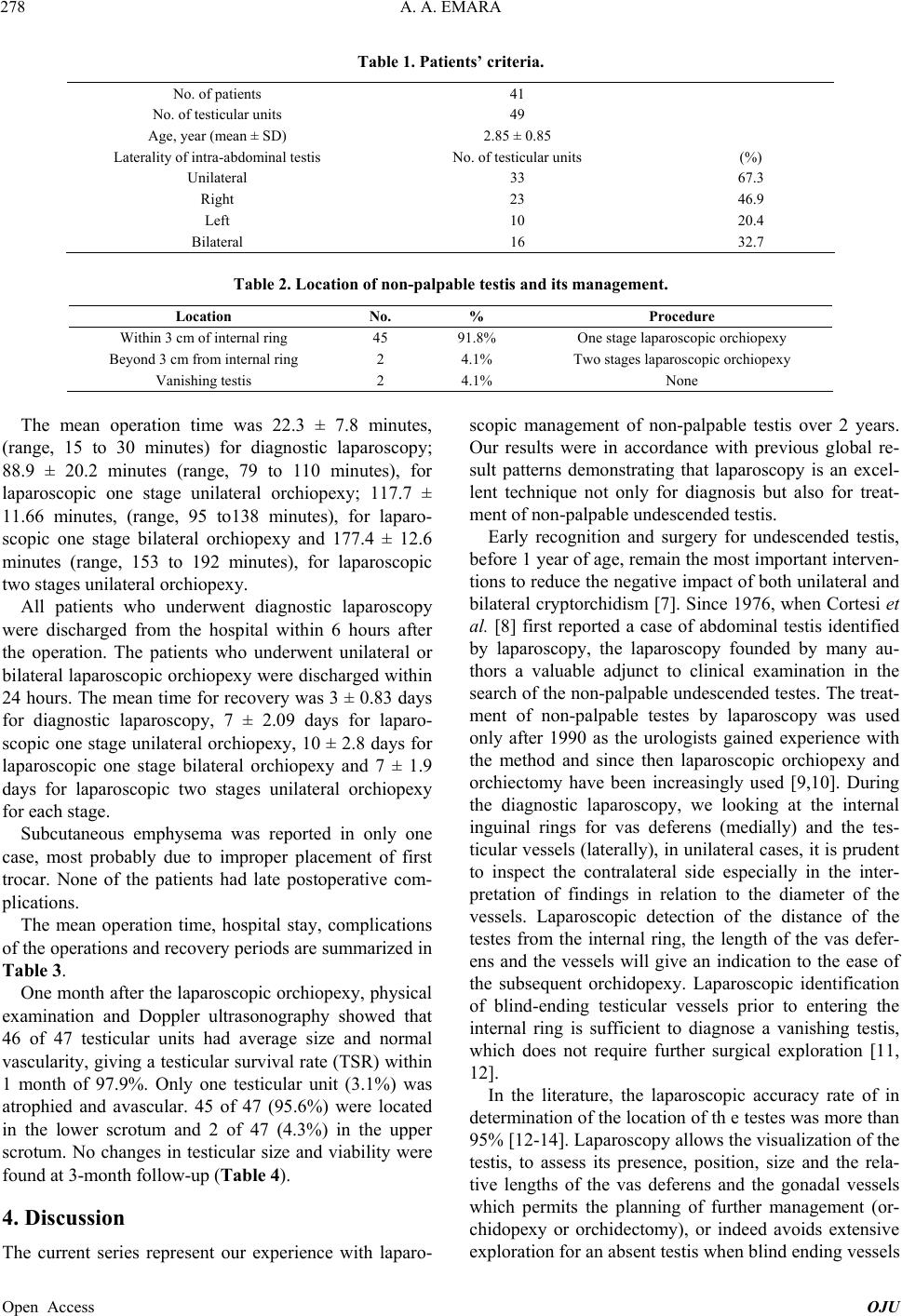 A. A. EMARA Open Access OJU 278 Table 1. Patients’ criteria. No. of patients 41 No. of testicular units 49 Age, year (mean ± SD) 2.85 ± 0.85 Laterality of intra-abdominal testis No. of testicular units (%) Unilateral 33 67.3 Right 23 46.9 Left 10 20.4 Bilateral 16 32.7 Table 2. Location of non-palpable testis and its management. Location No. % Procedure Within 3 cm of internal ring 45 91.8% One stage laparoscopic orchiopexy Beyond 3 cm from internal ring 2 4.1% Two stages laparoscopic orchiopexy Vanishing testis 2 4.1% None The mean operation time was 22.3 ± 7.8 minutes, (range, 15 to 30 minutes) for diagnostic laparoscopy; 88.9 ± 20.2 minutes (range, 79 to 110 minutes), for laparoscopic one stage unilateral orchiopexy; 117.7 ± 11.66 minutes, (range, 95 to138 minutes), for laparo- scopic one stage bilateral orchiopexy and 177.4 ± 12.6 minutes (range, 153 to 192 minutes), for laparoscopic two stages unilateral orchiopexy. All patients who underwent diagnostic laparoscopy were discharged from the hospital within 6 hours after the operation. The patients who underwent unilateral or bilateral laparoscopic orchiopex y were discharged within 24 hours. The mean time for recovery was 3 ± 0.83 days for diagnostic laparoscopy, 7 ± 2.09 days for laparo- scopic one stage unilateral orchiopexy, 10 ± 2.8 days for laparoscopic one stage bilateral orchiopexy and 7 ± 1.9 days for laparoscopic two stages unilateral orchiopexy for each stage. Subcutaneous emphysema was reported in only one case, most probably due to improper placement of first trocar. None of the patients had late postoperative com- plications. The mean operation time, hospital stay, complications of the operations and recovery periods are summarized in Table 3. One month after the laparoscopic orchiopexy, physical examination and Doppler ultrasonography showed that 46 of 47 testicular units had average size and normal vascularity, giving a testicular survival rate (TSR) within 1 month of 97.9%. Only one testicular unit (3.1%) was atrophied and avascular. 45 of 47 (95.6%) were located in the lower scrotum and 2 of 47 (4.3%) in the upper scrotum. No changes in testicular size and viability were found at 3-month follow-up (Table 4). 4. Discussion The current series represent our experience with laparo- scopic management of non-palpable testis over 2 years. Our results were in accordance with previous global re- sult patterns demonstrating that laparoscopy is an excel- lent technique not only for diagnosis but also for treat- ment of non-palpable undescended testis. Early recognition and surgery for undescended testis, before 1 year of age, remain the most important interven - tions to reduce the negative impact of both unilateral and bilateral cryptorchidism [7]. Since 1976, when Cortesi et al. [8] first reported a case of abdominal testis identified by laparoscopy, the laparoscopy founded by many au- thors a valuable adjunct to clinical examination in the search of the non-p alpable undescended testes. The treat- ment of non-palpable testes by laparoscopy was used only after 1990 as the urologists gained experience with the method and since then laparoscopic orchiopexy and orchiectomy have been increasingly used [9,10]. During the diagnostic laparoscopy, we looking at the internal inguinal rings for vas deferens (medially) and the tes- ticular vessels (laterally), in unilateral cases, it is prudent to inspect the contralateral side especially in the inter- pretation of findings in relation to the diameter of the vessels. Laparoscopic detection of the distance of the testes from the internal ring, the length of the vas defer- ens and the vessels will give an indication to the ease of the subsequent orchidopexy. Laparoscopic identification of blind-ending testicular vessels prior to entering the internal ring is sufficient to diagnose a vanishing testis, which does not require further surgical exploration [11, 12]. In the literature, the laparoscopic accuracy rate of in determination of the location of th e testes was more than 95% [12-14]. Laparoscopy allows the visualization of the testis, to assess its presence, position, size and the rela- tive lengths of the vas deferens and the gonadal vessels which permits the planning of further management (or- chidopexy or orchidectomy), or indeed avoids extensive exploration for an absent testis when blind ending vessels 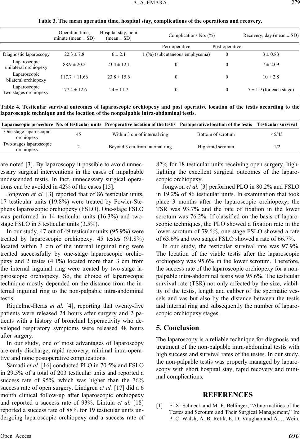 A. A. EMARA 279 Table 3. The mean operation time, hospital stay, complications of the operations and recovery. Operation time, minute (mean ± S D) Hospital stay, hour (mean ± SD) Complications No. (%) Recovery, da y (mean ± SD) Peri-operative Post-operative Diagnostic laparoscopy 22.3 ± 7.8 6 ± 2.1 1 (%) (subcutaneous emphysema)0 3 ± 0.83 Laparoscopic unilateral orchiopexy 88.9 ± 20.2 23.4 ± 12.1 0 0 7 ± 2.09 Laparoscopic bilateral orchiopexy 117.7 ± 11 .66 23.8 ± 15.6 0 0 10 ± 2.8 Laparoscopic two stages orchiopexy 177.4 ± 12.6 24 ± 11.7 0 0 7 ± 1.9 (for each stage) Table 4. Testicular survival outcomes of laparoscopic orchiopexy and post operative location of the testis according to the laparoscopic technique and the loc a tion of the nonpalpable intra-abdominal te stis. Laparoscopic procedure No. of testicular units Preoperative location of the testisPostoperative location of the testis Testicular survival One stage laparoscopic orchiopexy 45 Within 3 cm of inter na l ring Bottom of scrotum 45/45 Two stages laparoscopic orchiopexy 2 Beyond 3 cm from internal ring High/mid scrotum 1/2 are noted [3]. By lapar oscopy it possible to avoid unne c- essary surgical interventions in the cases of impalpable undescended testis. In fact, unnecessary surgical opera- tions can be avoided in 42% of the cases [15]. Jongwon et al. [3] reported that of 86 testicular units, 17 testicular units (19.8%) were treated by Fowler-Ste- phens laparoscopic orchiopexy (FSLO). One-stage FSLO was performed in 14 testicular units (16.3%) and two- stage FSLO in 3 testicular units (3.5%). In our study, 47 ou t of 4 9 testicu lar un its (95.9 %) were treated by laparoscopic orchiopexy. 45 testes (91.8%) located within 3 cm of the internal inguinal ring were treated successfully by one-stage laparoscopic orchio- pexy and 2 testes (4.1%) located more than 3 cm from the internal inguinal ring were treated by two-stage la- paroscopic orchiopexy. So, the choice of laparoscopic technique mostly depended on the distance from the in- ternal inguinal ring to the non-palpable intra-abdominal testis. Riquelme-Heras et al. [4], reporting that twenty-five patients were released 24 hours after surgery and 2 pa- tients with a history of bronchial hyperactivity who de- veloped respiratory symptoms were released 48 hours after surgery. In our study, one of most advantages of laparoscopy are early discharge, rapid recovery, minimal intra-opera- tive and none postoperative complications. Samadi et al. [16] conducted PLO in 70.5% and FSLO in 29.5% of a total of 203 testicular units and reported a success rate of 95%, which was higher than the 76% success rate of open surgery. Lindgren et al. [17] did a 6 month clinical follow-up after laparoscopic orchiopexy and reported a success rate of 93%. Lintula et al. [18] reported a success rate of 88% for 19 testicular units un- dergoing laparoscopic orchiopexy and a success rate of 82% for 18 testicular units receiving open surgery, high- lighting the excellent surgical outcomes of the laparo- scopic orchiopexy. Jongwon et al. [3] performed PLO in 80.2% and FSLO in 19.2% of 86 testicular units. In examination that took place 3 months after the laparoscopic orchiopexy, the TSR was 93.7% and the rate of fixation in the lower scrotum was 76.2%. If classified on the basis of laparo- scopic techniques, the PLO showed a fixation rate in the lower scrotum of 79.6%, one-stage FSLO showed a rate of 63.6% and two stages FSLO showed a r a te of 66.7%. In our study, the testicular survival rate was 97.9%. The location of the viable testis after the laparoscopic orchiopexy was 95.6% in the lower scrotum. Therefore, the success rate of the laparoscopic orchiopexy for a non- palpable intra-ab dominal testis was 95.6%. The testicular survival rate (TSR) not only affected by the size, viabil- ity of the testis, length and caliber of the spermatic ves- sels and vas but also by the distance between the testis and internal ring and subs equently the number of laparo- scopic orchiopexy stages. 5. Conclusion The laparoscopy is a reliable technique for diagnosis and treatment of the non-palpable intra-abdominal testis with high success and survival rates of the testes. In our study, the non-palpable testis was properly managed by laparo- scopy with short hospital stay, rapid recovery and mini- mal complications. REFERENCES [1] F. X. Schneck and M. F. Bellinger, “Abnormalities of the Testes and Scrotum and Their Surgical Management,” In: P. C. Walsh, A. B. Retik, E. D. Vaughan and A. J. Wein, Open Access OJU  A. A. EMARA 280 Eds., Campbell’s Urology, 8th Edition, Saunders Com- pany, Philadelphia, 2002, pp. 2353-2394. [2] K. Choi, T. Park and K. S. Kim, “Laparoscopic Orchio- pexy for Intra-Abdominal Te stis: Complications a nd Tec h- nical Aspects,” Korean Journal of Urology, Vol. 41, 2000, pp. 420-424. [3] K. Jongwon, E. M. Gyeong and S. K. Kun, “Laparoscopic Orchiopexy for a Nonpalpable Testis,” Korean Journal of Urology, Vol. 51, No. 2, 2010, pp. 106-110. [4] M. A. Riquelme-Heras, S. Landa-Juárez, D. Miguel-Gó- mez, H. Meneses-Juárez and R. Andraca-Dumit, “Laparo- scopic Orchidopexy in Palpable and Nonpalpable Un- descended Testis,” Revista Mexicana de Urología, Vol. 69, No. 5, 2009, pp. 215-218. [5] C. H. Chui and A. S. Jacobsen, “Laparoscopy in the Eva- luation of the Non-Palpable Undescended Testes,” Sin- gapore Medical Journal, Vol. 41, No. 5, 2000, pp. 206- 208. [6] S. G. Docimo, R. G. Moore, J. Adams and L. R. Kavoussi, “Laparoscopic Orchiopexy for the High Palpable Unde- scended Testis: Preliminary Experience,” Journal of Uro- logy, Vol. 154, No. 4, 1995, pp. 1513-1515. http://dx.doi.org/10.1016/S0022-5347(01)66919-2 [7] P. A. Lee and C. P. Houk, “Cryptorchidism,” Current Opinion in Endocrinology, Diabetes & Obesity, Vol. 20, No. 3, 2013, pp. 210-216. http://dx.doi.org/10.1097/MED.0b013e32835ffc7d [8] N. Cortesi, A. Baldini, P. Ferrari, et al., “Diagnosis of Bilateral Abdominal Cryptorchidism by Laparoscopy,” En- doscopy, Vol. 8, 1976, pp. 33-34. http://dx.doi.org/10.1055/s-0028-1098372 [9] I. S. Gill, J. H. Ross, G. T. Sung and R. Kay, “Needle- scopic Surgery for Cryptorchidism: The Initial Series,” Journal of Pediatric Surgery, Vol. 35, No. 10, 2000, pp. 1426-1430. http://dx.doi.org/10.1053/jpsu.2000.16405 [10] B. W. Lindgren, E. C. Darby, L. Faiella, et al., “Laparo- scopic Orchiopexy: Procedure of Choice for the Nonpal- pable Testis,” Journal of Urology, Vol. 159, No. 6, 1998, pp. 2132-2135. http://dx.doi.org/10.1016/S0022-5347(01)63294-4 [11] A. B. Retik and C. A. Peters, “Laparoscopic Management of the Impalpable Abdominal Testis, Editorial Comment,” Urology, Vol. 42, 1993, pp. 578-579. http://dx.doi.org/10.1016/0090-4295(93)90279-J [12] G. W. Holcomb, J. W. Brock, W. W. Neblett, J. B. Pietsch and W. M. Morgan, “Laparoscopy for the Nonpalpable Testis,” The American Journal of Surgery, Vol. 60, 1994, pp. 143-147. [13] M. K. Moslemi, “Evaluation of Orchiopexy Practice Pat- terns in Patients with Cryptorchidism: A Single-Centre Study,” Journal of Pediatric Urology, Vol. 15, No. 13, 2013. [14] J. W. Brock, G. W. Holcomb and W. M. Morgan, “The Use of Laparoscopy in the Management of the Nonpalpa- ble Testis,” Journal of Laparoendoscopic Surgery, Vol. 6, 1996, pp. 35-39. [15] P. P. Godbole, J. A. Morecroft and A. E. Mackinon, “La- paroscopy for the Impalpable Testis,” British Journal of Surgery, Vol. 84, No. 10, 1997, pp. 1430-1432. http://dx.doi.org/10.1002/bjs.1800841024 [16] A. A. Samadi, L. S. Palmer and I. Franco, “Laparoscopic Orchiopexy: Report of 203 Cases with Review of Diag- nosis, Operative Technique, and Lessons Learned,” Jour- nal of Endourology, Vol. 17, 2003, pp. 365-368. http://dx.doi.org/10.1089/089277903767923128 [17] B. W. Lindgren, E. C. Darby, L. Faiella, W. A. Brock, E. F. Reda, S. B. Levitt, et al., “Laparoscopic Orchiopexy: Procedure of Choice for the Nonpalpable Testis,” Journal of Urology, Vol. 159, No. 6, 1998, pp. 2132-2135. http://dx.doi.org/10.1016/S0022-5347(01)63294-4 [18] H. Lintula, H. Kokki, M. Eskelinen and K. Vanamo, “Laparoscopic versus Open Orchidopexy in Children with Intra-Abdominal Testes,” Journal of Laparoendoscopic & Advanced Surgical Techniques, Vol. 18, No. 3, 2008, pp. 449-456. http://dx.doi.org/10.1089/lap.2007.0176 Open Access OJU |