

Paper Menu >>
Journal Menu >>
 Open Journal of Orthopedics, 2013, 3, 296-299 Published Online November 2013 (http://www.scirp.org/journal/ojo) http://dx.doi.org/10.4236/ojo.2013.37054 Open Access OJO Evaluation of Conservative Treatment of Acute Fracture of the Odontoid Process of Axis with a Halo-Vest* Yuichiro Yokoyama, Hiroshi Takahashi#, Yasuaki Iida, Yasuhiro Inoue, Keiji Hasegawa, Daisuke Suzuki, Shintaro Tsuge, Wataru Shishikura, Katsunori Fukutake, Ryo Takamatsu, Kazumasa Na kamura, Akihito Wada Department of Orthopaedic Surgery, Toho University School of Medicine, Tokyo, Japan. Email: #[email protected] Received September 13th, 2013; revised October 25th, 2013; accepted October 5th, 2013 Copyright © 2013 Yuichiro Yokoyama et al. This is an open access article distributed under the Creative Commons Attribution Li- cense, which permits unrestricted use, distribution, and reproduction in any medium, provided the original work is properly cited. ABSTRACT We fitted a halo-vest on patients with a fracture of the odontoid process of the axis and evaluated the effects of conser- vative treatment on cervical muscular atrophy and the number and type of patient complaints after bone union. Four patients had Anderson Type II fractures and 6 patients had Type III fractures. Bone union was observed in all patients within 3 months after injury. After halo-vest fitting, cervical muscular atrophy increased with time, however, it gradu- ally improved after removal of the halo-vest. Patients fixed with a halo-vest for more than 10 weeks complained of cer- vical discomfort and limited range of motion. No patients with fixation for less than 10 weeks had problems in bone union or had cervical complaints. Thus, less than a 10-week fixation period was considered to be appropriate. Keywords: Fracture; Odontoid Process of the Axis; Halo-Vest; Conservative Treatment 1. Introduction Surgery is recommended for patients with a fresh frac- ture of the odontoid process of the axis because it allows for early discharge and successful rehabilitation. How- ever, the only option is the conservative therapy if a pa- tient has poor systemic conditions or does not desire sur- gical therapy. We examined the appropriate fixation period based on the percentage of bone union and complaints after re- moval of the halo-vest. We also evaluated the degree of atrophy of the posterior cervical muscle that underwent conservative therapy using a halo-vest. 2. Materials and Method The subjects included 10 patients (6 males and 4 females) aged from 17 to 82 years (mean: 47 yrs) with a fresh fracture of the odontoid process of the axis who under- went conservative therapy using a halo-vest in our hos- pital between June 2004 and January 2013. The causes of injury were falling down the stairs (5 patients), traffic accident (3 patients), rugby accident (1 patient), and suicide attempt (1 patient). The follow-up period was from 7 months to 4 years and 1 month (mean: 1 year and 5 months). Patients were classified into Anderson Type II [1] (4 patients) and Type III (6 patients). Eight of them had no impaired consciousness or neurological deficits at the initial visit, however, the remaining two were intubated and we could not confirm if they had any impairment (Table 1). The area of muscular atrophy was determined by di- viding the CT cross-sections of the sternocleidomastoid and posterior cervical muscles at C5/6 level into seg- ments a, b, c, and d using the RapideyeTM (Toshiba Medical Systems Corporation, Japan). The degree of muscular atrophy was measured over time, and we set the degree at the time of injury as 100% (Figure 1). 3. Results The fixation period of halo-vest ranged from 6 to 11 weeks (mean: 8 weeks). Bone union was confirmed in all patients within 3 months after injury, however, 2 patients with fixation for more than 10 weeks still had cervical *Conflict of interest: None Financial support: None #Corresponding author. 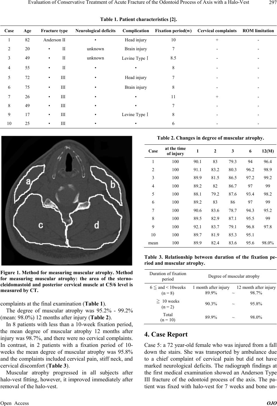 Evaluation of Conservative Treatment of Acute Fracture of the Odontoid Process of Axis with a Halo-Vest 297 Table 1. Patient characteristics [2]. Case Age Fracture type Neurological deficitsComplicationFixation period(w)Cervical complaints ROM limitation 1 82 Anderson II • Head injury 10 + - 2 20 • II unknown Brain injury 7 - - 3 49 • II unknown Levine TypeⅠ8.5 - - 4 55 • II • • 8 - - 5 72 • III • Head injury 7 - - 6 75 • III • Brain injury 8 - - 7 26 • III • • 11 + - 8 49 • III • • 7 - - 9 17 • III • Levine TypeⅠ8 - - 10 25 • III • • 6 - - Figure 1. Method for measuring muscular atrophy. Method for measuring muscular atrophy: the area of the sterno- cleidomastoid and posterior cervical muscle at C5/6 level is measured by CT. complaints at the final examination (Table 1). The degree of muscular atrophy was 95.2% - 99.2% (mean: 98.0%) 12 months after injury (Table 2). In 8 patients with less than a 10-week fixation period, the mean degree of muscular atrophy 12 months after injury was 98.7%, and there were no cervical complaints. In contrast, in 2 patients with a fixation period of 10- weeks the mean degree of muscular atrophy was 95.8% and the complaints included cervical pain, stiff neck, and cervical discomfort (Table 3). Muscular atrophy progressed in all subjects after halo-vest fitting, however, it improved immediately after removal of the halo-vest. Table 2. Changes in degree of muscular atrophy. Case at the time of injury 1 2 3 6 12(M) 1 100 90.1 83 79.3 94 96.4 2 100 91.1 83.2 80.3 96.2 98.9 3 100 89.9 81.5 86.5 97.2 99.2 4 100 89.2 82 86.7 97 99 5 100 88.1 79.2 87.6 93.4 98.2 6 100 89.2 83 86 97 99 7 100 90.6 83.6 78.7 94.3 95.2 8 100 89.5 82.9 87.1 95.5 99 9 100 92.1 83.7 79.1 96.8 97.8 10 100 89.7 81.9 85.3 95.1 mean100 89.9 82.4 83.6 95.6 98.0% Table 3. Relationship between duration of the fixation pe- riod and muscular atrophy. Duration of fixation period Degree of muscular atrophy 6 and < 10weeks (n = 8) 1 month after injury 89.9% ~ 12 month after injury 98.7% 10 weeks (n = 2) 90.3% ~ 95.8% Total (n = 10) 89.9% ~ 98.0% 4. Case Report Case 5: a 72 year-old female who was injured from a fall down the stairs. She was transported by ambulance due to a chief complaint of cervical pain but did not have marked neurological deficits. The radiograph findings at the first medical examination showed an Anderson Type III fracture of the odontoid process of the axis. The pa- tient was fixed with halo-vest for 7 weeks and bone un- Open Access OJO 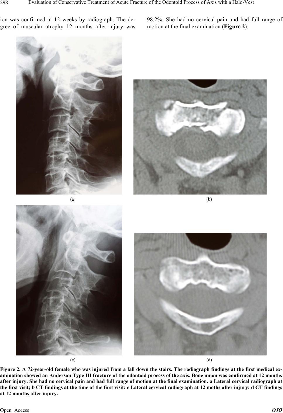 Evaluation of Conservative Treatment of Acute Fracture of the Odontoid Process of Axis with a Halo-Vest 298 ion was confirmed at 12 weeks by radiograph. The de- gree of muscular atrophy 12 months after injury was 98.2%. She had no cervical pain and had full range of motion at the final examination (Figure 2). (a) (b) (c) (d) Figure 2. A 72-year-old female who was injured from a fall down the stairs. The radiograph findings at the first medical ex- amination showed an Anderson Ty pe III frac ture of the odontoid pr ocess of the axis. Bone union w as confirme d at 12 months after injury. She had no cervical pain and had full range of motion at the final examination. a Lateral cervical radiograph at the first visit; b CT findings at the time of the first visit; c Lateral cervical radiograph at 12 moths after injury; d CT findings at 12 months after injury. Open Access OJO  Evaluation of Conservative Treatment of Acute Fracture of the Odontoid Process of Axis with a Halo-Vest 299 5. Discussion Surgical therapy is recommended for patients with a fresh fracture of the odontoid process of the axis because it allows for early discharge and successful rehabilitation. However, surgery cannot always be conducted. If a pa- tient has poor systemic conditions or does not desire sur- gical therapy, and if there are problems with access to the appropriate equipment, the only option is conservative therapy. Therefore, the percentage of bone union in con- servative therapy was evaluated in patients treated in our hospital. The mean percentage of bone union of halo- vest-fixed odontoid process fractures was 72.6% in Anderson Type II patients [1,3-7]. Greene et al. [3] showed that the percentage of bone union in 340 Ander- son Type II patients treated with conservative therapy was 79%. They also found that the percentage of bone union of patients with a ≥6 mm fracture dislocation at the time of injury was significantly different, consequently, injury to the soft tissues was affected it. It was confirmed that the percentage of bone union in Type III patients was better than that of Type II patients [1,3,4,5,7] and the mean percentage of bone union was 97.5% [1,3,4,5,7] (Table 4). Therefore, an Anderson Type II patient with ≥6 mm fracture dislocation was considered to be a can- didate for surgery in our hospital based on the results of Greene et al. [3]. Patients in this study were Anderson Type II with <6 mm and III fracture dislocations who could not undergo surgery due to multiple trauma or did not desire surgery. Bone union was observed in all of our patients at 3 months after injury. Muscular atrophy in the sternocleidomastoid and pos- terior cervical muscles was evaluated based on the dura- tion of the fixation period. Callus formation in radio- graph and CT findings was detectable, however, it was difficult to evaluate when the halo-vest was removed. The longer the fixation is, the better the bone union ad- vances. However, muscular atrophy occurs, possibly re- sulting in cervical pain, stiff neck, cervical discomfort, or limited range of motion in the cervical spine. Therefore, muscular atrophy was measured over time. Ono et al. [8] examined cervical muscular atrophy after halo-vest fit- ting and showed that atrophy was observed in approxi- mately 15% of the sternocleidomastoid muscles and in 22% of the posterior cervical muscles, and cervical dis- comfort and limited range of motion were found when atrophy was observed in more than 20% of the posterior cervical muscles. Atrophy in the sternocleidomastoid and posterior cervical muscles in this study was less than that in Ono’s study because no patients were fixed with a halo-vest for as long as 3 months. However, complaints, including stiff neck and cervical discomfort in patients with fixation for 10 weeks or more, were more frequent than those in patients with cervical muscular atrophy for less than 10 weeks. Table 4. Bone union rates. Fracture type Bone union rates No. of cases Odontoid process fracture Anderson Type II 72.6% 386/531 cases Type III 97.5 241/247 Based on these results, no patient with fixation for less than 10 weeks had problems with bone union. Thus, fixation with a halo-vest for less than 10 weeks is appro- priate for a fresh fracture of the odontoid process of the axis since cervical muscular atrophy and complaints as- sociated with muscular atrophy are caused by long-term fixation. 6. Conclusion Halo-vest is useful for treatment of a fresh fracture of the odontoid process in patients who cannot have surgery, and the appropriate fixation period should be less than 10 weeks. REFERENCES [1] L. D. Anderson and R. T. D’Alonzo, “Fracture of the Odontoid Process of the Axis,” The Journal of Bone & Joint Surgery, Vol. 56, No. 8, 1974, pp. 1663-1674. [2] A. M. Levine and C. C. Edwards, “The Management of Traumatic Spondylolisthesis of the Axis,” The Journal of Bone & Joint Surgery, Vol. 67, No. 2, 1985, pp. 217-226. [3] K. A. Greene, C. A. Dickman, F. F. Marciano, J. B. Drabier, M. N. Hadley and V. K. Sonntag, “Acute Axis Fractures. Analysis of Management and Outcome in 340 Consecutive Cases,” Spine, Vol. 22, 1997, pp. 1843-1852. http://dx.doi.org/10.1097/00007632-199708150-00009 [4] M. N. Hadley, C. Browner and V. K. Sonntag, “Axis Fractures: A Comprehensive Review of Management and Treatment in 107 Cases,” Neurosurgery, Vol. 17, 1985, pp. 281-290. http://dx.doi.org/10.1227/00006123-198508000-00006 [5] P. J. Lennarson, H. Mostafavi, V. C. Traynelis and B. C. Walters, “Management of type Ⅱ Dens Fractures: A Case- Control Study,” Spine, Vol. 25, No. 10, 2000, pp. 1234- 1237. [6] E. A. Seybold and J. C. Bayley, “Functional Outcome of Surgically and Conservatively Managed Dens Fractures,” Spine, Vol. 23, No. 17, 1998, pp. 1837-1846. http://dx.doi.org/10.1097/00007632-199809010-00006 [7] U. Vieweg and R. Schultheiss, “A Review of Halo Vest Treatment of Upper Cervical Spine Injuries,” Archives of Orthopaedic and Trauma Surgery, Vol. 125, 2001, pp. 50-55. http://dx.doi.org/10.1007/s004020000182 [8] A. Ono, M. Amano, Y. Okamura, T. Numazawa, K. Ueyama, S. Nishizawa and S. Toh, “Muscle Atrophy after Treatment with Halovest,” Spine, Vol. 30, 2005, pp. E8- 12. Open Access OJO |