

Paper Menu >>
Journal Menu >>
 Advances in Nanoparticles, 2013, 2, 378-383 Published Online November 2013 (http://www.scirp.org/journal/anp) http://dx.doi.org/10.4236/anp.2013.24052 Open Access ANP Synthesis and Characterization of Ni-Zn Ferrite Nanoparticles (Ni0.25Zn0.75Fe2O4) by Thermal Treatment Method Poh Lin Leng1, Mahmoud Goodarz Naseri2*, Elias Saion1, Abdul Halim Shaari1, Mazaliana Ahmad Kamaruddin1 1Department of Physics, Faculty of Science, Universiti Putra Malaysia, Serdang, Malaysia 2Department of Physics, Faculty of Science, Malayer University, Malayer, Iran Email: *[email protected] Received August 14, 2013; revised October 2, 2013; accepted October 16, 2013 Copyright © 2013 Poh Lin Leng et al. This is an open access article distributed under the Creative Commons Attribution License, which permits unrestricted use, distribution, and reproduction in any medium, provided the original work is properly cited. ABSTRACT Cubic structured nickel-zinc ferrite nanoparticles (Ni0.25Zn0.75Fe2O4) have been synthesized by thermal treatment method. In this procedure, an aqueous solution containing metal nitrates as precursors, polyvinyl pyrrolidone as a cap- ping agent, and deionized water as a solvent were thoroughly stirred, dried at 353 K for 24 h, and crushed into powder before calcination to remove organic matters and crystallize the particles. The structure and particle size were charac- terized by X-ray powder diffraction and transmission electron microscopy. The average particle size increased from 7 to 25 nm with increase of calcination temperature from 723 to 873 K respectively. The magnetic properties were deter- mined by vibrating sample magnetometer and electron paramagnetic resonance electron paramagnetic resonance at room temperature. By increasing the calcinations temperatures from 723 to 873 K it showed an increase of the mag- netization saturation from 11 to 26 emu/g and the g-factor from 2.0670 to 2.1220. The Fourier transform infrared spec- troscopy was used to confirm the presence of metal oxide bands at all temperatures and the removal of organic matters at 873 K. Keywords: Thermal Treatment; Nickel Zinc Ferrite; Nanoparticles; Magnetic Property 1. Introduction The last two decades have seen a remarkable progress in the synthesis of spinel ferrites nanocrystals, aiming at a better material with excellent chemical stability, low magnetic coercivity, moderate saturation magnetization, high permeability, high electrical resistivity and low eddy current. In particular, the nickel-zinc (Ni-Zn) ferrite nanocrystals have been extensively studied for their su- per-paramagnetic properties, which are suitable for high- frequency applications such as rod antennas and cores of inductors and transformers [1,2]. The magnetic proper- ties of ferrites of spinel structural formula AB2O4 are mainly controlled by the divalent cations, which occupy the tetrahedral A sites and the trivalent cation, which has high degree affinity for octahedral B sites [3,4]. Zn fer- rite bulk material has a normal spinel structure, where all divalent cations are located on the tetrahedral sites and trivalent cations all located on the octahedral sites. Ni ferrites bulk materials on the other hand have an inverse spinel structure, where half of trivalent cations occupy the tetrahedral sites while the other half remain on octa- hedral, while divalent cations all migrate to octahedral positions. It has been shown that for the Ni-Zn ferrite nanoparticles, octahedral sites prefer for Ni and tetrahe- dral prefers for Zn [5]. Substituting Zn for Ni cations leads to the formula (2 x Zn 3 1x Fe ) (1x N i 3 1x Fe )O4 (0 ≤ x ≤ 1), in which the first and second brackets indicate oc- cupancy of the A and B sub-lattices respectively. Beside the distribution of divalent and trivalent cations in the spinel structure, the properties of Ni-Zn ferrite nanopar- ticles are highly sensitive to the quantum confinement effect of particle size, which in turn depends on the method of preparation of the nanoparticles [6]. A variety of methods have been proposed for the syn- thesis of Ni-Zn ferrite nanoparticles with controllable size, shape, and chemical stability such as sol-gel methods [7], *Corresponding author. 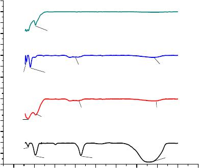 P. L. LENG ET AL. 379 thermal combustion method [8], citrate precursor route [9], co-precipitation method [6,10], thermal plasma syn- thesis [11], reverse micelle [12,13], hydrothermal [14], micro-emulsion [15] and sonochemical reaction [16]. Some disadvantages of these methods include compli- cated procedure, high reaction temperature, long reaction time, and use of reduction agents, which are potential upsetting the environment. In the present study, Ni-Zn ferrite nanoparticles (Ni0.25Zn0.75Fe2O4) were synthesized from an aqueous solution containing only metal nitrates as precursors, polyvinyl pyrrolidone (PVP) as a capping agent and deionized water as solvent by a simple thermal treatment at moderate calcinations temperatures. No other chemicals were added, thus this method offers the advantages of simplicity, low reaction temperatures, a low cost, and an environmentally friendly operation since it produces no by-product effluents [17-20]. 2. Experimental Procedure 2.1. Preparation Iron (III) nitrate, Fe(NO3)3·9H2O, nickel(II) nitrate, Ni(NO3)2·6H2O, and zinc nitrate, Zn(NO3)2·6H2O were purchased from Acros Organics and PVP (MW = 10,000) was supplied by Sigma Aldrich. All the chemical re- agents were of research grade and used without further purification. 3 g of PVP was dissolved in 100 ml of de- ionized water at 343 K before mixing 0.2 mmol of iron (III) nitrate, 0.025 mmol of nickel nitrate and 0.075 mmol zinc nitrate and the solution stirred for 2 hours. No precipitation occurred in the solution. The brown solu- tion was poured into a glass Petri dish and heated at 353 K in an oven for 24 hours to release most of the water. The brown solid material was crushed into powder and the samples were heated for 3 hours in alumina boat at different calcination temperatures of 723, 773, 823 and 873 K to decompose the organic matters and crystallize the nanoparticles. 2.2. Characterization The textural and morphological characteristics of the pre- pared Ni0.25Zn0.75Fe2O4 nanoparticles were studied with several techniques to verify the particle size, shape and size distribution as well as to explore the parameters of interest. The structure was characterized by the XRD technique using a Shimadzu diffract meter model XRD 6000 employing Cu Kα (0.1542 nm) radiation to generate diffraction patterns from powder crystalline samples at room temperature in 2θ range of 10˚ - 70˚. The infrared spectra in the range 280 - 4000 cm−1 were recorded using FTIR spectrometer (Perkin Elmer model 1650). The FTIR spectra are used to confirm the presence of metal oxide bands and the removal of organic matters at 873 K. The structural and particle size of the calcined Ni0.25 Zn0.75Fe2O4 nanoparticles were determined at room tem- perature by using transmission electron micrograph (TEM) (JEOL 2010 UHR version microscopy) at an ac- celerating electron voltage of 200 kV. The magnetization was measured using a vibration sample magnetometer (VSM) (Lake Shore 4700) at room temperature with a maximum magnetic field of 15 kOe. The electron para- magnetic resonance (EPR) spectra were recorded on a JEOL JES-FA200 EPR spectrometer (JEOL, Tokyo, Ja- pan) at room temperature. 3. Results and Discussion 3.1. Structural Studies The FTIR spectra recorded for Ni0.25Zn0.75Fe2O4 nanopar- ticles in the range between 280 cm−1 and 4000 cm−1 are shown in Figure 1. The spectra give information about the chemical and molecular structure changes in the syn- thesized ferrites after calcination treatments. For all cal- cined samples at calcination temperatures from 673 to 873 K, shown in Figures 1(a)-(d), two assigned absorp- tion bands appear at 528 cm−1, which is attributed to stretching vibration of octahedral group Fe-O stretching band and that at 351 cm−1 are attributed to the tetrahedral group Zn-O and Ni-O stretching bands. All absorption peaks above 1000 cm−1 were attributed to covalent bonds of PVP. The bending mode around 1602 - 1637 and 33,274 - 3390 cm−1 were associated with C = O stretch- ing and O-H stretching vibration respectively. The ab- sence of these peaks at 873 K confirmed the organic sources were removed from the calcined samples and pure Ni0.25Zn0.75Fe2O4 nanoparticles were obtained. Figure 2 shows EDX pattern of the sample calcined at 873 K, which confirms the Ni, Zn, Fe and O peaks ap- pearing along with the C substrate peak. No contaminat- 05001000 1500 2000 2500 3000 3500 4000 4500 0 50 100 150 200 250 300 350 400 450 500 550 600 650 700 750 Transmittance wave number (cm-1) (a) (b) (c) (d) 351 528 1635 3274 351 528 1637 3390 351 5281602 3300 351 528 Figure 1. FTIR spectra of Ni0.25Zn0.75Fe2O4 nanoparticles calcined at (a) 723, (b) 773, (c) 823 and (d) 873 K. Open Access ANP 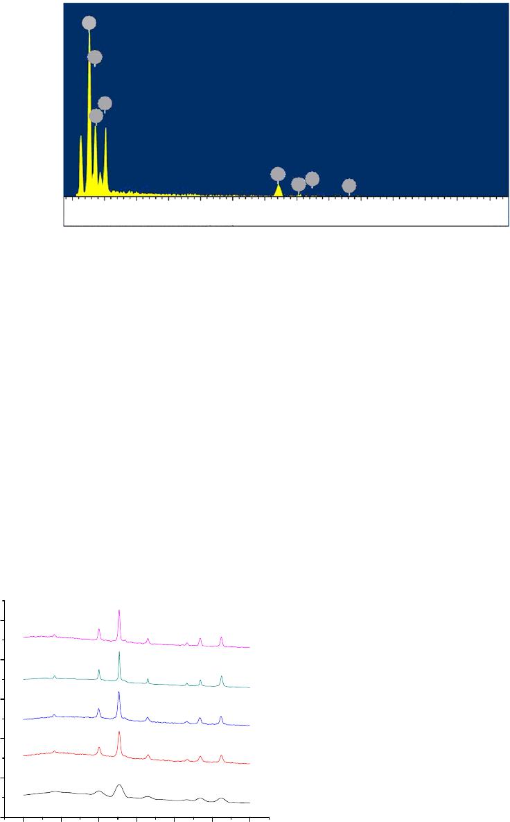 P. L. LENG ET AL. Open Access ANP 380 0 1 2 3 4 5 6 7 8 9 10 11 12 13 Spectrum 3 O Fe Zn N i Full Scale 785 cts Cursor: 13.525 (0 cts)keV Fe Fe N i Zn Figure 2. The EDX spectra of Ni0.25Zn0.75Fe2O4 nanoparticles grown on carbon at calcination temperature of 873 K. ing elements from organic reagents, such as hydrogen or nitrogen were detected. Figure 3 shows the XRD diffraction patterns of the Ni0.25Zn0.75Fe2O4 nanoparticles at 353 K and at calcina- tion temperatures from 723 to 873 K. The diffraction peaks show the reflection planes (111), (220), (311), (400), (422), (333) and (440) which are consistent with the standard powder diffraction reported from XRD li- brary code (00-052-0279) and no other metal oxides could be identified. The diffraction peaks become shar- per when the calcinations temperature increased. It can be said that all samples formed the spinal phase with a face centered cubic structure (f.c.c). The particle size of the ferrite nanoparticles has been estimated from the XRD plane (311) by the Scherrer’s formula: d = 0.9 λ/β cosθ, where d is the average particle size in nm, β is the FWHM of the intensity measured in radians, λ is the X-ray wavelength and θ is the Bragg angle. The average particle size increased from 9 nm to 23 nm at calcination 10 20 30 40 50 60 70 0 2000 4000 6000 8000 10000 (e) (d) (c) (b) (a) (440) (333) (422) (400) (311) (220) (111) Intensity 2 Theta (degree) Figure 3. XRD pattern of Ni0.25Zn0.75Fe2O4 nanoparticles at (a) 353 K, and calcined at (b) 723, (c) 773, (d) 823 and (e) tempera 873 K. ture from 723 K to 873 K, as listed in Table 1. d si 3.2. Magnetic Studies s of magnetization measured at The TEM images in Figure 4 show the size, shape an ze distribution of the Ni0.25Zn0.75Fe2O4 nanoparticles at different calcination temperatures. The particle size in- creased from 7 nm at 723 K to 25 nm at 873 K (Table 1). The reason for the increase in the particle size is that the surface of the nanoparticles melted and fused with the neighboring particles at higher calcination temperatures [17-20]. Thus it is possible to control particle size by the calcination temperature. Smaller particle size has been reported for Ni0.25Zn0.75Fe2O4 nanoparticles prepared by wet co-precipitation routes with the average particle size of about 4 nm [21] and 7 nm for Ni0.3Zn0.7Fe2O4 prepared by co-precipitating aqueous solutions in alkaline medium [6]. By ball-milling on Ni0.2Zn0.8Fe2O4 nanoparticles fab- ricated by mechanochemical method, particle size of about 11 nm was obtained but following annealing at 773 K the particle size increased to about 26 nm [22]. Figure 5 shows the curve room temperature. Table 1 depicts the values of satura- tion magnetization (Ms) and coercivity (Hc) of different samples. Increasing the calcination temperatures from 723 K to 873 K, the Ms value increased from 11 emu/g to 26 emu/g and the coercivity value also increased from 7 to 29 G, when the particle size increased from 7 to 25 nm, as listed in Table 1. The magnetization curves demon- strate a typical superparamagnetic behavior. The Hc is in direct proportional to the volume of single domain grains. Therefore, Hc increased when the particles size increased. Higher Ms values of about 30 emu/g have been achieved for 15 nm Ni0.25Zn0.75Fe2O4 nanoparticles prepared by co- precipitation [21], 22 emu/g for 6 - 7 nm Ni0.2Zn0.8Fe2O4 synthesized by forced hydrolysis in diethylenglycol [23] and 28 emu/g for 6 - 8 nm Ni0.35Zn0.65Fe2O4 obtained by thermal decomposition [6]. 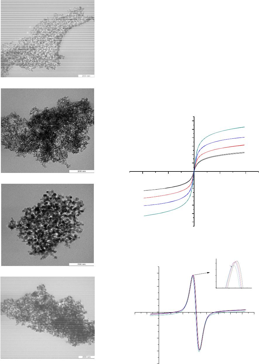 P. L. LENG ET AL. 381 (a) (b) (c) (d) Figure 4. TEM images of Zn0.75Fe2O4 nanoparticles calcined at (a)723, (b) 773, (c) 823 and (d) 873 K. ed a thermal treatment method for the Ni0.25 Figure 6 shows the EPR spectra of the samples cal- cined at (a) 723, (b) 773, (c) 823 and (d) 873 K. Peak-to- peak line width (ΔHpp), resonant magnetic field(H),and g-factor are three parameters that characterize the mag- netic properties. The g-factor can be calculated according to the equation: g = hν/βH where h is Planck’s constant, ν is the microwave frequency, β is the Bohr magneton (9.274 × 10⁻²¹ erg·G−1), and H is resonant magnetic field. The values of g-factor increased from 2.0670 to 2.1220 correspond to the decrease of the resonance magnetic field from 3170 to 3090 G with the increase of calcina- tion temperature from 723 K to 873 K. 4. Conclusion This paper present 30 -10000 -50000500010000 -30 -25 -20 -15 -10 -5 0 5 10 15 20 25 M (emu/g) H (G) (a) (b) (c) (d) Figure 5. VSM curves of Ni0.25Zn0.75Fe2O4 nanoparticles ca cined at (a)723, (b) 773, (c) 823 and (d) 873 K. l- -2000-100001000 2000 3000 40005000 6000 7000 800 0 -2000 -1500 -1000 -500 0 500 1000 1500 2400 2700 3000 1000 1200 1400 B A (a) (b) (d)(c) intensity M agnetic field (G) Figure 6. EPR spectra of Ni0.25Zn0.75Fe2O4 nanoparticl calcined at (a) 723, (b) 773, (c) 823 and (d) 873 K. es Open Access ANP  P. L. LENG ET AL. Open Access ANP 382 na magnetic Ni0.25Zn0.75Fe2O4 Calcinations Average particle Average particle Saturation magnetization,Coercivity Peak to peak line factorResonance Table 1. The average particle sizes (nm) of Ni0.25Zn0.75Fe2O4oparticles determined from XRD and TEM andn properties observed from VSM and EPR at room temperature. nanoparticles temperature (K) size XRD (nm) size TEM (nm)Ms (emu/g) field Hc (G)width ΔHpp (G) g- field, Hr (G) Ni-2.0670Zn ferrite 1 723 9 7 11.414 7 758 3170 Ni-Zn ferrite 2 773 11 9 15.785 10 755 2.08853140 Ni-Zn ferrite 3 823 14 13 20.320 26 753 2.10503118 Ni-Zn ferrite 4 873 23 20 26.447 29 750 2.12203090 nthesis of Ni0.25Zn0.75Fe2O4 nanocrystals with grain by the Ministry of Higher [1] A. Goldman, “logy,” 2nd Edition, 6/j.jmmm.2006.02.031 sy size ranging from 7 to 25 nm at the calcination tempera- tures from 723 to 873 K as measured by XRD and TEM. The PVP stabilized the particles and prevented them from agglomerating. The FTIR measurement confirmed the removal of all organic matters and leaving pure metal oxides at 873 K. The VSM results showed that the satu- ration magnetization increased from 11 emu/g to 26 emu/ g and the coercivity value increased from 7 to 29 G at calcination temperatures from 723 K to 873 K due to the increasing of the volume of single domain grains. The values of g-factor increases with increase of calcination temperature and particle size were increased. This simple method, which is cost-effective and environmentally friendly, produces no toxic byproduct effluents and can be used to fabricate pure, crystalline spinel ferrite nano- crystals. 5. Acknowledgements This work was supported Education of Malaysia under the FRGS grant. The au- thors would also like to thank staff of the Faculty of Sci- ence and the Bioscience Institute of University Putra Malaysia, who had contributed to this work. REFERENCES Modern Ferrite Techno Springer Science Business Media, Inc., New York, 2006. [2] S. W. Lee and C. S. Kim, “Superparamagnetic Properties Ni-Zn Ferrite for Nano-Bio Fusion Applications,” Jour- nal of Magnetism and Magnetic Materials, Vol. 304, No. 1, 2006, pp. 418-420. http://dx.doi.org/10.101 assa[3] G. R. Amiri, M. H. Yousefi, M. R. Abolhni, S. Manouchehri, M. H. Keshavarz and S. Fatahian, “Mag- netic Properties and Microwave Absorption in Ni-Zn and Mn-Zn Ferrite Nanoparticles Synthesized by Low-Tem- perature Solid-State Reaction,” Journal of Magnetism and Magnetic Materials, Vol. 323, No. 6, 2011, pp. 730-734. http://dx.doi.org/10.1016/j.jmmm.2010.10.034 [4] M. Mohapatra and S. Anand, “Synthesis and Applications nology, Vol. 2, No. 8, 2010, pp. 127-146. anodimensional of Nano-Structured Iron Oxides/Hydroxides—A Review,” International Journal of Engineering, Science and Tech- [5] E. Manova, D. Paneva, B. Kunev, E. Rivière, C. Estour- nès and I. Mitov, “Characterization of N Ni-Zn Ferrite Prepared by Mechanochemical and Thermal Methods,” Journal of Physics: Conference Series, Vol. 217, No. 1, 2010, Article ID: 012102. http://dx.doi.org/10.1088/1742-6596/217/1/012102 [6] K. Velmurugan, V. S. K. Venkatachalapathy and S. hilnathan, “Synthesis of Nickel Zinc Iron Nanopar Send- ticles by Coprecipitation Technique,” Materials Research, Vol. 13, No. 3, 2010, pp. 299-303. http://dx.doi.org/10.1590/S1516-14392010000300005 [7] B. T. Naughton, P. Majewski and D. R. Clarke, “Mag- netic Properties of Nickel-Zinc Ferrite Toroids Prepar ed from Nanoparticles,” Journal of the American Ceramic Society, Vol. 90, No. 11, 2007, pp. 3547-3553. http://dx.doi.org/10.1111/j.1551-2916.2007.01981.x [8] E. E. Sileo, R. Rotelo and S. E. Jacobo, “Nickel Ferrites Prepared by the Citrate Precursor Method,” Zinc Phy- sica B: Condensed Matter, Vol. 320, No. 1-4, 2002, pp. 257-260. http://dx.doi.org/10.1016/S0921-4526(02)00705-6 [9] R. K. Singh, C. Upadhyay, S. Layek and A. Yadav “Cation Distribution of NiZnFe O Nanopart , icles,” ed Nickel Zinc 0.50.524 International Journal of Engineering, Science and Tech- nology, Vol. 2, No. 8, 2010, pp. 104-109. [10] A. Kumar, Annveer, M. Arora, M. S. Yadav and R. P. Panta, “Induced Size Effect on Ni Dop Ferrite Nanoparticles,” Physics Procedia, Vol. 9, 2010, pp. 20-23. http://dx.doi.org/10.1016/j.phpro.2010.11.006 [11] M. Shigeta and A. B. Murphy, “Thermal Plasmas for Nanofabrication,” Journal of Physics D: Applied Physics, Vol. 44, No. 17, 2011, Article ID: 174025. http://dx.doi.org/10.1088/0022-3727/44/17/174025 [12] S. A. Morrison, C. L. Cahill, E. E. Carpenter, S. Calvin Swaminathan, M. E. McHenry and V. G. Harris, “ , R. Mag- netic and Structural Properties of Nickel Zinc Ferrite Nanoparticles Synthesized at Room Temperature,” Jour- nal of Applied Physics, Vol. 95, No. 11, 2004, Article ID: 6392. http://dx.doi.org/10.1063/1.1715132 [13] S. Thakur, S. C. Katyal, A. Gupta, V. R. Reddy, S. K. Sharma, M. Knobel and M. Singh, “Nickel-Zinc Ferrite from Reverse Micelle Process: Structural and Magnetic Properties, Mössbauer Spectroscopy Characterization,” Journal of Physical Chemistry C, Vol. 113, No. 49, 2009, pp. 20785-20794. http://dx.doi.org/10.1021/jp9050287  P. L. LENG ET AL. 383 [14] P. E. Meskin, V. K. Ivanov, A. E. Barantchikov, B. R. Churagulov and Y. D. Tretyakov, “Ultrasonically As- sisted Hydrothermal Synthesis of Nanocrystalline ZrO2, TiO2, NiFe2O4 and Ni0.5Zn0.5Fe2O4 Powders,” Ultrasonics Sonochemistry, Vol. 13, No. 1, 2006, pp. 47-53. http://dx.doi.org/10.1016/j.ultsonch.2004.12.002 [15] D. Mathew and R. Juang, “An Overview of the S and Magnetism of Spinel Ferrite Nanoparticles an tructur d e Their Synthesis in Microemulsions,” Chemical Engineering Journal, Vol. 129, No. 1-3, 2007, pp. 51-65. http://dx.doi.org/10.1016/j.cej.2006.11.001 [16] M. Sivakumar, T. Takami, H. Ikuta, A. Towata T. Tuziuti, T. Kozuka, D. Bhattacharya , K. Yasui, and Y. Lida, “Fabrication of Zinc Ferrite Nanocrystals by Sonochemi- cal Emulsification and Evaporation: Observation of Mag- netization and Its Relaxation at Low Temperature,” Jour- nal of Physical Chemistry B, Vol. 110, No. 31, 2006, pp. 15234-15243. http://dx.doi.org/10.1021/jp055024c [17] M. G. Naseri, E. B. Saion, M. Hashim, A. H. Shaari and H. A. Ahangard, “Synthesis and Characterization of Zinc Ferrite Nanoparticles by Thermal Treatment Method,” Solid State Communications, Vol. 151, No. 14-15, 2011, pp. 1031-1035. http://dx.doi.org/10.1016/j.ssc.2011.04.018 [18] M. G. Naseri, E. and A. H. Shaari, “Synthesis and Chara B. Saion, H. A. Ahangar, M. Hashim cterization of Manganese Ferrite Nanoparticles by Thermal Treatment Method,” Journal of Magnetism and Magnetic Materials, Vol. 323, No. 13, 2011, pp. 1745-1749. http://dx.doi.org/10.1016/j.jmmm.2011.01.016 [19] M. G. Naseri, E. B. Saion, H. A. Ahangar, M. Hashim . Saion, H. A. Ahangar, A. H. Shaari Bueno-Baqués, F. Paraguay-Delgado, and A. H. Shaari, “Simple Preparation and Characteriza- tion of Nickel Ferrite Nanocrystals by a Thermal Treat- ment Method,” Powder Technology, Vol. 212, No. 1, 2011, pp. 80-88. [20] M. G. Naseri, E. B and M. Hashim, “Simple Synthesis and Characterization of Cobalt Ferrite Nanoparticles by a Thermal Treatment Method,” Journal of Nanomaterials, Vol. 2010, 2010, Article ID: 907686. [21] V. Corral-Flores, D. C. E. Botez, R. Ibarra-Gómez and R. Ziolo, “Magnetic Properties of Nickel-Zinc Ferrite Nanoparticles Synthe- sized by Coprecipitation,” Physica Status Solidi (A), Vol. 204, No. 6, 2007, pp. 1742-1745. http://dx.doi.org/10.1002/pssa.200675358 [22] E. Manova, D. Paneva, B. Kunev, E. Rivière, C. Estournès, I. Mitov and E. Manov, “Characterization of Nanodimen- sional Ni-Zn Ferrite Prepared by Mechanochemical and Thermal Methods,” Journal of Physics: Conference Se- ries, Vol. 217, No. 1, 2010, Article ID: 012102. http://dx.doi.org/10.1088/1742-6596/217/1/012102 [23] Z. Beji1, T. B. Chaabane, S. L. Smiri, S. Ammar, F. Fiévet, N. Jouini and J. M. Grenèche, “Synthesis of Nickel-Zinc Ferrite Nanoparticles in Polyol: Morphological, Structural and Magnetic Studies,” Physica Status Solidi (A), Vol. 203, No. 3, 2006, pp. 504-512. http://dx.doi.org/10.1002/pssa.200521454 Open Access ANP |