

Paper Menu >>
Journal Menu >>
 World Journal of Cardiovascular Diseases, 2013, 3, 512-518 WJCD http://dx.doi.org/10.4236/wjcd.2013.38081 Published Online November 2013 (http://www.scirp.org/journal/wjcd/) Carrier rate of hepatitis B surface antigen (HBsAg) among urban pregnant women in a secondary health facility in Maiduguri, Northeastern Nigeria David Nadeba Bukbuk1, Maxwell Bayo Samaila2, Akawu Denue Ballah3, Ibrahim Musa Kida3, Chima Iwuoha2 1Department of Microbiology, Faculty of Science, Essien Udom Court, University of Maiduguri, Maiduguri, Nigeria 2Department of Biological Sciences, Faculty of Science, Essien Udom Court, University of Maiduguri, Maiduguri, Nigeria 3Department of Medicine, College of Medical Sciences, University of Maiduguri, Maiduguri, Nigeria Email: [email protected], [email protected] Received 22 August 2013; revised 29 September 2013; accepted 12 October 2013 Copyright © 2013 David Nadeba Bukbuk et al. This is an open access article distributed under the Creative Commons Attribution License, which permits unrestricted use, distribution, and reproduction in any medium, provided the original work is properly cited. ABSTRACT A serological survey conducted among 196 pregnant women resident in and around Maiduguri, an urban town in north eastern Nigeria showed that 33 (16.8%) were asymptomatic carriers of hepatitis B virus sur- face antigen (HBsAg). They are within the age range of 14 - 40 years with mean (± SD) age of 24.7 (± 6.3). The women attend antenatal clinic at the State Spe- cialist Hospital, a secondary health facility in Maiduguri, Borno state. The women are all hetero- sexuals. The distribution of HBsAg carrier rate ac- cording to age of women studied showed a significant (p = 0.0061) increase of the prevalence of HBsAg from 4 (8.7%) in the lower age group (<20 years) to 1 (50%) among those in the higher age group (>39 years). Ac- cording to the number of pregnancies, the prevalence of HBsAg infection increased significantly from 6 (9.2%) among primiparous to 13 (13.7%) and 14 (38.9%) among multiparous and grand multiparous respectively. The carrier rate of HBsAg infection among the pregnant women studied based on their duration of pregnancies and their social class showed no significant differences. However, according to the factors known to be commonly associated with the highest risk of transmission of HBV such as history of blood transfusion, Tribal marks/tattooing, use of sharps, sharing of articles, history of jaundice in the women and husbands, history of sexually transmitted infections (STIs) of the women and husban ds and the type of marriage (monogamous or polygamous), num- ber of sexual relationships per week, only the history of blood transfusion was significantly associated with HBsAg carrier rate (RR = 3.71., 95% Confidence In- terval (C.I.), 1.89 - 7.30, p = 0.0078). The study con- firms that pregnant women who attend antenatal clinic in this secondary health facility are at higher risk of being infected with the HBV, which could lead to both prenatal and post natal transmission to their newborns. Keywords: Carrier Rate; HBsAg; Urban; Pregnant Women; SSH; Borno State; Nigeria 1. INTRODUCTION Hepatitis B is one of the commonest and most wide- spread infections of humans [1]. It is estimated that world- wide more than two billion people have been infected by HBV and 350 million people have chronic infection [2]. Worldwide, HBV infections account for 1 million deaths/ year, most of which occur in the developing world [3]. The prevalence of infection with the virus varies greatly from country to country and even within the same country or continent. This variation depends upon a com- plex of behavioural, environmental and host or genetic factors [1]. The variation in prevalence of the infection is significant within the different geographic areas of the world and can be divided into areas of high, intermediate and low endemicity based on the prevalence of HBV markers and carriers [4]. The infection appears to be highly endemic in areas such as Southeast Asia, the Amazon Basin, sub-Saharan Africa and areas of China, where an estimated 70% - 90% of the population shows serologic evidence of previous or current hepatitis B in- fection and high carrier rates of 8% - 20% [4]. In the intermediate endemicity areas such as Middle East, Ja- pan, North Africa, Central and Latin America, Russia and parts of eastern and southern Europe, 20% - 55% of the OPEN ACCESS  D. N. Bukbuk et al. / World Journal of Cardiovascular Diseases 3 (2013) 512-518 513 populations show markers and 2% - 7% are carriers [5]. North America, western and northern Europe and Austra- lia are classified as low endemicity areas with a preva- lence of HBV markers ranging from 4% - 6% and a car- rier rate of 0.5% - 2%. In high endemicity areas, neo- nates are most at risk, through infection from carrier mothers during pregnancy or just after birth [6,7]. Be- cause of the high carrier rate, those in the general popu- lation who have not had the infection remain at risk of being infected throughout their lives from those carriers in their midst [8]. In areas of intermediate endemicity, however, young children are mostly at risk through hori- zontal transmission [9]. While in low endemicity areas, heterosexual transmission makes adolescent and young adults the highest risk group [10]. Travellers to areas of high endemicity are also at increased risk [11]. Nigeria is classified among the group of countries highly endemic for HBV infection, where about 75% of the populace is reported likely to have been exposed to HBV at one time or the other in their life [12]. The clas- sification of high endemicity for HBV has been defined as HBsAg greater than 7% in an adult population [13]. In regions where hepatitis B is endemic, high inci- dence of hepatoma (liver cancer) has also been recorded and a strong association between HBV infection and liver cancer established [14]. Worldwide, it has been re- ported that those who become carriers in adulthood have a 15-40-fold increased risk of developing chronic liver disease and primary liver cancer [15]. HBV may be the cause of up to 8% of all cases of hepatocellular carci- noma worldwide second only to tobacco among known human carcinogens. This cancer is very common in the Maiduguri area [16] where hepatitis B virus in the popu- lation is also known to be highly prevalent [17-19]. Pregnant women are a very important group to study with regards to HBV transmission, since infection with HBV is mainly transmitted through the mother-to-neo- nate route in endemic countries, where in most cases, chronic infection results and the transmission will there- fore occur from generation to generation [20]. Perinatal and postnatal transmission of the virus had been stated to be important in some societies in South East Asia [21]. Ghana [22] and Nigeria [23,24]. Many children who ac- quired the virus in the early years of life may eventually become carriers with its attendant complications. There- fore, prevention of early childhood infection will eventu- ally reduce the prevalence of HBsAg in early childhood and coupled with the advent of an effective HBV vaccine, such preventive measures and treatment options with drugs such as peg-interferon and lamivudine among others are possible. The present study aims to determine the possible risk factors associated with HBV transmission among preg- nant women and the prevalence of one of the markers of hepatitis B virus infection (HBsAg). And also to suggest possible preventive and control measures for hepatitis B virus infection among pregnant women in our commu- nity. 2. MATERIALS AND METHODS 2.1. Study Area and Study Population The study was carried out at the Antenatal Clinic of the State Specialist Hospital, Maiduguri, Borno State, Nige- ria. The clinic has the capacity of attending to over 500 pregnant women monthly. Pregnant women from within Maiduguri metropolis and from other parts of the state attend the clinic. A total of 196 pregnant women in the age range of 14 - 40 years, mean (SD) age of 24.7 (6.3) years and a median age of 23 years were recruited and tested for the presence of HBsAg. They were studied be- cause of the increased risk of either acquiring or spread- ing hepatitis B virus to their newborns/spouses. The so- cial class of the women involved in the study was assess- ed using the scoring method earlier proposed [25]. This is obtained through a scoring index combining a wo- man’s level of education with the occupation of her hus- band, which allocates each woman to a social class I to V, social class V being at the bottom of the social stratifica- tion. The suitability and application of this social classi- fication system for our environment has been well tested and presented elsewhere [26,27]. 2.2. Sample Collection After explaining the purpose of the study to the women, from those that expressed verbal or written consent to participate about 5ml of blood were aseptically drawn by venepuncture into sterile clean test tube containers. The blood were allowed to clot at room temperature and sera separated by centrifugation at 1,500 rpm for 5 min. The separated sera were aspirated into clean cryogenic vials, labelled and stored frozen at −20˚C until tested. 2.3. Screening and Confirmatory Assay on the Serum Samples for HBsAg All sera samples were thawed once and thereafter screened for the presence of hepatitis B surface antigen using KEMRI HEP CELL II B (HBsAg) a reversed pas- sive haemagglutination test kit for the detection of hepa- titis B surface antigen produced by the Kenya Medical Research Institute (KEMRI) and developed at the centre for virus Research, Hepatitis Division. In this system, a serum is considered positive if the coated sheep erythro- cytes in the microplate wells forms an agglutination with the test serum and is considered negative if the coated sheep erythrocytes settle and form a button-like precipi- tate in the bottom of the well of the microplate. The prin- ciple of the assay is that when the test serum or plasma Copyright © 2013 SciRes. OPEN ACCESS  D. N. Bukbuk et al. / World Journal of Cardiovascular Diseases 3 (2013) 512-518 514 are mixed with anti-HBs coated sheep erythrocytes in a microplate and incubated at room temperature, the sam- ples that contain HBsAg will form an agglutination pat- tern during the incubation period. On the other hand, those samples that contain no HBsAg will not form any agglutination instead the coated sheep erythrocyte will settle to form a button-like precipitate. The presence of HBsAg is thereafter determined by the appearance of the agglutination patterns. All the positive sera samples were later confirmed as positive by inhibition of agglutination using confirmation inhibition assay according to instruc- tions of the kit manufacturers. In this system a positive result for the presence of HBsAg showed a decrease of agglutinating titre. Those samples that showed no fall in agglutinating titre were presumed as negatives for HBsAg. A positive control and negative control sera are included in each assay. 2.4. Data Analysis All data generated from the study were entered analyzed using Epi Info version 6.04d [Centers for Disease Con- trol and Prevention, Atlanta, GA, USA (www.cdc.gov/epiinfo /) and World Health Organization, Geneva, Switzerland (www.who.int)] and GraphPad Prism version 5.00 for Windows (GraphPad Software, San Diego California USA, www.graphpad.com). Socio- demographic characteristic or variables of the women studied were calculated and expressed as means, stan- dard deviation (SD), median, range, frequencies and per- centages. Chi-square and linear chi-square for trend were calculated for categorical variables such as age groups, parity, duration of pregnancy, duration of marriage, ave- rage number of sexual relationships per week and social class and compared with the carrier rate of HBsAg in the group, while fisher exact test was used as appropriate. Relative risks (RR) and 95% confidence intervals (95% C.I.) were also calculated to assess the association be- tween the different risk factors for HBsAg transmission and HBV infection in the study group. The data on the occupation of the women and their spouses studied were represented as charts. A p-value of less than or equal to 0.05 was considered as significant. 3. RESULTS 3.1. Socio-demographic Characteristics of the Pregnant Women S tu died A total of 196 pregnant women at various stages of ges- tation were recruited/studied at the State Specialist Hos- pital Maiduguri, Borno State for the prevalence and risk factors to HBV infection. Table 1 shows some socio- demographic characteristics of the pregnant women stu- died. They fall within the age range of 14 to 40 years with mean (SD) age of 24.7 (6.3) years and a median age Table 1. Socio-demographic characteristics of the pregnant women studied. Characteristics Mean ± SD MedianRange Age (years) 24.7 ± 6.3 23 14 - 40 No. of pregnancies 3.4 ± 2.5 3 1 - 12 Duration of pregnancy (months)7.2 ± 1.4 7 3 - 9 Duration of marriage (years) 6.6 ± 5.9 5 0.5 - 30 Mean no. sexual relationships (week) 2.3 ± 1.0 2 1 - 5 Social class Frequency Percentage I 8 4.1 II 22 11.2 III 58 29.6 IV 48 24.5 V 60 30.6 SD, Standard deviation, No., Number. of 23 years. The mean (SD) duration of pregnancies were 7.2 (1.4) months with a range of 3 to 9 months while the number of pregnancies were of the mean (SD) of 3.4 (2.5) and range of 1 to 12. The mean (SD) duration of mar- riage were 6.6 (5.9) years with range of 6 months to 30 years and median of 5 years, while mean (SD) number of sexual relationships with their husbands are of the risk factors earlier found to be associated with HBV infection was 2.3 (1.0), range of 1 to 5. The socio-economic clas- sification of the women shows that very few of them belonged to the higher social classes of I and II, with 4.1% and 11.2% respectively. About 30.6% belonged to class V and 29.6%, 24.5% in classes III and IV respec- tively. 3.2. Distribution of the Occupation of the Wo men and Their Husbands Figures 1 and 2 are pie charts showing the distribution of the occupation of the women and that of their husbands. Farming appears to be the least occupation engaged by the two groups with only 1% and 2% among wives and husbands respectively. Those attending various schools are higher in the women (6% vs. 2%) than in the hus- bands, while most husbands are traders/ Business men and civil servants (40% vs. 37%), most of the women are full time house wives (FTHW) (64%) and only 17% are Traders/Business women. 3.3. Distribution of HBsAg According to Maternal Age The prevalence of HBsAg according to maternal age is as shown in Ta b l e 2 . The prevalence rate increases sig- nificantly with the age of the mothers studied from 8.7% Copyright © 2013 SciRes. OPEN ACCESS 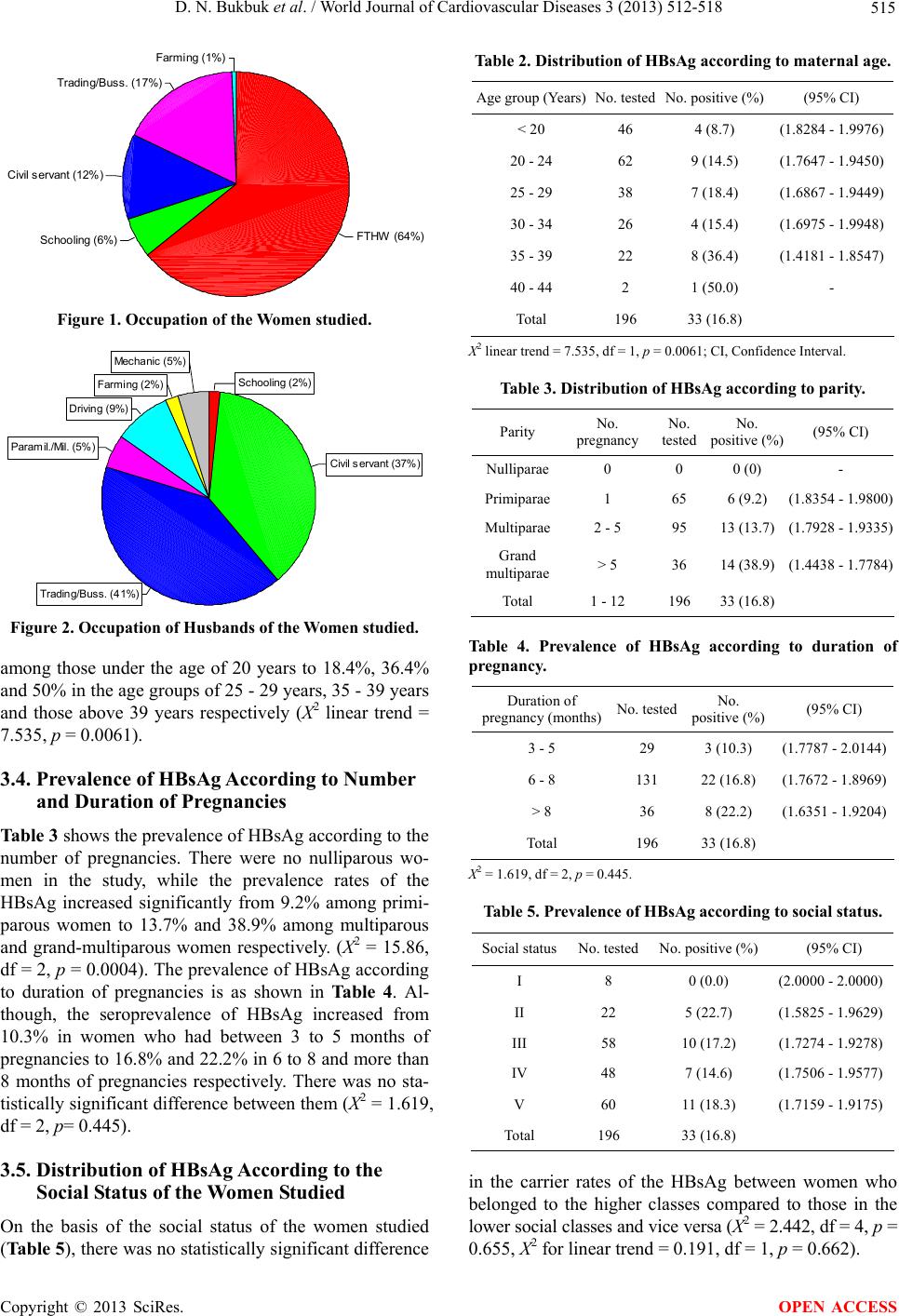 D. N. Bukbuk et al. / World Journal of Cardiovascular Diseases 3 (2013) 512-518 515 Farming (1%) Trading/Buss. (17%) Civil servant (12%) Schooling (6%)FTHW (64 %) Figure 1. Occupation of the Women studied. Mechanic (5%) Farming (2%) Dri ving (9% ) Paramil ./Mil. (5%) Trading/Buss. (41%) Civil servant (3 7%) Schooling (2%) Figure 2. Occupation of Husbands of the Wome n studie d. among those under the age of 20 years to 18.4%, 36.4% and 50% in the age groups of 25 - 29 years, 35 - 39 years and those above 39 years respectively (X2 linear trend = 7.535, p = 0.0061). 3.4. Prevalence of HBsAg According to Number and Duration of Pregnancies Table 3 shows the prevalence of HBsAg according to the number of pregnancies. There were no nulliparous wo- men in the study, while the prevalence rates of the HBsAg increased significantly from 9.2% among primi- parous women to 13.7% and 38.9% among multiparous and grand-multiparous women respectively. (X2 = 15.86, df = 2, p = 0.0004). The prevalence of HBsAg according to duration of pregnancies is as shown in Table 4. Al- though, the seroprevalence of HBsAg increased from 10.3% in women who had between 3 to 5 months of pregnancies to 16.8% and 22.2% in 6 to 8 and more than 8 months of pregnancies respectively. There was no sta- tistically significant difference between them (X2 = 1.619, df = 2, p= 0.445). 3.5. Distribution of HBsAg According to the Social Status of the Women Studied On the basis of the social status of the women studied (Table 5), there was no statistically significant difference Table 2. Distribution of HBsAg according to maternal age. Age group (Years)No. testedNo. positive (%) (95% CI) < 20 46 4 (8.7) (1.8284 - 1.9976) 20 - 24 62 9 (14.5) (1.7647 - 1.9450) 25 - 29 38 7 (18.4) (1.6867 - 1.9449) 30 - 34 26 4 (15.4) (1.6975 - 1.9948) 35 - 39 22 8 (36.4) (1.4181 - 1.8547) 40 - 44 2 1 (50.0) - Total 196 33 (16.8) X2 linear trend = 7.535, df = 1, p = 0.0061; CI, Confidence Interval. Table 3. Distribution of HBsAg according to parity. Parity No. pregnancy No. tested No. positive (%) (95% CI) Nulliparae0 0 0 (0) - Primiparae1 65 6 (9.2) (1.8354 - 1.9800) Multiparae2 - 5 95 13 (13.7) (1.7928 - 1.9335) Grand multiparae > 5 36 14 (38.9) (1.4438 - 1.7784) Total 1 - 12 196 33 (16.8) Table 4. Prevalence of HBsAg according to duration of pregnancy. Duration of pregnancy (months)No. testedNo. positive (%) (95% CI) 3 - 5 29 3 (10.3) (1.7787 - 2.0144) 6 - 8 131 22 (16.8) (1.7672 - 1.8969) > 8 36 8 (22.2) (1.6351 - 1.9204) Total 196 33 (16.8) X2 = 1.619, df = 2, p = 0.445. Table 5. Prevalence of HBsAg according to social status. Social statusNo. testedNo. positive (%) (95% CI) I 8 0 (0.0) (2.0000 - 2.0000) II 22 5 (22.7) (1.5825 - 1.9629) III 58 10 (17.2) (1.7274 - 1.9278) IV 48 7 (14.6) (1.7506 - 1.9577) V 60 11 (18.3) (1.7159 - 1.9175) Total 196 33 (16.8) in the carrier rates of the HBsAg between women who belonged to the higher classes compared to those in the lower social classes and vice versa (X2 = 2.442, df = 4, p = 0.655, X2 for linear trend = 0.191, df = 1, p = 0.662). Copyright © 2013 SciRes. OPEN ACCESS  D. N. Bukbuk et al. / World Journal of Cardiovascular Diseases 3 (2013) 512-518 516 3.6. Risk Factors and Their Possible Association with HBsAg Transmission The factors that are commonly associated with the high- est risk of transmission of the HBV are as shown in Ta- ble 6. About 75% (147 out of 196) of the women are mostly from monogamous marriages compared to only 25% (49 out of 196) who are polygamous. Few of the women had a positive history of blood transfusion 4.6% (9 out of 196), and a history of sexually transmitted in- fections (STIs) in their husbands, 5.6% (11 out of 196). Most of the women however, gave a positive history of tribal makes/tattooing, 62.8% (123 out of 196), use of sharps, 70.4% (138 out of 196), and self history of jaun- dice and in their husbands. 20.9% (41 out of 196), 18.4% (36 out of 196) respectively. Sharing of articles such as toothbrushes, spoons, cups etc with their spouses showed 96.9% (190 out of 196). The only risk factor that has been positively and signi- ficantly associated with HBsAg carrier rate is those with the history of blood transfusion (RR = 3.71, 95% Confi- dence Interval C.I., 1.89 to 7.30, p = 0.008). 4. DISCUSSION This study reports an overall high carrier rate of 16.8% HBsAg among pregnant women attending antenatal cli- nic at state specialist Hospital Maiduguri, Borno State. The group studied fairly represents part of the entire po- pulation and particularly those at an increased risk of either acquiring or spreading the infection in our commu- nity. A prevalence rate of 16.8% HBsAg in pregnant wo- men is high which shows that the virus (HBV) infection is endemic in our community and in Nigeria generally as in areas such as south-east Asia, sub-Saharan Africa, etc. [4,22,28]. It can be suggested that within our community the infection occurring early in childhood could be through vertical transmission from mother to child [29]. Pregnant women are very important group to study with regards to HBV infection or transmission. This is because both perinatal and postnatal transmission of the virus had been stated to be important in some societies in south-east Asia [28]. This higher carrier rate of 16.8% for the HBsAg among pregnant women agrees with the study in the same area [17] who reported a rate of 11.6% for HBsAg among pregnant women. Other reports in Nigeria among pregnant women had observed a similarly higher carrier rate. A higher value of 11.2% was reported in Ibadan [23] and 13.8% among pregnant women at- tending antenatal clinic in Lagos [30]. The high carrier rate among the pregnant women in the present study could be due to the increased suscepti- bility of women to infection during pregnancy. They could hence serve as an important reservoir or potential Table 6. Risk factors and their possible association with HBsAg transmission. Risk factor Tested No. (%) Positive No. (%) RR (95% CI) Type of marriage Monogamous 147 (75.0) 24 (16.3) 0.89 (0.44 - 1.78) Polygamous 49 (25.0) 9 (18.4) History of blood transfusion* Yes 9 (4.6) 5 (55.6) 3.71 (1.89 - 7.30) No 187 (95.4) 28 (15.0) Tribal marks/Tattooing Yes 123 (62.8) 21 (17.1) 1.04 (0.54 - 1.99) No 73 (37.2) 12 (16.4) Use of sharps Yes 138 (70.4) 23 (16.7) 0.97 (0.49 - 1.90) No 58 (29.6) 10 (17.2) History of jaundice/hepatitis Yes 41 (20.9) 6 (14.6) 0.84 (0.37 - 1.90) No 155 (79.1) 27 (17.4) History of jaundice/ hepatitis in husband 36 (18.4) 4 (11.1) 0.61 (0.23 - 1.64) Yes 160 (81.6) 29 (18.1) No Sharing of articles 190 (96.9) 32 (16.8) 1.01 (0.16 - 6.22) Yes 6 (3.1) 1 (16.7) No No. of sexual relationships 1 - 3 173 (88.3) 29 (16.8) 0.96 (0.37 - 2.49) > 3 23 (11.7) 4 (17.4) History of STIs Yes 34 (17.3) 4 (11.8) 2.48 (1.11 - 5.53) No 162 (82.7) 29 (17.9) History of STIs in husband Yes 11 (5.6) 2 (18.2) 0.91 (0.25 - 3.31) No 185 (94.4) 31 (16.8) RR, Relative risk, STIs, Sexually transmitted Infections; *Fisher exact, p = 0.008. pockets of infection to their newborns in utero or through subsequent exposure to unsterilized and contaminated materials used during deliveries and breast feeding. Blood transfusion which was shown to be significantly associated with a higher carrier rate of HBsAg, is an im- Copyright © 2013 SciRes. OPEN ACCESS  D. N. Bukbuk et al. / World Journal of Cardiovascular Diseases 3 (2013) 512-518 517 portant risk factor in Nigeria as pregnancy-related haem- orrhage among others are known to increase the possibil- ity of transmission of HBV (and other blood-borne pa- thogens) through contaminated blood as reported by Uni- ted Nations System in Nigeria (UNSN) [31]. 5. CONCLUSIONS From the result of this study which showed a higher car- rier rate 16.8% of HBsAg among pregnant women, who are said to be representatives of our society, it can be said that Maiduguri is an endemic area for HBV infection. Blood transfusion appears to be the only major risk fac- tor of HBV infection among pregnant women in this secondary health facility in Maiduguri, Borno state. This might be a reflection of the situation in other health fa- cilities across the country (Nigeria), possibly as a result of increased demand for blood to treat certain pregnancy related emergencies such as the different gynaecology bleeding disorders. There is therefore, the need to know the prevalence of HBV markers such as HBsAg in pregnancy and the level of perinatal transmission in the planning of preventive measures. Hence routine screening for HBV infection in pregnant women attending antenatal clinics is necessary, followed by health enlightenment campaigns on the risk and mode of transmission of the virus which can be linked to the ongoing campaigns against HIV-AIDS. This is coupled with the fact that an effective recombinant vaccine against HBV has been developed, the govern- ment and employers of labour should therefore supple- ment the cost and make it available and affordable for selective vaccination of those associated with high risk exposures in our society. 6. ACKNOWLEDGEMENTS We are greatly indebted to Mzee Muli of the Kenya Medical Research Institute, Nairobi for providing us with the hepatitis test kit (HEP CELL kit). We also thank the doctors, nurses and all staff of the Ante- natal Clinic, Obstetrics & Gynaecology department of the State Spe- cialist Hospital, Maiduguri for allowing us access to the patients. REFERENCES [1] Gust, I.D. (1985) Viral hepatitis. In: Warren, K.S. and Mohammed, A.A.F. Eds., Tropical Geographical Medi- cines, McGraw-Hill Books Company, New York, 572-585. [2] Drosten, C., Nippraschk, T., Manegold, C., Meisel, H., Brixner, V., Roth, W.K. Apedjinou, A. and Gunther, S. (2004) Prevalence of hepatitis B virus DNA in anti- HBC-positive/HBsAg-negative sera correlates with HCV but not HIV serostatus. Journal of Clinical Virology, 29, 59-68. http://dx.doi.org/10.1016/S1386-6532(03)00090-8 [3] Mulders, M.N., Venard, V., Njayou, M., Edorh, A.P., Oyefolu, A.O.B., Kehinde, M.O., Tamfum, J.J.M., Nebie, Y.K., Maiga, I., Ammerlaan, W., Fack, F., Omilabu, S.A., Fau, le A. and Muller, C.P. (2004) Low genetic diversity despite hyperendemicity of hepatitis B virus genotype E throughout West Africa. The Journal of Infectious Disea- ses, 190, 400-408. http://dx.doi.org/10.1086/421502 [4] Maynard, J.E., Kane, M., Alter, M.J. and Hadlex, S. (1988) Control of hepatitis B by immunization. In: Zuc- kerman, A.J. Ed., Global Perspective on Viral Hepatitis and Liver Disease, Alan R. Liss, New York, 967-969. [5] Toukan, A. (1990) Strategy for the control of hepatitis B virus infection in the Middle East and North Africa. Vac- cine, 8, S117-S128. http://dx.doi.org/10.1016/0264-410X(90)90231-A [6] Ghendon Y. (1987) Perinatal transmission of hepatitis B virus in high incidence countries. Journal of Virological Methods, 17, 69-79. http://dx.doi.org/10.1016/0166-0934(87)90070-X [7] Yeoh, E.K. (1990) Hepatitis B virus infection in children. Vaccine, 8, S29-S30. http://dx.doi.org/10.1016/0264-410X(90)90212-5 [8] Chainuvati, T. (1988) Prevention of hepatitis B infection in Thailand. Handbook of the International Symposium on Hepatocellular Carcinoma, Bangkok, 9-10 February 1988, 55-63. [9] Kiire, G.F. (1990) Hepatitis B infection in sub-Saharan Africa. Vaccine, 8, S107-S112. http://dx.doi.org/10.1016/0264-410X(90)90229-F [10] West, D.J., Calandra, G.B. and Ellis, R.W. (1990) Vacci- nation of infants and children against hepatitis B. Pedia- tric Clinics of North America, 37, 585-601. [11] Steffen, R. (1991) The epidemiological basis for the prac- tice of travel medicine. 2nd Conference on International Travel Medicine, Altanta, 9-12 May 1991. [12] Sirisena, N.D., Njoku, M.O., Idoko, J.A., Isamade, E., Barau, C., Jelpe, D., Zamani, A. and Otowo, S. (2002) Carriage rate of hepatitis B surface antigen (HBsAg) in an urban community in Jos, Plateau state, Nigeria. Nige- rian Postgraduate Medical Journal, 9, 7-10. [13] Hodges, M., Sanders, E. and Aitken, C. (1998) Seropre- valence of hepatitis markers: HAV, HBV, HCV and HEV amongst primary school pupils in Freetown, West African Journal of Medicine, 17, 36-37. [14] Beasley, R.P and Hwang, L.Y. (1984) Hepatocellular car- cinoma and hepatitis B virus. Seminars in Liver Disease, 4, 113-121. http://dx.doi.org/10.1055/s-2008-1040651 [15] World Health Organization (1990) Consultation on heap- titis B as a sexually transmitted disease. Report of a WHO consultation, 1990, WHO/MIM/VDT/91.457. [16] Gashau, M. (1988) Hepatocellular carcinoma in Maidu- guri: A prospective clinical study. Thesis submitted for the Fellowship of the West African College of Physicians, 1988. [17] Harry, T.O., Bajani, M.D. and Moses, A.E. (1994) Hepa- titis B virus infection among blood donors and pregnant women in Maiduguri, Nigeria. East African Medical Journal, 71, 32-33. [18] Bukbuk, D.N. and Udoh, E. (1997) The relationship of Copyright © 2013 SciRes. OPEN ACCESS  D. N. Bukbuk et al. / World Journal of Cardiovascular Diseases 3 (2013) 512-518 Copyright © 2013 SciRes. 518 OPEN ACCESS blood groups and hepatitis B virus surface antigen carrier state among blood donors in Maiduguri, Nigeria. Nige- rian Medical Practitioner, 33, 335-337. [19] Bukbuk, D.N., Bassi, A.P. and Mangoro, Z.M. (2005) Seroprevalence of hepatitis B surface antigen among pri- mary school pupils in Hawal valley, Borno state, Nigeria. Journal of Community Medicine & Primary Health Care, 17, 20-23. [20] Denny, P. (2006) Hepatitis Delta virus genetic variability: From genotypes I, II, III to eight major clades? Current Topics in Microbiology and Immunology, 307, 151-171. [21] Stevens, C.E., Beasley, R.P., Tsui, J. et al. (1975) Verti- cal transmission of hepatitis B antigen in Taiwan. The New England Journal of Medicine, 292, 771-774. http://dx.doi.org/10.1056/NEJM197504102921503 [22] Acquaye, J.K and Mingle, J.A. (1994) Hepatitis B viral markers in Ghanaian pregnant women. West African Journal of Medicine, 13, 134-137. [23] Ayoola, E.A., Ogunbode, O and Odelola, H.A. (1981) Congenital transmission of hepatitis B antigen in Nige- rians. Archives of Virology, 67, 97-99. http://dx.doi.org/10.1007/BF01314607 [24] Abiodun, P.O., Olomu, A.O., Okolo, S.N., Obasohan, A and Freeman, O. (1994) The prevalence of hepatitis B an- tigen and anti-HBe in adults in Benin City. West African Journal of Medicine, 13, 171-174. [25] Olusanya, O. and Amiegheme, N. (1989) Biosocial fac- tors in maternal mortality. A study from a Nigerian Mis- sion Hospital. West African Journal of Medicine, 8, 160- 165. [26] Olusanya, O. (1984) An original system of social classi- fication for use in Nigeria and other developing countries. 24th Annual Conference of the West African College of Surgeons Freetown, Sierra Leone, 1984. [27] Olusanya, O., Okpere, E. and Ezimokhai, M. (1985) The importance of social class in voluntary fertility control in a developing country. West African Journal of Medicine, 4, 205-212. [28] Stevens, C.E., Neurath, R.A., Beasley, R.P. and Szmu- ness, W. (1979) HBeAg and anti-HBe detection by radio- immunoassay: Correlation with vertical transmission of hepatitis B virus in Taiwan. Journal of Medical Virology, 3, 237-241 [29] Fakunle, Y.M., Addulrahman, M.B. and Whittle, H.C. (1980) Hepatitis B. virus infection in children and adults in Northern Nigeria: A preliminary survey. Transactions of the Royal Society of Tropical Medicine and Hygiene, 75, 626-629. http://dx.doi.org/10.1016/0035-9203(81)90133-4 [30] Nasidi, A., Harry, T.O., Yazou, S.O., Munube, G.M.R., Azzan, B.B. and Ananier, V.A. (1983) Prevalence of he- patitis B infection markers in two different geographical areas of Nigeria. Proceedings of the First International Conference on Viral and Bacterial Vaccines, Paris, De- cember 1983, 213-217. [31] United Nations System in Nigeria (2001) Nigerian com- mon country assessment. World Health Organization, Ge- neva. |