

Paper Menu >>
Journal Menu >>
 World Journal of Cardiovascular Diseases, 2013, 3, 483-486 WJCD http://dx.doi.org/10.4236/wjcd.2013.38076 Published Online November 2013 (http://www.scirp.org/journal/wjcd/) Acute myocardial infarction with normal coronary artery (MINCA) in a patient with multiple sclerosis Murat Celik1*, Mehmet Ata Akil2, Mustafa Tuncer2 1Department of Cardiology, School of Medicine, Gulhane Military Medical Academy, Ankara, Turkey 2Department of Cardiology, Van Medicalpark Hospital, Van, Turkey Email: *[email protected] Received 24 August 2013; revised 25 September 2013; accepted 9 October 2013 Copyright © 2013 Murat Celik et al. This is an open access article distributed under the Creative Commons Attribution License, which permits unrestricted use, distribution, and reproduction in any medium, provided the original work is properly cited. ABSTRACT Interestingly, some patients with ST elevation myo- cardial infarction have normal coronary artery ana- tomy at cooronary angiography. This situation is call- ed as MINCA (acute myocardial infarction with nor- mal coronary artery). The incidence of MINCA va- ries between 1.0% and 8.5%. Although some reasons have been propounded, the exact underlying mecha- nism remains unclear. Nevertheless, thoracic myelitis can be as a possible cause of myocardial infarction. Herein, we report a case of acute myocardial infarc- tion with normal coronary artery in a patient with multiple sclerosis. Keywords: Acute Myocardial İnfarction; Normal Coronary Artery; Multiple Sclerosis 1. INTRODUCTION Although it is known that rupture of atherosclerotic plaque and subsequent intra-luminal thrombus formation is the main cause of acute myocardial infarction, some patients have normal coronary arteries at coronary an- giography in the setting of acute myocardial infarction. The existence of acute myocardial infarction despite an- giographically normal coronary arteries (MINCA) was recognized for more than 30 years and its incidence varied between 1.0% and 8.5% [1-5]. The aetiology and patho- genesis of the condition are still a source of debate. Some reasons such as cocaine ingestion, coronary vasospasm, endothelial dysfunction, thrombosis, embolization and inflammation, coagulopathies (inherited or acquired), perimyocarditis and stress cardiomyopathy (Takotsubo syndrome) may explain some of these occurrences [3]. Additionally, myocardial infarctions may have been caused by vasospastic reactions secondary to spinal cord pathology such as thoracic myelitis. Abnormal cardiac function has been described after upper-spinal-cord lesions due to acute autonomic imbal- ance by the disruption of sympathetic pathways [6]. Nevertheless, coronary vasospastic reactions secondary to spinal cord pathology may lead to myocardial infarc- tion [7-9]. However, the number of case report de- scriging the relationship between myocardial ischemia and multiple sclerosis (MS) is limited and pathogenesis of this condition is still being debated. We hereby wanted to draw attention to this rare togetherness by reporting a case of MINCA in a patient with MS and tried to explain the relationship between MINCA and MS. 2. CASE REPORT A 40-year-old woman without a history of any cardio- vascular disease was admitted to our hospital because of typical chest pain for 1 hour. She had a history of MS for 5 years. An admission 12-lead electrocardiogram (ECG) showed ST elevation in leads II, III, aVF and reciprocal changes in leads V1,V2, suggesting an acute inferior myocardial infarction (Figure 1). Vital parameters and standart biochemical tests at admission were with in nor- mal ranges. Coronary angiography (CAG) was per- formed. Interestingly, CAG did not reveal any stenosis, luminal irregularities, thrombus or coronary spasm in any of 3 coronary arteries (Figures 2(a) and (b)). Addi- tionally, we performed aortography for the suspicion of occlusion of any anomalous coronary artery, but there was not a coronary artery anomaly. The patient took in coronary intensive care unit and treated with infusion of unfractioned heparin and glyceryl trinitrate at recom- mended dose for the possibility of coronary vasopasm and/or spontenous resolution of thrombus and emboliza- tion into the microvascular integrity. Cardiac enzymes were elevated 2.5 hours later: peak creatine kinase 3045 IU/L; peak creatine kinase MB 405 IU/L; and troponin, *Corresponding author. OPEN ACCESS 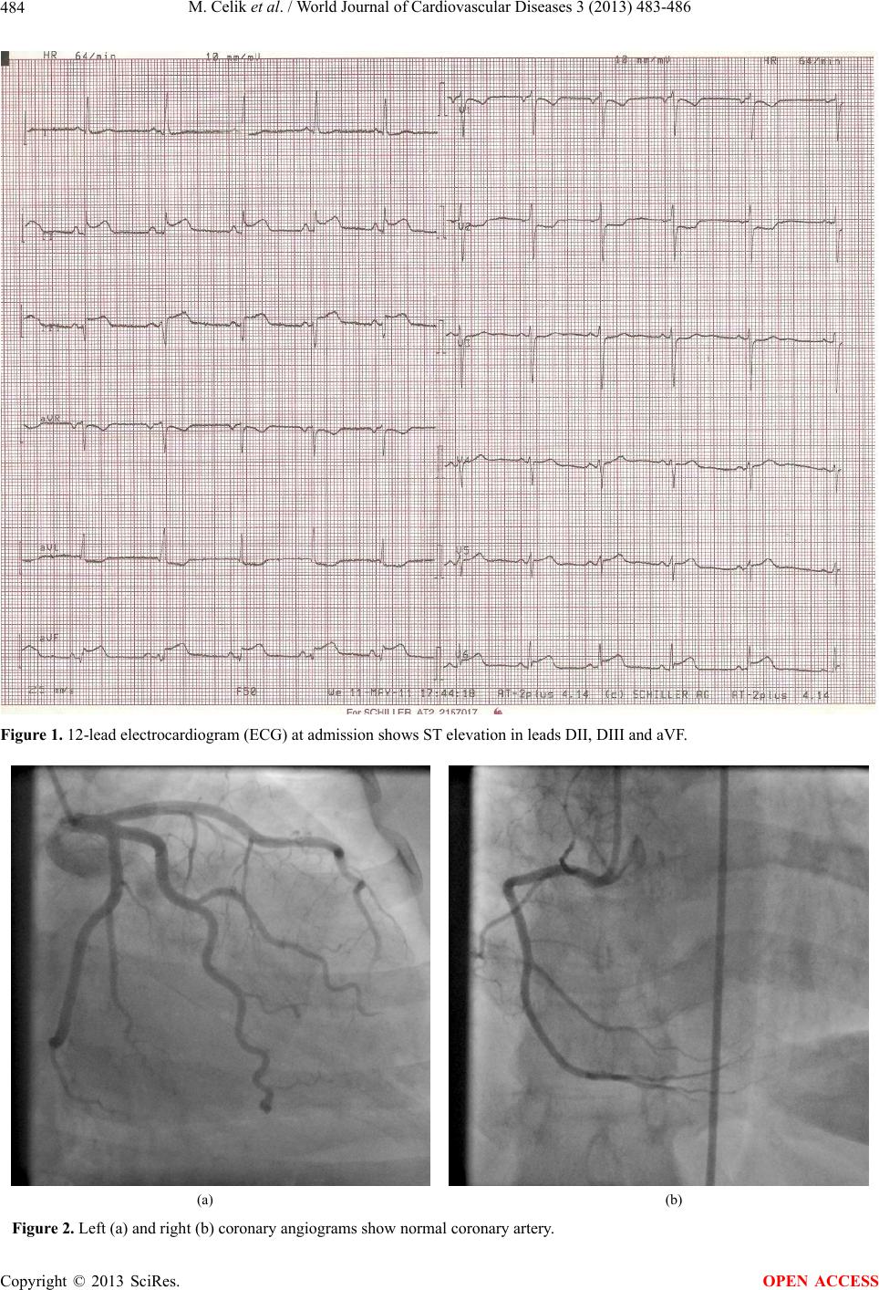 M. Celik et al. / World Journal of Cardiovascular Diseases 3 (2013) 483-486 484 Figure 1. 12-lead electrocardiogram (ECG) at admission shows ST elevation in leads DII, DIII and aVF. (a) (b) Figure 2. Left (a) and right (b) coronary angiograms show normal coronary artery. Copyright © 2013 SciRes. OPEN ACCESS  M. Celik et al. / World Journal of Cardiovascular Diseases 3 (2013) 483-486 485 7.22 ng/mL. The erythrocyte sedimentation rate was 16 mm/h, total leukocyte count was 9600 cells/μL, C-reac- tive protein level was 0.3 mg/dl. Echocardiogram show- ed inferior hypokinesis without any finding of pericardi- tis. Her chest pain recurred and same ECG finding sug- gesting an acute inferior myocardial infarction were found at the 2nd day of hospitalization. CAG repeated and revealed normal coronary arteries once more. At that time, she also complained of dizziness, nausea and vom- iting. Magnetic resonance imaging (MRI) was performed and revealed multiple foci of increased T2 signal inten- sity, but there was not any finding of new foci suggesting MS exacerbation. The patient was treated with infusion of methylprednisolone at recommended dose for five days. Her all complaints including chest pain did not occurred again during hospitalization. On the 4th day of hospitalization, creatine kinase was 164 IU/L; creatine kinase MB was 32 IU/L; and troponin was 2.63 ng/mL. Then, the patient was discharged in a very good condi- tion on the 7th day of hospitalization. We presumed that the patient had an acute myocardial infarction. We thought about the possibility of coronary vasospasm or intra-luminal thrombosis resolved sponta- neously. However, either clear atherosclerosis or throm- bus formation was not present at CAG. Thus, we could not point out the underlying mechanism exactly. 3. DISCUSSION MINCA is a rare but fair clinical entity. Patients with MINCA are younger and less likely to have traditional risk factors for atherosclerosis than those of identifiable CAD [3]. The incidence and the underlying etiology of MINCA remain unclear. A number of possible mecha- nisms have been suggested. The most known are coro- nary vasospasm and thromboembolic disease. Changes in autonomic neural control, changes in contractile function, locally released vasoactive mediators in contact with dysfunctional endothelium, history of smoking, mental stress and oral contraceptive use may be responsible for coronary artery vasospasm [7]. In their study, Bulkley et al. [10] described the autopsy findings of nine patients with progressive systemic sclerosis and evidence of ischemic heart disease but morphologically normal coro- nary arteries. They observed contraction band necrosis, as a form of reperfusion lesion, in seven of nine patients, and suggested that intermittent vascular spasm at some level of the microcirculation would account for the con- traction band type of necrosis and cause myocardial in- farction with morphologically normal coronary arteries in patients with progressive systemic sclerosis. Larsen et al. [3] showed a trend toward a higher prevalence of ma lignancy in patients with MINCA, and suggested that malignancy—induced hypercoagulability or inflammation —might be the underlying mechanism of MINCA. Also, myocarditis is one of the possible causes of MINCA, es- pecially in patients below the age of 35 years [11]. Other potential mechanisms are cocaine ingestion, coagulo- pathies (inherited or acquired), stress cardiomyopathy or Takotsubo syndrome, myocardial bridging and sponta- neous coronary artery dissection [1,12,13]. Sympathetic innervation of the heart originates from the cervical and the upper four or five thoracic segments of the spinal cord and has an important role in regulat- ing cardiac function and myocardial blood flow [14]. Abnormalities of cardiac function have been described after acute injuries to the cervical spinal cord and/or spi- nal cord pathologies such as thoracic myelitis. Lehmann et al. [6] evaluated cardiac functions after acute injury to the spinal cord in 71 consecutive patients and they found that acute severe injury to the cervical spinal cord is regularly accompanied by arrhythmias (persistent bra- dycardia, marked sinus slowing, supraventricular ar- rhythmias) and hemodynamic abnormalities (hypoten- sion) resolved spontaneously within 2 to 6 weeks. They suggested the primary mechanism underlying these ob- servations appears to involve the acute autonomic im- balance created by the disruption of sympathetic path- ways located in the cervical cord [6]. Multiple sclerosis (MS), also known as disseminated sclerosis or encephalomyelitis disseminate, is an inflam- matory autoimmune disorder in which the body’s own immune system destroys the fatty myelin sheath protects and insulates the neurons of the brain and the spinal cord. MS is typically a relapsing-remitting neurologic disorder and usually occurs in young adults, and it is more common in women [15]. Nevertheless, patients with MS have been shown to be at risk for various forms of car- diovascular dysfunction related to involvement of reflex pathways in the brainstem and parasympathetic dysfunc- tion [16]. Additionally, coronary vasospasm and/or myocardial ischemia can be seen in patients with MS and normal coronary arteries. A search of the literature re- veals only 3 case reports of indicating the relationship between myocardial infarction and spinal cord pathology. Lalouschek et al. [8] reported a 53-year-old woman with normal coronary arteries who had angina at rest, which was associated with myelitis of the thoracic spinal cord. Joing et al. [7] reported a case of 38-year-old woman who experienced Prinzmetal variant angina during a multiple sclerosis relapse. Lappegard et al. [9] reported myocardial infarction in a 45-year-old male with acute and documented inflammatory changes in the spinal cord. In all of the 3 case reports, the thoracic spinal cord lesion led to coronary vasoconstriction due to the fact that in- tense activation of cardiac sympathetic nerves had been suggested as the most plausible explanation by authors. In our patients, the coronary arteries were angiographically completely normal indicating the fact that patients’ Copyright © 2013 SciRes. OPEN ACCESS  M. Celik et al. / World Journal of Cardiovascular Diseases 3 (2013) 483-486 486 symptoms might be due to vasospastic coronary occlu- sion and no obvious cause could be found except MS. We did not observe any significant neurological finding related to patient’s lower or upper extremities. So, we did not perform MRI of spinal cord. Although we did not perform a MRI of spinal cord, a lesion in the stated seg- ments of the spinal cord might be the cause of the find- ings of myocardial ischemia in our patient. Patients with MINCA have a significantly better prognosis than those of angiographically documented coronary artery disease (CAD) [3]. Long-term outcomes appear favourable in the majority of patients [1]. It was reported that there was no recurrence of anginal symp- toms in any of the MINCA patients and no patient un- derwent a revascularization procedure within 1 year [1, 3]. However, patients’ chest patin was repeated 2 days later in our case indicating acute myocardial infarctions in the same coronary territories. The optimal treatment of MINCA is currently controversial. There is no consensus about the use of heparin, aspirin, antiplatelet drugs and statins, even after the angiographic finding of normal coronary arteries. It is reasonable to make a cause-spesi- fic treatment. It was shown that no further episodes of angina pectoris were observed after administration of high dose methylprednisolone treatment in patient with MS [8]. In conclusion, MINCA is a rare entity, but its inci- dence seems to be more increased than expected by the frequent use of CAG. The prognosis of this condition is much better than myocardial infarction with coronary artery disease, and prognosis is likely variable according to the underlying mechanism. Cardiologist should be aware of the possible underlying mechanism in the sett- ing of the MINCA, because treatment is likely variable according to the underlying mechanism. Multiple sclero- sis may be one of these possible explanations. REFERENCES [1] Ahmar, W. and Lefkovits, J. (2008) Acute ST elevation myocardial infarction with angiographically normal cor- onary arteries: Causes and outcomes. International Jour- nal of Cardiology, 128, 131-133. http://dx.doi.org/10.1016/j.ijcard.2007.05.053 [2] Ammann, P., Marschall, S., Kraus, M., et al. (2000) Cha- racteristics and prognosis of myocardial infarction in patients with normal coronary arteries. Chest, 117, 333- 338. http://dx.doi.org/10.1378/chest.117.2.333 [3] Larsen, A.I., Galbraith, P.D., Ghali, W.A., et al. (2005) Characteristics and outcomes of patients with acute my- ocardial infarction and angiographically normal coronary arteries. American Journal of Cardiology, 95, 261-263. http://dx.doi.org/10.1016/j.amjcard.2004.09.014 [4] Sharifi, M., Frohlich, T.G. and Silverman, I.M. (1995) Myocardial infarction with angiographically normal cor- onary arteries. Chest, 107, 36-40. http://dx.doi.org/10.1378/chest.107.1.36 [5] Gehani, A.A., Al-Mulla, A.W., Chaikhouni, A., et al. (2001) Myocardial infarction with normal coronary an- giography compared with severe coronary artery disease without myocardial infarction: The crucial role of smoking. Journal of Cardiovascular Risk, 8, 1-8. http://dx.doi.org/10.1097/00043798-200102000-00001 [6] Lehmann, K.G., Lane, J.G., Piepmeier, J.M., et al. (1987) Cardiovascular abnormalities accompanying acute spinal cord injury in humans: Incidence, time course and severity. Journal of the American College of Cardiology, 10, 46-52. http://dx.doi.org/10.1016/S0735-1097(87)80158-4 [7] Joing, S., Casey, R. and Forgosh, L. (2004) Prinzmetal variant angina associated with multiple sclerosis. Journal of the American Board of Family Medicine, 17, 71-73. http://dx.doi.org/10.3122/jabfm.17.1.71 [8] Lalouschek, W., Muller, C., Gamper, G., et al. (1997) Myocardial ischemia with normal coronary arteries as- sociated with thoracic myelitis. The New England Jour- nal of Medicine, 337, 1920. http://dx.doi.org/10.1056/NEJM199712253372612 [9] Lappegard, K.T., Justad, G., Prytz, J.F., et al. (2005) Thoracic myelitis as a possible cause of myocardial infarction. Journal of Internal Medicine, 257, 558-560. http://dx.doi.org/10.1111/j.1365-2796.2005.01487.x [10] Bulkley, B.H., Klacsmann, P.G. and Hutchins, G.M. (1978) Angina pectoris, myocardial infarction and sudden cardiac death with normal coronary arteries: A clinico- pathologic study of 9 patients with progressive systemic sclerosis. American Heart Journal, 95, 563-569. http://dx.doi.org/10.1016/0002-8703(78)90297-1 [11] Agewall, S., Eurenius, L., Hofman-Bang, C., et al. My- ocardial infarction with angiographically normal coronary arteries. Atherosclerosis, 95, 398-400. [12] Kardasz, I. and De Caterina, R. (2007) Myocardial in- farction with normal coronary arteries: A conundrum with multiple aetiologies and variable prognosis: An update. Journal of Internal Medicine, 261, 330-348. http://dx.doi.org/10.1111/j.1365-2796.2007.01788.x [13] Wittstein, I.S., Thiemann, D.R., Lima, J.A., et al. (2005) Neurohumoral features of myocardial stunning due to sudden emotional stress. The New England Journal of Medicine, 352, 539-548. http://dx.doi.org/10.1056/NEJMoa043046 [14] Di Carli, M.F., Tobes, M.C., Mangner, T., et al. (1997) Effects of cardiac sympathetic innervation on coronary blood flow. The New England Journal of Medicine, 336, 1208-1215. http://dx.doi.org/10.1056/NEJM199704243361703 [15] Noseworthy, J.H., Lucchinetti, C., Rodriguez, M., et al. (2000) Multiple sclerosis. The New England Journal of Medicine, 343, 938-952. http://dx.doi.org/10.1056/NEJM200009283431307 [16] Acevedo, A.R., Nava, C., Arriada, N., et al. (2000) Car- diovascular dysfunction in multiple sclerosis. Acta Neu- rologica Scandinavica, 101, 85-88. http://dx.doi.org/10.1034/j.1600-0404.2000.101002085.x Copyright © 2013 SciRes. OPEN ACCESS |