

Paper Menu >>
Journal Menu >>
 Open Journal of Stomatology, 2013, 3, 447-451 OJST http://dx.doi.org/10.4236/ojst.2013.38074 Published Online November 2013 (http://www.scirp.org/journal/ojst/) Is there a connection between synthetic bone grafts and sisters chromatide exchange? Banu Gürkan Köseoğlu1, Amila Brkić2*, Mehmet Ali Erdem1, Şükrü Öztürk3, Şükrü Palanduz3, Kivanç Çefle3 1Department of Oral and Maxillofacial Surgery, School of Dentistry, Istanbul University, Istanbul, Turkey 2Department of Oral Surgery, School of Dentistry, Sarajevo University, Sarajevo, Bosnia and Herzegovina 3Division of Medical Genetics, Department of Internal Medicine, School of Medicine, Istanbul University,Istanbul, Turkey Email: *[email protected] Received 24 September 2013; revised 25 October 2013; accepted 7 November 2013 Copyright © 2013 Banu Gürkan Köseoğlu et al. This is an open access article distributed under the Creative Commons Attribution License, which permits unrestricted use, distribution, and reproduction in any medium, provided the original work is properly cited. ABSTRACT Background: In oral and maxillofacial surgery, syn- thetic bone grafts are most widely used as bone sub- stitutes, due to the limited sources of autologous bone. The aim of this study was to examine the influence of three different synthetic bone grafts (Cerasorb, For- toss and Perioglass) on sisters chromatide exchanges (SCEs) in peripheral lymphocytes. Materials and Methods: Peripheral blood samples taken from 68 patients (45 females and 23 males), who underwent oral surgery procedures, such as apical resection, cyst enucleation or periodontal curretage, were obtained for SCE a day before and two months after the sur- geries. A control group included 30 patients, while the study group was made of the patients who underwent bone grafting with Cerasorb® (11 patients), Fortoss® VITAL (10 patients) or Perioglass® (17 patients). Re- sults: Comparing with the results of the study group before and after the treatment, it was concluded that the results were statistically significant (p = 0.001). In the Perioglass® subgroup, a greater statistical signifi- cance (p = 0.003) was noted, than that in either the Cerasorb® (p = 0.620) or Fortoss® (p = 0.210) sub- groups, in which there was no statistical significance. Conclusions: Although further investigations may be necessary, our results suggest that the synthetic bone grafts might have an influence on SCE in peripheral lymphocytes. Keywords: Bone Graft; Sisters Chromatide Exchange; Genotoxic; Bone Defect 1. INTRODUCTION Bone grafts present different spectrum of organic and syntethic materials that are necessary to provide struc- tural stability and linkage by stimulating osteogenesis, which leads to biological repair of sceletal defects. In oral and maxillofacial surgery, the use of bone grafts is limited to reconstruction of bone defects after trauma, cyst or tumor removals in implant and periodon- tal surgery. The gold standard of bone grafts is consid- ered to be an autologous bone (bone harvested from the patient’s own body), due to the retaining of cell viability [1,2]. However, limited autograft quantities and biologic performances, extra donor site surgery including morbid- ity, as well as infection risk and other complaints, have brought some other materials in use such as alloplasts [1,3-5]. These synthetic materials are biocompatible, re- sorbable, easily and rapidly replaceable by authentic bone tissue [1,6,7]. The resorption rate is synchronous with bone remodelling [1]. Some of these materials are bioactive glasses and calcium phosphates which can be effective for reparation of bone defects in orthopedic and maxillofacial surgery. Bioactive glasses are composed of SIO2, CaO, Na2O, P2O5 and bond to bone through the development of a surface layer of carbonated hydrohylapatit (HA) [3,8,9]. Some other authors suggest that the mechanism of activ- ity of Bioactive glasses have an effect of cortical con- centrations of soluble Si, Ca, P and Na iones, inducing favorable intracellular and extracellular responses, lead- ing to rapid bone formation [8]. One of representatives of these materials is PerioGlass® or Bioglass, which has proven to be an effective graft material owing to the apa- tit layer on the surface of the particles, which attracts osteoprogenitor cells and osteoblasts, thus stimulating bone formation [3,10]. This material is not only a osteo- conductive scaffold for new bone formation, it also may act as a biological membrane retarding epithelial down- growth [11]. *Corresponding author. OPEN ACCESS  B. G. Köseoğlu et al. / Open Journal of Stomatology 3 (2013) 447-451 448 Calcium phosphates are synthetic materials in the form of isolated alpha or beta-tri calcium phosphate (beta-TCP) (Cerasorb®) or in combination with Hydroxyl sulphate matrix (Fortoss® VITAL). The TCP mostly behaves as osteoconductive material, which allows bone osteopro- genitor cells to grow on its surface or in its pores, chan- nels or pipes, and to differentiate into osteoblast, thus bringing bone deposition [12]. This phenomenon might also be called “osteostimulation”, which implies upregu- lation of osteoblast precursors [13]. However, due to the fact that this differentiation does not take place in an ectopic site, the phenomenon does not meet the strict standard of osteoinduction [14]. Synthetic bone grafts were a subject of various studies in which their biocompatibility and osteoinductive and osteoconductive qualities were examinated [1,3,8,10,15]. So far, potential genotoxic effects caused by synthetic bone grafts are almost unknown. Sisters chromatide exchange (SCE) is one of the me- thods used for examination of chromosomes and ge- netic damages, caused by exposure to potential environ- mental mutagens and carcinogens [16-19]. It represents the interchange of DNA replication at apparently ho- mologous chromosomal loci, involving DNA breakage and reunion [18]. The aim of this study was to compare in vivo geno- toxic effects of three commercially available synthetic bone graft materials: beta-tri calcium phosphate (Cera- sorb®), beta-tricalcium phosphate in a hydroxyl sulphate matrix (Fortoss® VITAL) and bioactive glass ceramic (Perioglass®) by analyzing the frequency of sisters chromatide exchange of peripheral lymphocytes. 2. MATERIAL AND METHODS The study was approved by the Istanbul University Eth- ics Committee for Scientific Research in Humans (NO: 2006/507). Written and oral consents were obtained from all patients. 2.1. Patient Selection Sixty eight patients (45 females and 23 males), age range from 24 to 58 years, who were referred to the Istanbul University, School of dentistry, Department of Oral and Maxillofacial Surgery, for oral surgery procedures, such as apical resection, cyst enucleation or periodiodontal curettage were included into a study. All participans in the study were medically healthy, were nonsmokers, and were not taking any drugs for at least 2 months before surgery. Work histories excluded relation to chemical mutagens and/or exposure to ionizing radiation. The pa- tients were randomly separated into a two groups. The first group was control group and included 30 patients (16 females and 14 males). The second group, subdi- vided into three subgroups, included 38 patients who underwent oral surgery procedures in combination with one of the three synthetic bone grafts; Beta-tricalcium phosphate (Cerasorb®), Beta-tricalcium phosphate in a hydroxyl sulphate matrix (Fortoss® VITAL) or bioac- tive glass ceramic (Perioglass®). The Cerasorb sub- group included eleven patients (7 females and 4 males), the Fortoss® subgroup comprised ten patients (7 fe- males and 3 males), while 17 patients (15 females and 2 males) were included into the Perioglass® subgroup. The quantity of the graft materials, used in all of the patients from the second group was 2 gr. Following surgical procedures, the patients were under antibiotic (Amoxicillin 2000 mg/day for 7 days) and anal- gesic (Naproxen sodium 1100 mg/day, as needed) therapy. 2.2. Sisters Chromatid Exchange (SCE) Analysis Procedure Sisters Chromatid Exchange (SCE) analysis was per- formed in the cytogenetic laboratory of the Istanbul University, School of Medicine, Department of Internal Medicine, Division of Medical Genetics. Heparinized blood samples (3 ml) taken from all of the patients one day before and two months after the oral surgery procedures, were cultured and examined for sis- ters chromatid exchanges. The peripheral lymphocytes were cultured in medium containing RPMI 1640 (Bio- chrom KG, Berlin, Germany) supplemented with 1% phytohaemagglutinin-M (Biological Industries, Kibbutz- Beit Haemek, Israel), 20% fetal calf serum, 1% peni- cilin-streptomycin and 1% L-glutamine (PAA Laborato- ries GmbH, Pasching, Austria). At 24 hours 0.5 μg/ml 5’-bromodeoxyuridine (BrdU, Sigma Chemical Com- pany, USA) was added to the medium and it was further incubated in the dark. Later at the 70th hours a Colchi- cine (0.2 μ/ml) (Colchicine powder, Sigma Chemical Co, USA) was added. Two hours later the cells were col- lected and treated 10 minutes with 0.075 mol/L KCl at 37˚C, then fixed with methanol-acetic acid (Merck, Darmstadt, Germany) and standard harvesting procedure was performed. Slides were stained by flourescein plus Giemsa technique (FPG). The slides were examined by a Leitz-Ortoplan microscope (100×) with respect to SCE frequency per metaphases. 30 metaphases were analyzed for each patient before and after treatment. 2.3. Statistical Analyses Analyses in this study were performed by NCSS 2007 packet program. Kruskal-Wallis test was used for group comparison, while Dunn’s multiple comparison test was performed for subgroups comparison. For repeated-measures Wil- coxon test was used. The results were evaluated in a con- Copyright © 2013 SciRes. OPEN ACCESS 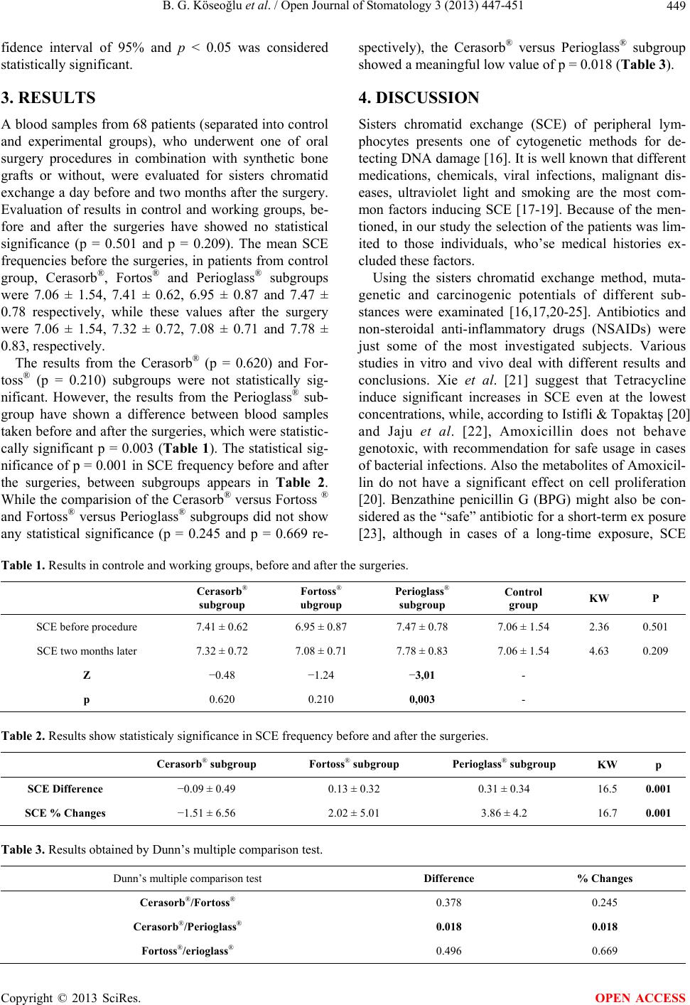 B. G. Köseoğlu et al. / Open Journal of Stomatology 3 (2013) 447-451 Copyright © 2013 SciRes. 449 OPEN ACCESS fidence interval of 95% and p < 0.05 was considered statistically significant. 3. RESULTS A blood samples from 68 patients (separated into control and experimental groups), who underwent one of oral surgery procedures in combination with synthetic bone grafts or without, were evaluated for sisters chromatid exchange a day before and two months after the surgery. Evaluation of results in control and working groups, be- fore and after the surgeries have showed no statistical significance (p = 0.501 and p = 0.209). The mean SCE frequencies before the surgeries, in patients from control group, Cerasorb®, Fortos® and Perioglass® subgroups were 7.06 ± 1.54, 7.41 ± 0.62, 6.95 ± 0.87 and 7.47 ± 0.78 respectively, while these values after the surgery were 7.06 ± 1.54, 7.32 ± 0.72, 7.08 ± 0.71 and 7.78 ± 0.83, respectively. The results from the Cerasorb® (p = 0.620) and For- toss® (p = 0.210) subgroups were not statistically sig- nificant. However, the results from the Perioglass® sub- group have shown a difference between blood samples taken before and after the surgeries, which were statistic- cally significant p = 0.003 (Table 1). The statistical sig- nificance of p = 0.001 in SCE frequency before and after the surgeries, between subgroups appears in Table 2. While the comparision of the Cerasorb® versus Fortoss ® and Fortoss® versus Perioglass® subgroups did not show any statistical significance (p = 0.245 and p = 0.669 re- spectively), the Cerasorb® versus Perioglass® subgroup showed a meaningful low value of p = 0.018 (Table 3). 4. DISCUSSION Sisters chromatid exchange (SCE) of peripheral lym- phocytes presents one of cytogenetic methods for de- tecting DNA damage [16]. It is well known that different medications, chemicals, viral infections, malignant dis- eases, ultraviolet light and smoking are the most com- mon factors inducing SCE [17-19]. Because of the men- tioned, in our study the selection of the patients was lim- ited to those individuals, who’se medical histories ex- cluded these factors. Using the sisters chromatid exchange method, muta- genetic and carcinogenic potentials of different sub- stances were examinated [16,17,20-25]. Antibiotics and non-steroidal anti-inflammatory drugs (NSAIDs) were just some of the most investigated subjects. Various studies in vitro and vivo deal with different results and conclusions. Xie et al. [21] suggest that Tetracycline induce significant increases in SCE even at the lowest concentrations, while, according to Istifli & Topaktaş [20] and Jaju et al. [22], Amoxicillin does not behave genotoxic, with recommendation for safe usage in cases of bacterial infections. Also the metabolites of Amoxicil- lin do not have a significant effect on cell proliferation [20]. Benzathine penicillin G (BPG) might also be con- sidered as the “safe” antibiotic for a short-term ex posure [23], although in cases of a long-time exposure, SCE Table 1. Results in controle and working groups, before and after the surgeries. Cerasorb® subgroup Fortoss® ubgroup Perioglass® subgroup Control group KW P SCE before procedure 7.41 ± 0.62 6.95 ± 0.87 7.47 ± 0.78 7.06 ± 1.54 2.36 0.501 SCE two months later 7.32 ± 0.72 7.08 ± 0.71 7.78 ± 0.83 7.06 ± 1.54 4.63 0.209 Z −0.48 −1.24 −3,01 - p 0.620 0.210 0,003 - Table 2. Results show statisticaly significance in SCE frequency before and after the surgeries. Cerasorb® subgroup Fortoss® subgroup Perioglass® subgroup KW p SCE Difference −0.09 ± 0.49 0.13 ± 0.32 0.31 ± 0.34 16.5 0.001 SCE % Changes −1.51 ± 6.56 2.02 ± 5.01 3.86 ± 4.2 16.7 0.001 Table 3. Results obtained by Dunn’s multiple comparison test. Dunn’s multiple comparison test Difference % Changes Cerasorb®/Fortoss® 0.378 0.245 Cerasorb®/Perioglass® 0.018 0.018 Fortoss®/erioglass® 0.496 0.669  B. G. Köseoğlu et al. / Open Journal of Stomatology 3 (2013) 447-451 450 frequency changes might be expected [24]. Non-steroidal anti-inflammatory drugs (NSAIDs) in- clude variety of chemicals with analgesic, antipyretic and anti-inflammatory effects [16]. In one of our previous studies, we examined genotoxic effects of the etodolac, nimesulid and naproxen sodium, and concluded that short term use of selective and non-selective NSAIDs was not associated with a significant genotoxic effect that could be detected using the SCE method in pe- ripheric lymphocytes [16]. However, findings from Özkul et al. [25], althought statistically insignificant were opposite to ours in case of naproxen sodium. The patients included in our study were under antibi- otic and analgesic therapy after the surgeries. Following the results of previously mentioned studies, Amoxicillin 2 gr/day (for 7 days) and Naproxen sodium 1100 mg/day (as needed) were prescribed. Due to the fact that all of the patients were under the same therapy, with changes of SCE frequency in working subgroups, the results of the study suggests that synthetic bone graft materials might be genotoxic. The results before and after the treatment have shown the statistical significance (p = 0.003) in the Perioglass® subgroup. Also, multiple subgroup comparison tests were statistically significant p = 0.001, while compari- son between subgroups have shown that a meaningful low value was in the Cerasorb® versus Perioglass® sub- group (p = 0.018). This finding suggests, that the bone augmentation with synthetic bone grafts might have an influence on the sisters chromatid exchange frequency. Although studies of bone grafts are limited to exami- nations of their biocompatibility, osteoinductive and os- teoconductive qualities by radiolographic, histologic or histomorphometric analysis, a review of the English lit- erature suggests that this could be the first study investi- gating a genotoxic influence of the synthetic bone grafts by sisters chromatide exchange method. Due to the fact that the examined materials in the previous studies have shown good results as the bone substitutes [26,27], we wonder whether the resorption rate of the materials play a role in their genotoxicity. A TCPs show bioresorbabil- ity during bone regeneration and completely substitute for a bone tissue after bone stimulation, while hydroxyl apatite (HA) remains in the body for a long time after replantation, due to a low grade of bioresorbability [26]. Low grade of resorption is also characteristic of bioac- tive glasses, where radiographic density and volume are more expressed in bone sites treated by these materials [27]. These senses might explain a meaningful low val- ues (p = 0.018) in the Cerasorb® (TCP) versus Pe- rioglass® (bioactive glass) and Cerasorb® versus For- toss® (TCP + HA) (p = 0.245), as compared with the results with the Fortoss® versus Perioglass® subgroups (p = 0.669). Although this is just hypothesis, because we did not look at the resorption rate of the materials, we are of the opinion that the bioresorption of the synthetic bone sub- stitutes did not have any influence on sisters chromatide exchange frequency, due to the fact that the blood sam- ples were taken two months after the bone augmentation, which is not long enough a period for the graft replace- ment with the authentic bone tissue. We hope that with these findings, we will open a gate to some other researchers and studies that will examinate the genotoxic influence of other bone synthetic substi- tutes. 5. CONCLUSION Although further investigations may be necessary, our results suggests that some alloplasts such as bioactive glass ceramic might have an influence on SCE in periph- eral lymphocytes. REFERENCES [1] Rojbani, H., Nyan, M., Ohya, K. and Kasugai, S. (2011) Evaluation of the osteoconductivity of α-tricalcium phos- phate, β-tricalcium phosphate, and hydroxyapatite com- bined with or without simvastatin in rat calvarial defect. Journal of Biomedical Materials Research Part A, 98A, 488-498. http://dx.doi.org/10.1002/jbm.a.33117 [2] Brugnami, F., Caiazzo, A. and Leone, C. (2009) Local intraoral autologous bone harvesting for dental implant treatment: Alternative sources and criteria of choice. The Keio Journal of Medicine, 58, 24-28. http://dx.doi.org/10.2302/kjm.58.24 [3] Wheeler, D.L., Stokes, K.E., Hoellrich, R.G., Chamber- land, D.L. and McLoughlin, S.W. (1998) Effect of bio- active glass particle size on osseous regeneration of cancellous defects. Journal of Biomedical Materials Re- search, 41, 527-533. http://dx.doi.org/10.1002/(SICI)1097-4636(19980915)41: 4<527::AID-JBM3>3.0.CO;2-E [4] Kaptein, M.L., de Putter, C., de Lange, G.L. and Blijdorp, P.A. (1998) Survival of cylindrical implants in composite grafted maxillary sinuses. Journal of Oral and Maxillo- facial Surgery, 56, 1376-1380. http://dx.doi.org/10.1016/S0278-2391(98)90396-2 [5] Tadjoedin, E.S., de Lange, G.L., Lyaruu, D.M., Kuiper, L. and Burger, E.H. (2002) High concentrations of bioactive glass material (BioGran) vs. autogenous bone for sinus floor elevation. Clinical Oral Implants Research, 13, 428-436. http://dx.doi.org/10.1034/j.1600-0501.2002.130412.x [6] Wetzel, A.C., Stich, H. and Caffesse, R.G. (1995) Bone apposition onto oral implants in the sinus area filled with different grafting materials. A histological study in beagle dogs. Clinical Oral Implants Research, 6, 155-163. http://dx.doi.org/10.1034/j.1600-0501.1995.060304.x [7] Hürzeler, M.B., Kirsch, A., Ackermann, K.L. and Qui- ñones, CR. (1996) Reconstruction of the severely re- Copyright © 2013 SciRes. OPEN ACCESS  B. G. Köseoğlu et al. / Open Journal of Stomatology 3 (2013) 447-451 451 sorbed maxilla with dental implants in the augmented maxillary sinus: A 5-year clinical investigation. Interna- tional Journal of Oral & Maxillofacial Implants, 11, 466- 475. [8] Xynos, I.D., Edgar, A.J., Buttery, L.D.K., Hench, L.L. and Polak, M. (2001) Gene expression profiling of human osteoblasts following treatment with the ionic products of BioglassR 45S5 dissolution. Journal of Biomedical Ma- terials Research, 55, 151-157. http://dx.doi.org/10.1002/1097-4636(200105)55:2<151:: AID-JBM1001>3.0.CO;2-D [9] Hench, L.L. and Paschall, H.A. (1973) Direct chemical bond of bioactive glass-ceramic materials to bone and muscle. Journal of Biomedical Materials Research, 7, 25-42. http://dx.doi.org/10.1002/jbm.820070304 [10] Hench, L.L., Andersson, O.H. and LaTorre, G.P. (1991) The kinetics of bioactive ceramics part III: Surface reactions for bioactive glasses compared with an inactive glass. Bioceramics, 4, 156-162. [11] Aichelmann-Reidy, M.E. and Yukna, R.A. (1998) Bone replacement grafts. The bone substitutes. Dental Clinics of North America, 42, 491-503. [12] Albrektsson, T. and Johansson, C. (2001) Osteoinduction, osteoconduction and osseointegration. European Spine Journal, 10, S96-101. http://dx.doi.org/10.1007/s005860100282 [13] Schepers, E., Declercq, M., Ducheyne, P. and Kem- peneers, R. (1991) Bioactive glass particulate material as a filler for bone lesions. Journal of Oral Rehabilitation, 18, 439-452. http://dx.doi.org/10.1111/j.1365-2842.1991.tb01689.x [14] Ducheyne, P. (1999) Effect of bioactive glass particle size on osseous regeneration. Journal of Biomedical Materials Research, 46, 301-304. http://dx.doi.org/10.1002/(SICI)1097-4636(199908)46:2< 301::AID-JBM20>3.0.CO;2-# [15] Nyan, M., Sato, D., Kihara, H., Machida, T., Ohya, K. and Kasugai, S. (2009) Effects of the combination with alpha-tricalcium phosphate and simvastatin on bone regeneration. Clinical Oral Implants Research, 20, 280- 287. http://dx.doi.org/10.1111/j.1600-0501.2008.01639.x [16] Koseoglu, B.G., Ozturk, S., Kocak, H., Palanduz, S. and Cefle, K. (2008) The effects of etodolac, nimesulid and naproxen sodium on the frequency of sister chromatid ex- change after enclused third molars surgery. Yonsei Medi- cal Journal, 49, 742-747. http://dx.doi.org/10.3349/ymj.2008.49.5.742 [17] Oztürk, S., Köseoglu, B.G., Koçak, H., Palanduz, S., Cefle, K. and Erkal, H. (2004) In vitro effects of selective and non-selective nonsteroidal anti-inflammatory drugs on the frequency of sister chromatid exchanges. Drugs in R&D, 5, 327-330. http://dx.doi.org/10.2165/00126839-200405060-00003 [18] Carbonell, E., Peris, F., Xamena, N., Creus, A. and Marcos, R. (1995) SCE analysis in human lymphocytes of a Spanish control population. Drugs in R&D, 335, 35-46. http://dx.doi.org/10.1016/0165-1161(95)90062-4 [19] Sahin, A., Tatar, A., Oztas, S., Seven, B., Varoglu, E., Yesilyurt, A. and Ayan, AK. (2009) Evaluation of the genotoxic effects of chronic low-dose ionizing radiation exposure on nuclear medicine workers. Nuclear Medicine and Biology, 36, 575-578. http://dx.doi.org/10.1016/j.nucmedbio.2009.02.003 [20] Istifli, E.S. and Topaktaş, M. (2010) Cytogenetic gen- otoxicity of amoxicillin. Environmental and Molecular Mutagenesis, 51, 222-228. [21] Xie, X., Zhou, Q., Bao, Q., He, Z. And Bao, Y. (2011) Genotoxicity of tetracycline as an emerging pollutant on root meristem cells of wheat (Triticum aestivum L.). Environmental Toxicology, 26, 417-423. http://dx.doi.org/10.1002/tox.20567 [22] Jaju, M., Jaju, M. and Ahuja, Y.R. (1984) Evaluation of genotoxicity of ampicillin and carbenicillin on human lymphocytes in vitro: Chromosome aberrations, mitotic index, cell cycle kinetics, satellite associations of acro- centric chromosomes and sister chromatid exchanges. Human Toxicology, 3, 173-191. http://dx.doi.org/10.1177/096032718400300303 [23] Köseoglu, V., Kismet, E., Soysal, Y., Ulucan, H., Dün- daröz, R., Imirzalioglu, N. and Gökçay, E. (2004) Investi- gation of DNA damage in lymphocytes exposed to ben- zathine penicillin G. Pediatrics International, 46, 415- 418. http://dx.doi.org/10.1111/j.1442-200x.2004.01930.x [24] Dündaröz, R., Ozisik, T., Baltaci, V., Karapinar, K., Aydin, H.I. and Denli, M. (2001) Sister-chromatid ex- change analysis on long-term benzathine penicillin for secondary prophylaxis of rheumatic fever. Indian Journal of Pediatrics, 68, 121-122. http://dx.doi.org/10.1007/BF02722027 [25] Ozkul, Y., Erenmemisoglu, A., Ekecik, A., Saatci, C., Ozdamar, S. and Demirtas, H. (1996) Do non-steroidal anti-inflammatory drugs induce sister chromatid exchanges in T lymphocytes? Journal of International Medical Re- search, 24, 84-87. [26] Kamitakahara, M., Ohtsuki, C. and Miyazaki, T. (2008) Review paper: Behavior of ceramic biomaterials derived from tricalcium phosphate in physiological condition. Journal of Biomaterials Applications, 23, 197-212. http://dx.doi.org/10.1177/0885328208096798 [27] Subbaiah, R. and Thomas, B. (2011) Efficacy of a bio- active alloplast, in the treatment of human periodontal osseous defects-a clinical study. Medicina Oral, Pato- logía Oral y Cirugía Bucal, 16, e239-244. http://dx.doi.org/10.4317/medoral.16.e239 Copyright © 2013 SciRes. OPEN ACCESS |