

Paper Menu >>
Journal Menu >>
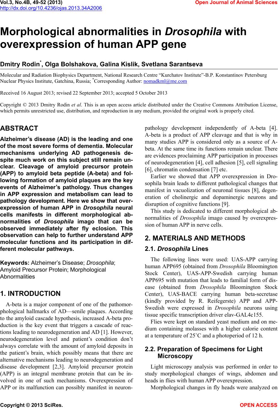 Vol.3, No.4B, 49-52 (2013) Open Journal of Animal Sciences http://dx.doi.org/10.4236/ojas.2013.34A2006 Morphological abnormalities in Drosophila with overexpression of human APP gene Dmitry Rodin*, Olga Bolshakova, Galina Kislik, Svetlana Sarantseva Molecular and Radiation Biophysics Department, National Research Centre “Kurchatov Institute”-B.P. Konstantinov Petersburg Nuclear Physics Institute, Gatchina, Russia; *Corresponding Author: [email protected] Received 16 August 2013; revised 22 September 2013; accepted 5 October 2013 Copyright © 2013 Dmitry Rodin et al. This is an open access article distributed under the Creative Commons Attribution License, which permits unrestricted use, distribution, and reproduction in any medium, provided the original work is properly cited. ABSTRACT Alzheimer ’s disease (AD) is the leading and one of the most severe forms of dementia. Molecular mechanisms underlying AD pathogenesis de- spite much work on this subject still remain un- clear. Cleavage of amyloid precursor protein (APP) to amyloid beta peptide (A-beta) and fol- lowing formation of amyloid plaques are the key events of Alzheimer’s pathology. Thus changes in APP expression and metabolism can lead to patholog y development. Here we show that over- expression of human APP in Drosophila neural cells manifests in different morphological ab- normalities of Drosophila imago that can be observed immediately after fly eclosion. This observation can help to further understand APP molecular functions and its participation in dif- ferent molecular pathways. Keywords: Alzheimer’s Disease; Drosophila; Amyloid Precursor Protein; Morphological Abnormalities 1. INTRODUCTION A-beta is a major component of one of the pathomor- phological hallmarks of AD—senile plaques. According to the amyloid cascade hypothesis, increased A-beta pro- duction is the key event that triggers a cascade of reac- tions leading to neurodegeneration and AD [1]. However, neurodegeneration level and patient’s condition don’t always correlate with the amount of amyloid deposits in the patient’s brain, which possibly means that there are alternative mechanisms leading to neurodegeneration and disease development [2,3]. Amyloid precursor protein (APP) is an integral membrane protein that can be in- volved in one of such mechanisms. Overexpression of APP or its malfunction can possibly manifest in neuron- pathology development independently of A-beta [4]. A-beta is a product of APP cleavage and that is why in many studies APP is considered only as a source of A- beta. At the same time its functions remain unclear. There are evidences proclaiming APP participation in processes of neurodegeneration [4], cell adhesion [5], cell signaling [6], chromatin condensation [7] etc. Earlier we showed that APP overexpression in Dro- sophila brain leads to different pathological changes that manifest in vacuolization of neuronal tissues [8], degen- eration of cholinergic and dopaminergic neurons and disruption of cognitive functions [9]. This study is dedicated to different morphological ab- normalities of Dros ophila imago caused by overexpres- sion of human APP in nerve cells. 2. MATERIALS AND METHODS 2.1. Drosophila Lines The following lines were used: UAS-APP carrying human APP695 (obtained from Drosophila Bloomington Stock Center), UAS-APP-Swedish carrying human APP695 with mutation that leads to familial form of dis- ease (obtained from Drosophila Bloomington Stock Center), UAS-BACE carrying human beta-secretase (kindly provided by R. Reifegerste) APP and APP- Swedish were expressed in Drosophila neurons using tissue specific transcription driver elav-GAL4c155. Flies were kept on standard yeast medium and on me- dium containing molasses with a higher calorie content at a temperature of 25˚C and a photoperiod of 12 h. 2.2. Preparation of Specimens for Light Microscopy Light microscopy analysis was performed in order to study morphological changes of wings, abdomen and heads in flies with human APP overexpression. Morphological changes in fly heads were analyzed on Copyright © 2013 SciRes. OPEN ACCESS 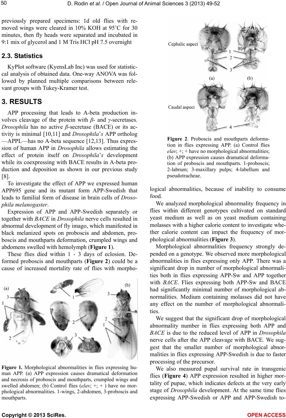 D. Rodin et al. / Open Journal of Animal Sciences 3 (2013) 49-5 2 50 previously prepared specimens: 1d old flies with re- moved wings were cleared in 10% KOH at 95˚C for 30 minutes, then fly heads were separated and incubated in 9:1 mix of glycerol and 1 M Tris HCl pH 7.5 overnight 2.3. Statistics KyPlot software (KyensLab Inc) was used for statistic- cal analysis of obtained data. One-way ANOVA was fol- lowed by planned multiple comparisons between rele- vant groups with Tukey-Kramer test. 3. RESULTS APP processing that leads to A-beta production in- volves cleavage of the protein with β- and γ-secretases. Drosophila has no active β-secretase (BACE) or its ac- tivity is minimal [10,11] and Dro sophila’s APP ortholog —APPL—has no A-beta sequence [12,13]. Thus expres- sion of human APP in Drosophila allows estimating the effect of protein itself on Drosophila’s development while its coexpressing with BACE results in A-beta pro- duction and deposition as shown in our previous study [8]. To investigate the effect of APP we expressed human APP695 gene and its mutant form APP-Swedish that leads to familial form of disease in brain cells of Droso- phila melanogaster. Expression of APP and APP-Swedish separately or together with BACE in Drosophila nerve cells resulted in abnormal development of fly imago, which manifested in black melanized spots on proboscis and abdomen, pro- boscis and mouthparts deformation, crumpled wings and abdomens swelled with hemolymph (Figur e 1). These flies died within 1 - 3 days of eclosion. De- formed proboscis and mouthparts (Figure 2) could be a cause of increased mortality rate of flies with morpho- Figure 1. Morphological abnormalities in flies expressing hu- man APP. (a) APP expression causes dramatical deformation and necrosis of proboscis and mouthparts, crumpled wings and swelled abdomen; (b) Control flies (elav; +; + ) have no mor- phological abnormalities. 1-wings, 2-abdomen, 3-proboscis and mouthparts. Figure 2. Proboscis and mouthparts deforma- tion in flies expressing APP. (a) Control flies elav; +; + have no morphological abnormalities; (b) APP expression causes dramatical deforma- tion of proboscis and mouthparts. 1-proboscis; 2-labrum; 3-maxillary pulps; 4-labellum and pseudotracheae. logical abnormalities, because of inability to consume food. We analyzed morphological abnormality frequency in flies within different genotypes cultivated on standard yeast medium as well as on yeast medium containing molasses with a higher calorie content to investigate whe- ther calorie content can impact the frequency of mor- phological abnormalities (Figure 3). Morphological abnormalities frequency strongly de- pended on a genotype. We observed more morphological abnormalities in flies expressing only APP. There was a significant drop in number of morphological abnormali- ties both in flies expressing APP-Sw and APP together with BACE. Flies expressing both APP-Sw and BACE had significantly minimal number of morphological ab- normalities. Medium containing molasses did not have any effect on the number of morphological abnormali- ties. We suggest that the significant drop of morphological abnormality number in flies expressing both APP and BACE is due to the reduced level of APP in Drosophila nerve cells after the APP cleavage with BACE. We sug- gest that the smaller number of morphological abnor- malities in flies expressing APP-Swedish is due to faster processing of the precursor. We also measured pupal survival rate in transgenic flies (Figure 4) APP expression resulted in higher mor- tality of pupae, which indicates defects at the very early stage of Drosophila development. At the same time flies expressing APP-Swedish or APP and APP-Swedish to- Copyright © 2013 SciRes. OPEN ACCESS 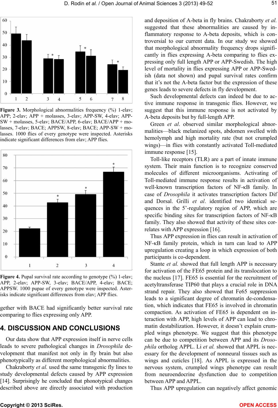 D. Rodin et al. / Open Journal of Animal Sciences 3 (2013) 49-5 2 51 Figure 3. Morphological abnormalities frequency (%) 1-elav; APP; 2-elav; APP + molasses, 3-elav; APP-SW, 4-elav; APP- SW + molasses, 5-elav; BACE/APP, 6-elav; BACE/APP + mo- lasses, 7-elav; BACE; APPSW, 8-elav; BACE; APP-SW + mo- lasses. 1000 flies of every genotype were inspected. Asterisks indicate significant differences from elav; APP flies. Figure 4. Pupal survival rate according to genotype (%) 1-elav; APP, 2-elav; APP-SW, 3-elav; BACE/APP, 4-elav; BACE; APPSW. 1000 pupae of every genotype were inspected. Aster- isks indicate significant differences from elav; APP flies. gether with BACE had significantly better survival rate comparing to flies expressing only APP. 4. DISCUSSION AND CONCLUSIONS Our data show that APP expression itself in nerve cells leads to severe pathological changes in Drosophila de- velopment that manifest not only in fly brain but also phenotypically as different morphological abnormalities. Chakraborty et al. used the same transgenic fly lines to study developmental defects caused by APP expression [14]. Surprisingly he concluded that phonotypical changes described above are directly associated with production and deposition of A-beta in fly brains. Chakraborty et al. suggested that these abnormalities are caused by in- flammatory response to A-beta deposits, which is con- troversial to our current data. In our study we showed that morphological abnormality frequency drops signifi- cantly in flies expressing A-beta comparing to flies ex- pressing only full length APP or APP-Swedish. The high level of mortality in flies expressing APP or APP-Swed- ish (data not shown) and pupal survival rates confirm that it’s not the A-beta factor but the expression of these genes leads to severe defects in fly development. Such developmental defects can indeed be due to ac- tive immune response in transgenic flies. However, we suggest that this immune response is not activated by A-beta deposits but by full-length APP. Green et al. observed similar morphological abnor- malities—black melanized spots, abdomen swelled with hemolymph and high mortality rate (but not crumpled wings)—in flies with constantly activated Toll-mediated immune response [15]. Toll-like receptors (TLR) are a part of innate immune system. Their main function is to recognize conserved molecules of different microorganisms. Activating of Toll-mediated immune response results in activation of well-known transcription factors of NF-κB family. In case of Drosophila it activates transcription factors Dif and Dorsal. Grilli et al. identified two identical se- quences in the 5’-regulatory region of APP, which are specific binding sites for transcription factors of NF-κB family. They also showed that activity of these sites cor- relates with APP expression [16]. Thus APP expression in flies can result in activation of NF-κB family protein, which in turn can lead to APP upregulation creating a loop in which expression of both participants is co-dependent. Stante et al. showed that full length APP is necessary for activation of the FE65 protein and its translocation to the nucleus [17]. FE65 is essential for the recruitment of acetyltransferase TIP60 that plays a crucial role in DNA strand repair. They also showed that Fe65 suppression leads to a significant degree of chromatin de-condensa- tion, which indicates that FE65 is involved in chromatin compaction. As activation of FE65 is dependent on in- teraction with APP, high levels of APP can lead to chro- matin destabilization. However, it doesn’t explain crum- pled wings phenotype. We suggest that this phenotype can be due to competition between APP and its Droso- phila ortholog APPL. Li et al. showed that APPL is nec- essary for the development of nonneural tissues such as wings and cuticles [18]. As APPL is expressed in the nervous system, crumpled wings phenotype can result from neuroendocrine dysfunction due to competition between APP and APPL. Thus APP upregulation can negatively affect genomic Copyright © 2013 SciRes. OPEN ACCESS 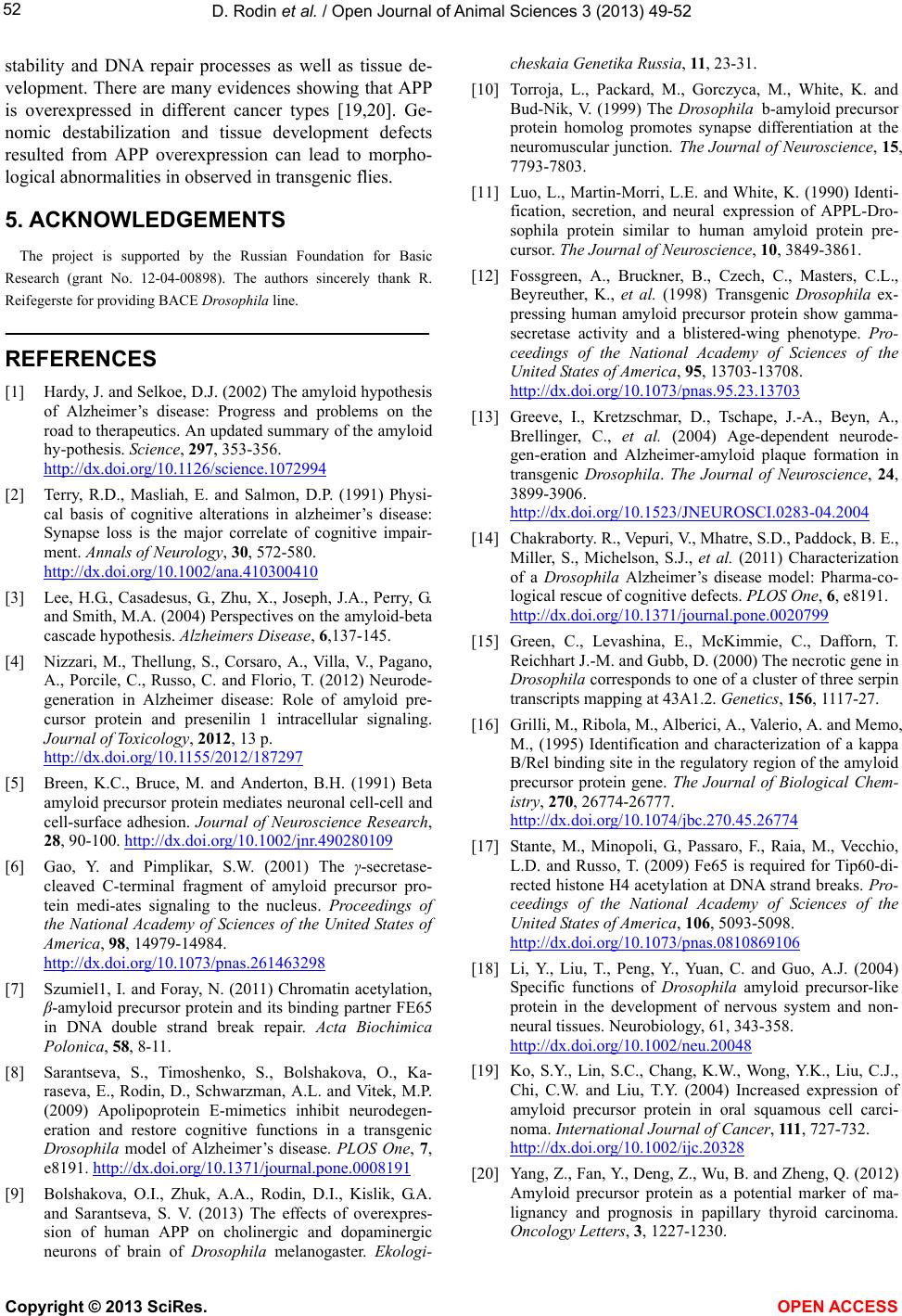 D. Rodin et al. / Open Journal of Animal Sciences 3 (2013) 49-5 2 Copyright © 2013 SciRes. 52 stability and DNA repair processes as well as tissue de- velopment. There are many evidences showing that APP is overexpressed in different cancer types [19,20]. Ge- nomic destabilization and tissue development defects resulted from APP overexpression can lead to morpho- logical abnormalities in observed in transgenic flies. OPEN A CCESS 5. ACKNOWLEDGEMENTS The project is supported by the Russian Foundation for Basic Research (grant No. 12-04-00898). The authors sincerely thank R. Reifegerste for providing BACE Drosophila line. REFERENCES [1] Hardy, J. and Selkoe, D.J. (2002) The amyloid hypothesis of Alzheimer’s disease: Progress and problems on the road to therapeutics. An updated summary of the amyloid hy-pothesis. Science, 297, 353-356. http://dx.doi.org/10.1126/science.1072994 [2] Terry, R.D., Masliah, E. and Salmon, D.P. (1991) Physi- cal basis of cognitive alterations in alzheimer’s disease: Synapse loss is the major correlate of cognitive impair- ment. Annals of Neurology, 30, 572-580. http://dx.doi.org/10.1002/ana.410300410 [3] Lee, H.G., Casadesus, G., Zhu, X., Joseph, J.A., Perry, G. and Smith, M.A. (2004) Perspectives on the amyloid-beta cascade hypothesis. Alzheimers Disease, 6,137-145. [4] Nizzari, M., Thellung, S., Corsaro, A., Villa, V., Pagano, A., Porcile, C., Russo, C. and Florio, T. (2012) Neurode- generation in Alzheimer disease: Role of amyloid pre- cursor protein and presenilin 1 intracellular signaling. Journal of Toxicology, 2012, 13 p. http://dx.doi.org/10.1155/2012/187297 [5] Breen, K.C., Bruce, M. and Anderton, B.H. (1991) Beta amyloid precursor protein mediates neuronal cell-cell and cell-surface adhesion. Journal of Neuroscience Research, 28, 90-100. http://dx.doi.org/10.1002/jnr.490280109 [6] Gao, Y. and Pimplikar, S.W. (2001) The γ-secretase- cleaved C-terminal fragment of amyloid precursor pro- tein medi-ates signaling to the nucleus. Proceedings of the National Academy of Sciences of the United States of America, 98, 14979-14984. http://dx.doi.org/10.1073/pnas.261463298 [7] Szumiel1, I. and Foray, N. (2011) Chromatin acetylation, β-amyloid precursor protein and its binding partner FE65 in DNA double strand break repair. Acta Biochimica Polonica, 58, 8-11. [8] Sarantseva, S., Timoshenko, S., Bolshakova, O., Ka- raseva, E., Rodin, D., Schwarzman, A.L. and Vitek, M.P. (2009) Apolipoprotein E-mimetics inhibit neurodegen- eration and restore cognitive functions in a transgenic Drosophila model of Alzheimer’s disease. PLOS One, 7, e8191. http://dx.doi.org/10.1371/journal.pone.0008191 [9] Bolshakova, O.I., Zhuk, A.A., Rodin, D.I., Kislik, G.A. and Sarantseva, S. V. (2013) The effects of overexpres- sion of human APP on cholinergic and dopaminergic neurons of brain of Drosophila melanogaster. Ekologi- cheskaia Genetika Russia, 11, 23-31. [10] Torroja, L., Packard, M., Gorczyca, M., White, K. and Bud-Nik, V. (1999) The Drosophila b-amyloid precursor protein homolog promotes synapse differentiation at the neuromuscular junction. The Journal of Neuroscience, 15, 7793-7803. [11] Luo, L., Martin-Morri, L.E. and White, K. (1990) Identi- fication, secretion, and neural expression of APPL-Dro- sophila protein similar to human amyloid protein pre- cursor. The Journal of Neuroscience, 10, 3849-3861. [12] Fossgreen, A., Bruckner, B., Czech, C., Masters, C.L., Beyreuther, K., et al. (1998) Transgenic Drosophila ex- pressing human amyloid precursor protein show gamma- secretase activity and a blistered-wing phenotype. Pro- ceedings of the National Academy of Sciences of the United States of America, 95, 13703-13708. http://dx.doi.org/10.1073/pnas.95.23.13703 [13] Greeve, I., Kretzschmar, D., Tschape, J.-A., Beyn, A., Brellinger, C., et al. (2004) Age-dependent neurode- gen-eration and Alzheimer-amyloid plaque formation in transgenic Drosophila. The Journal of Neuroscience, 24, 3899-3906. http://dx.doi.org/10.1523/JNEUROSCI.0283-04.2004 [14] Chakraborty. R., Vepuri, V., Mhatre, S.D., Paddock, B. E., Miller, S., Michelson, S.J., et al. (2011) Characterization of a Drosophila Alzheimer’s disease model: Pharma-co- logical rescue of cognitive defects. PLOS One, 6, e8191. http://dx.doi.org/10.1371/journal.pone.0020799 [15] Green, C., Levashina, E., McKimmie, C., Dafforn, T. Reichhart J.-M. and Gubb, D. (2000) The necrotic gene in Drosophila corresponds to one of a cluster of three serpin transcripts mapping at 43A1.2. Genetics, 156, 1117-27. [16] Grilli, M., Ribola, M., Alberici, A., Valerio, A. and Memo, M., (1995) Identification and characterization of a kappa B/Rel binding site in the regulatory region of the amyloid precursor protein gene. The Journal of Biological Chem- istry, 270, 26774-26777. http://dx.doi.org/10.1074/jbc.270.45.26774 [17] Stante, M., Minopoli, G., Passaro, F., Raia, M., Vecchio, L.D. and Russo, T. (2009) Fe65 is required for Tip60-di- rected histone H4 acetylation at DNA strand breaks. Pro- ceedings of the National Academy of Sciences of the United States of America, 106, 5093-5098. http://dx.doi.org/10.1073/pnas.0810869106 [18] Li, Y., Liu, T., Peng, Y., Yuan, C. and Guo, A.J. (2004) Specific functions of Drosophila amyloid precursor-like protein in the development of nervous system and non- neural tissues. Neurobiology, 61, 343-358. http://dx.doi.org/10.1002/neu.20048 [19] Ko, S.Y., Lin, S.C., Chang, K.W., Wong, Y.K., Liu, C.J., Chi, C.W. and Liu, T.Y. (2004) Increased expression of amyloid precursor protein in oral squamous cell carci- noma. International Journal of Cancer, 111, 727-732. http://dx.doi.org/10.1002/ijc.20328 [20] Yang, Z., Fan, Y., Deng, Z., Wu, B. and Zheng, Q. (2012) Amyloid precursor protein as a potential marker of ma- lignancy and prognosis in papillary thyroid carcinoma. Oncology Letters, 3, 1227-1230. |