

Paper Menu >>
Journal Menu >>
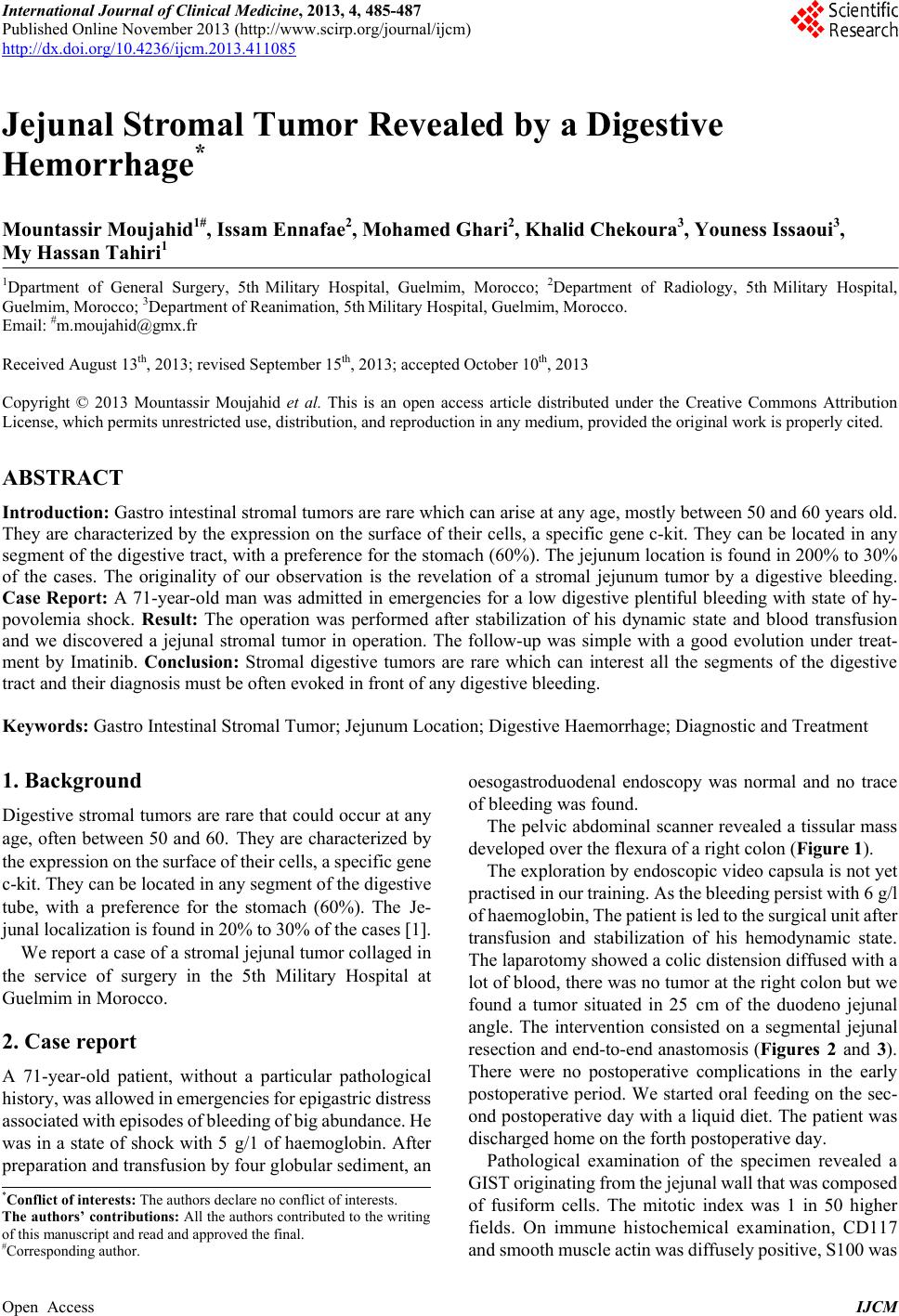 International Journal of Clinical Medicine, 2013, 4, 485-487 Published Online November 2013 (http://www.scirp.org/journal/ijcm) http://dx.doi.org/10.4236/ijcm.2013.411085 Open Access IJCM 485 Jejunal Stromal Tumor Revealed by a Digestive Hemorrhage* Mountassir Moujahid1#, Issam Ennafae2, Mohamed Ghari2, Khalid Chekoura3, Youness Issaoui3, My Hassan Tahiri1 1Dpartment of General Surgery, 5th Military Hospital, Guelmim, Morocco; 2Department of Radiology, 5th Military Hospital, Guelmim, Morocc o ; 3Department of Reanimation, 5th Military Hospital, Guelmim, Morocco. Email: #[email protected] Received August 13th, 2013; revised September 15th, 2013; accepted October 10th, 2013 Copyright © 2013 Mountassir Moujahid et al. This is an open access article distributed under the Creative Commons Attribution License, which permits unrestricted use, distribution, and reproduction in any medium, provided the original work is properly cited. ABSTRACT Introduction: Gastro intestinal stromal tumors are rare which can arise at any age, mostly between 50 and 60 years old. They are characterized by the expression on the surface of their cells, a specific gene c-kit. They can be located in any segment of the digestive tract, with a preference for the stomach (60%). The jejunum location is found in 200% to 30% of the cases. The originality of our observation is the revelation of a stromal jejunum tumor by a digestive bleeding. Case Report: A 71-year-old man was admitted in emergencies for a low digestive plentiful bleeding with state of hy- povolemia shock. Result: The operation was performed after stabilization of his dynamic state and blood transfusion and we discovered a jejunal stromal tumor in operation. The follow-up was simple with a good evolution under treat- ment by Imatinib. Conclusion: Stromal digestive tumors are rare which can interest all the segments of the digestive tract and their diagnosis must be often evoked in front of any digestive bleeding. Keywords: Gastro Intestinal Stromal Tumor; Jejunum Location; Digestive Haemorrhage; Diagnostic and Treatment 1. Background Digestive stromal tumors are rare that could occur at any age, often between 50 and 60. They are characterized by the expression on the s urface of their cells, a specific gene c-kit. They can be located in any segment of the digestive tube, with a preference for the stomach (60%). The Je- junal localization is found in 20% to 30% of the cases 1. We report a case of a stromal jejunal tumor collaged in the service of surgery in the 5th Military Hospital at Gu e lmim i n Mor occ o. 2. Case report A 71-year-old patient, without a particular pathological history, was allowed in emergencies for epigastric distress associated with episodes of bleedi ng of big ab undance. He was in a state of shock with 5 g/1 of haemoglobin. After preparation and transfusion by four globular sediment, an oesogastroduodenal endoscopy was normal and no trace of bleeding was found. The pelvic abdominal scanner revealed a tissular mass developed over the flexura o f a rig ht col o n (Figure 1). The exploration by endoscopic video capsula is not yet practised in our training. As the bleeding persist with 6 g/l of haemoglobin, The patient is led to the surgical unit after transfusion and stabilization of his hemodynamic state. The laparotomy showed a colic distension diffused with a lot of blood, there was no tumor at the right colon but we found a tumor situated in 25 cm of the duodeno jejunal angle. The intervention consisted on a segmental jejunal resectio n and en d-to-end anastomo sis ( Figures 2 and 3). There were no postoperative complications in the early postoperative period. We started oral feeding on the sec- ond postoperative day with a liquid diet. The patient was discharged home on the forth postoperative day. Pathological examination of the specimen revealed a GIST originating from the jejunal wall that was composed of fusiform cells. The mitotic index was 1 in 50 higher fields. On immune histochemical examination, CD117 and smooth muscle actin was diffusely positive, S100 was *Conflict of interests: The authors declare no conflict of interests. The authors’ contributions: All the authors contributed to the writing of this ma nuscript and read and a pproved the final. #Corresponding author. 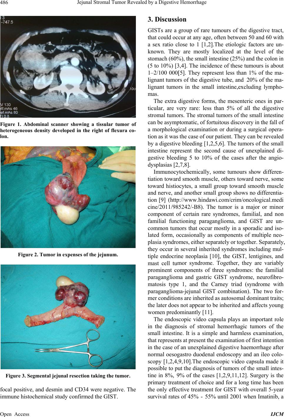 Jejunal Stromal Tumor Revealed by a Dige sti ve Hemorrha g e 486 Figure 1. Abdominal scanner showing a tissular tumor of heterogeneous density developed in the right of flexura co- lon. Figure 2. Tumor in expenses of the jejunum. Figure 3. Segmental jejunal resection taking the tumor. focal positive, and desmin and CD34 were negative. The immune histochemical study confirmed the GIST. 3. Discussion GISTs are a group of rare tumours of the digestive tract, that could occur at any age, often between 50 and 60 with a sex ratio close to 1 [1,2.The etiologic factors are un- known. They are mostly localized at the level of the stomach (60%), the small intestine (25%) and the colon in (5 to 10%) [3,4. The incidence of these tumours is about 1–2/100 0005. They represent less than 1% of the ma- lignant tumors of the digestive tube, and 20% of the ma- lignant tumors in the small intestine,excluding lympho- mas. The extra digestive forms, the mesenteric ones in par- ticular, are very rare: less than 5% of all the digestive stromal tumors. The stromal tumors of the small intestine can be asymptomatic, of fortuitous discovery in the fall of a morphological examination or during a surgical opera- tion as it was the case of our patient. They can be revealed by a digestive bleeding [1,2,5,6]. The tumors of the small intestine represent the second cause of unexplained di- gestive bleeding 5 to 10% of the cases after the angio- dysplasias [2,7,8]. Immunocytochemically, some tumours show differen- tiation toward smooth muscle, others toward nerve, some toward histiocytes, a small group toward smooth muscle and nerve, and another small group shows no differentia- tion [9] (http://www.hindawi.com/crim/oncological.medi cine/2011/985242/-B8). The tumor is a major or minor component of certain rare syndromes, familial, and non familial functioning paraganglioma, and GIST are un- common tumors that occur mostly in a sporadic and iso- lated form, occasionally as components of multiple neo- plasia syndromes, either separately or together. Separately, they occur in several inherited syndromes including mul- tiple endocrine neoplasia [10], the GIST, lentigines, and mast cell tumor syndrome. Together, they are variably prominent components of three syndromes: the familial paraganglioma and gastric GIST syndrome, neurofibro- matosis type 1, and the Carney triad (syndrome with paraganglioma-jejunal GIST combination). The two for- mer conditions are inherited as autosomal dominant traits; the later does not appear to be inherited and affects young women predominantly [11]. The endoscopic video capsula plays an important role in the diagnosis of stromal hemorrhagic tumors of the small intestine. It is a simple and harmless examination, that represents at present the examination of first intention in the case of an unexplain ed digestiv e h aemorrhage after normal oesogastro duodenal endoscopy and an ileo colo- scopy 1,2,4,9,10.The endoscopic video capsula made it possible to put the diagnosis of tumors of the small intes- tine in 8%, 9% of the cases [1,2,9,11,12]. Surgery is the primary treatment of choice and for a long time has been the only effective treatment for GIST with overall 5-year survival rates of 45% - 55% until 200 1 when Imatinib, a Open Access IJCM 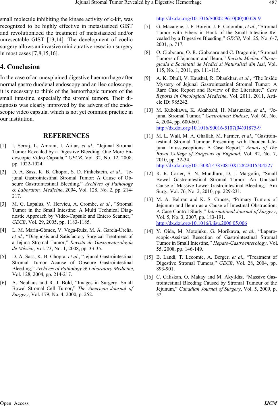 Jejunal Stromal Tumor Revealed by a Dige sti ve Hemorrha g e Open Access IJCM 487 small molecule inhibiting the kinase activity of c-k it, was recognized to be highly effective in metastasized GIST and revolutionized the treatment of metastasized and/or unresectable GIST [13,14]. The development of coelio surgery allows an invasive mini curative resection surgery in most cases [7,8,15,16]. 4. Conclusion In the case of an unexplained digestive haemorrhage after norma l ga str o du o denal e nd os co py and a n il eo col osc o py, it is necessary to think of the hemorrhagic tumors of the small intestine, especially the stromal tumors. Their di- agnosis was clearly improved by the advent of the endo- scopic vide o capsula, which i s not yet c ommon practice in our institution. REFERENCES [1] I. Serraj, L. Amrani, I. Atitar, et al., “Jejunal Stromal Tumor Revealed by a Digestive Bleeding: One More En- doscopic Video Capsula,” GECB, Vol. 32, No. 12, 2008, pp. 1022-1024. [2] D. A. Sass, K. B. Chopra, S. D. Finkelstein, et al., “Je- junal Gastrointestinal Stromal Tumor: A Cause of Ob- scure Gastrointestinal Bleeding,” Archives of Pathology & Laboratory Medicine, 2004, Vol. 128, No. 2, pp. 214- 217. [3] M. G. Lapalus, V. Hervieu, A. Crombe, et al., “Stromal Tumor in the Small Intestine: A Multi Technical Diag- nostic Approach by Video-Capsule and Entero Scanner,” GECB, Vol. 29, 2005, pp. 1183-1185. [4] L. M. Marín-Gómez, V. Vega-Ruiz, M. A. García-Ureña, et al., “Diagnosis and Satisfactory Surgical Treatment of a Jejuna Stromal Tumor,” Revista de Gastroenterología de México, Vol. 73, No. 1, 2008, pp. 33-35. [5] D. A. Sass, K. B. Chopra, et al., “Jejunal Gastrointestinal Stromal Tumor Acause of Obscure Gastrointestinal Bleeding,” Archives of Pathology & Laboratory Medicine, Vol. 128, 2004, pp. 214-217. [6] A. Neuhaus and R. J. Bold, “Images in Surgery. Small Bowel Stromal Cell Tumor,” The American Journal of Surgery, Vol. 179, No. 4, 2000, p. 252. http://dx.doi.org/10.1016/S0002-9610(00)00329-9 [7] G. Macaigne, J. F. Boivin, J. P. Colombu, et al., “Stromal Tumor with Fibers in Hank of the Small Intestine Re- vealed by a Digestive Bleeding,” GECB, Vol. 25, No. 6-7, 2001, p. 717. [8] O. Ciobotaru, O. R. Ciobotaru and C. Dragomir, “Stromal Tumors of Jejunaum and Ileum,” Revista Medico Chirur- gicala a Societatii de Medici si Naturalisti din Iasi, Vol. 115, No. 1, 2011, pp. 111-115. [9] A. K. Dhull, V. Kaushal, R. Dhankhar, et al., “The Inside Mystery of Jejunal Gastrointestinal Stromal Tumor: A Rare Case Report and Review of the Literature,” Case Reports in Oncological Medicine, Vol. 2011, 2011, Arti- cle ID: 985242. [10] M. Kubokawa, K. Akahoshi, H. Matsuzaka, et al., “Je- junal Stromal Tumor,” Gastrointest Endosc, Vol. 60, No. 4, 2004, pp. 600-601. http://dx.doi.org/10.1016/S0016-5107(04)01875-9 [11] M. L. Wall, M. A. Ghallab, M. Farmer, et al., “Gastroin- testinal Stromal Tumour Presenting with Duodenal-Je- junal Intussusceptions: A Case Report,” Annals of The Royal College of Surgeons of England, Vol. 92, No. 7, 2010, pp. 32-34. http://dx.doi.org/10.1308/147870810X12822015504527 [12] R. R. Carter, S. N. Mundluru, D. J. Margolin, “Small Bowel Gastrointestinal Stromal Tumor: An Unusual Cause of Massive Lower Gastrointestinal Bleeding,” Am Surg., Vol. 76, No. 2, 2010, pp. 229-231. [13] M. A. Beltran and K. S. Cruces, “Primary Tumors of Jejunum and Ileum as a Cause of Intestinal Obstruction: A Case Control Study,” International Journal of Surgery, Vol. 5, No. 3, 2007, pp. 183-191. http://dx.doi.org/10.1016/j.ijsu.2006.05.006 [14] Y. Oida, M. Motojuku, G. Morikawa, et al., “Laparo- scopic-Assisted Resection of Gastrointestinal Stromal Tumor in Small Intestine,” Hepato-Gastroenterology, Vol. 55, 2008, pp. 146-149. [15] B. Landi, T. Lecomte, A. Berger, et al., “Treatment of Digestive Stromal Tumors,” GECB, Vol. 28, 2004, pp. 893-901. [16] C. Caliskan, O. Makay and M. Akyildiz, “Massive Gas- trointestinal Bleeding Caused by Stromal Tumour of the Jejunum,” Canadian Journal of Surgery, Vol. 5, 2009, p. 52. |