

Paper Menu >>
Journal Menu >>
 Vol.2, No.8, 490-493 (2013) Case Reports in Clinical Medicine http://dx.doi.org/10.4236/crcm.2013.28128 A decade without diagnosis: Sheehan’s syndrome Fábio A. Nascimento1*, Juliane Nery1, Gustavo Lenci Marques2, Felipe Dunin dos Santos2, Mauricio de Carvalho2 1Faculty of Medicine, Federal University of Paraná, Curitiba, Brazil; *Corresponding Author: [email protected] 2Department of Internal Medicine, Federal University of Paraná, Curitiba, Brazil Received 26 September 2013; revised 25 October 2013; accepted 5 November 2013 Copyright © 2013 Fábio A. Nascimento et al. This is an open access article distributed under the Creative Commons Attribution Li- cense, which permits unrestricted use, distribution, and reproduction in any medium, provided the original work is properly cited. ABSTRACT Sheehan’s Syndrome (SS) occurs as a result of ischemic pituitary necrosis due to severe post- partum hemorrhage. We report a 41-year-old woman with a history of severe postpartum hemorrhage 18 years prior to presentation. During this period of time, she experienced se- vere fatigue. Three months prior to being ad- mitted to our hospital, the patient started to no- tice alopecia, generalized edema and cognitive impairment. After proper investigation, labora- tory tests and clinical findings indicated pan- hypopituitarism. In addition to neuroimaging and past medical history, SS was diagnosed and treatment started. We emphasize the importance of thorough investigation, further diagnosis and management of this condition (especially in third world countries), since SS is a neurological and endocrinological emergency and potentially life threatening. Keywords: Sheehan’s Syndrome; P i t u i t a r y Insufficiency; Empty Sella 1. INTRODUCTION Sheehan’s Syndrome (SS) is a result of ischemic pi- tuitary necrosis due to severe postpartum hemorrhage. SS is characterized by varying degrees of anterior pitui- tary dysfunction. Some degrees of hypopituitarism oc- curs in nearly one third of patients with severe obstetric hemorrhage. Its epidemiology is discrepant depending on the location. The main reason for the different incidence relates to the quality of medical care a patient receives in her local hospital. Although this condition has been first described sev- enty-five years ago [1], it still is not promptly diagnosed by physicians. In fact, it is under-diagnosed in third world countries, given that in these nations the propor- tion of home baby deliveries is high [2]. On the other hand, the inciden ce of Sh eeh an’s Syndrome in first wor ld countries shows to have declined significantly. Regarding the etiology, the most frequent causes of postpartum hemorrhage are uterine atony, trauma, re- tained placenta and coagulopathy [2,3]. The aim of this presented transcript is to report a case of a patient with a history of obstetric hemorrhage until the diagnosis of panhypopituitarism. This was due to ischemic necrosis. We will review the main topics of how the diagnosis was made, the main complementary tests and reviewing the disease in general. We intend to alert clinicians on how important it is to remember that SS can occur in any patient with a history of postpartum bleeding, and may be preceded with a clinical presenta- tion of panhy p opi tuitarism. 2. CASE PRESENTATION A 41-year-old caucasian female was admitted to our hospital on October 1st, 2012. Her main complaint was weakness and edema (Figure 1). She presented with hair loss, bleeding gums and cognitive impairment. After further inquiry we found that she had been on amenor- rhea for 6 years and that she had been on iron supple- mentation for anemia, which did not resolve despite the treatment. Regarding her past medical history, she suffered from severe postpartum hemorrhage when she delivered her only child, 18 years ago. At this occasion, she needed to be admitted to the ICU and received blood transfusions. Furthermore, she was unable to breastfeed her child. There was no similar case in her family history. Physical examination showed generalized non-pitting edema, psychomotor lentification, altered level of consciousness and myxedema facies. Her blood pressure was 100 × 75 mmHg, heart rate of 80 beats per minute, respiratory rate of 16 respirations per minute, dry and coarse hair, alope- cia and thinning of the outer third of her eyebrows. Copyright © 2013 SciRes. OPEN ACCESS 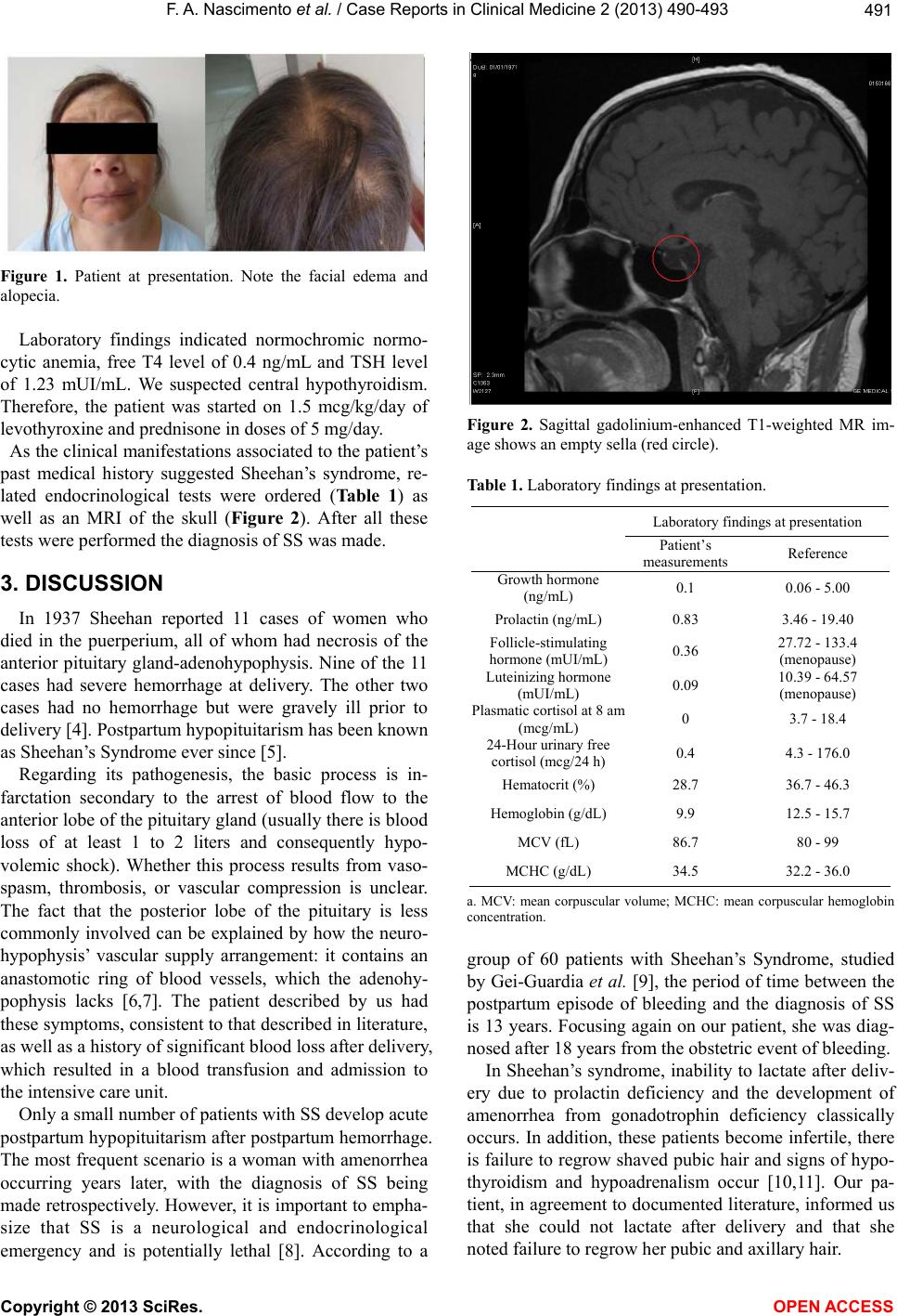 F. A. Nascimento et al. / Case Rep orts in Clinical Medicine 2 (2013) 490-493 491 Figure 1. Patient at presentation. Note the facial edema and alopecia. Laboratory findings indicated normochromic normo- cytic anemia, free T4 level of 0.4 ng/mL and TSH level of 1.23 mUI/mL. We suspected central hypothyroidism. Therefore, the patient was started on 1.5 mcg/kg/day of levothyro xine and pre dni sone in doses of 5 mg/day. As the clinical manifestations associated to the patient’s past medical history suggested Sheehan’s syndrome, re- lated endocrinological tests were ordered (Table 1) as well as an MRI of the skull (Figure 2). After all these tests were performed the diagnosis of SS was made. 3. DISCUSSION In 1937 Sheehan reported 11 cases of women who died in the puerperium, all of whom had necrosis of the anterior pituitary gland-adenohypophysis. Nine of the 11 cases had severe hemorrhage at delivery. The other two cases had no hemorrhage but were gravely ill prior to delivery [4]. Postpartum hypopituitarism has been known as Sheehan’s Syndrome ever since [5]. Regarding its pathogenesis, the basic process is in- farctation secondary to the arrest of blood flow to the anterior lobe of th e pituitar y gland (usu ally th ere is blood loss of at least 1 to 2 liters and consequently hypo- volemic shock). Whether this process results from vaso- spasm, thrombosis, or vascular compression is unclear. The fact that the posterior lobe of the pituitary is less commonly involved can be explained by how the neuro- hypophysis’ vascular supply arrangement: it contains an anastomotic ring of blood vessels, which the adenohy- pophysis lacks [6,7]. The patient described by us had these symptoms, consistent to that described in literature, as well as a history of significant blood loss after delivery, which resulted in a blood transfusion and admission to the intensive care unit. Only a small number of patients with SS develop acute postpartum hypopituitarism after postpartum hemorrhage. The most frequent scenario is a woman with amenorrhea occurring years later, with the diagnosis of SS being made retrospectively. Ho wever, it is impor tant to empha- size that SS is a neurological and endocrinological emergency and is potentially lethal [8]. According to a Figure 2. Sagittal gadolinium-enhanced T1-weighted MR im- age shows an empty sella (red circle). Table 1. Laboratory findings at presentation. Laboratory findings a t presentation Patient’s measurements Reference Growth hormone (ng/mL) 0.1 0.06 - 5.00 Prolactin (ng/m L) 0.83 3.46 - 19.40 Follicle-stimulating hormone (mUI/mL) 0.36 27.72 - 133.4 (menopause) Luteinizing hormone (mUI/mL) 0.09 10.39 - 64.57 (menopause) Plasmatic cortisol at 8 am (mcg/mL) 0 3.7 - 18.4 24-Hour urinary fr ee cortisol (mcg/24 h) 0.4 4.3 - 176.0 Hematocrit (%) 28.7 36.7 - 46.3 Hemoglobin (g/dL) 9.9 12.5 - 15.7 MCV (fL) 86.7 80 - 99 MCHC (g/dL) 34.5 32.2 - 36.0 a. MCV: mean corpuscular volume; MCHC: mean corpuscular hemoglobin concentration. group of 60 patients with Sheehan’s Syndrome, studied by Gei-Guardia et al. [9], the period of time between the postpartum episode of bleeding and the diagnosis of SS is 13 years. Focusing again on our patient, she was diag- nosed after 18 years from the obstetric event of bleeding. In Sheehan’s syndrome, inability to lactate after deliv- ery due to prolactin deficiency and the development of amenorrhea from gonadotrophin deficiency classically occurs. In addition, these patients become infertile, there is failure to regrow shaved pubic hair and signs of hypo- thyroidism and hypoadrenalism occur [10,11]. Our pa- tient, in agreement to documented literature, informed us that she could not lactate after delivery and that she noted failure to regrow her pubic and axillary hair. Copyright © 2013 SciRes. OPEN ACCESS 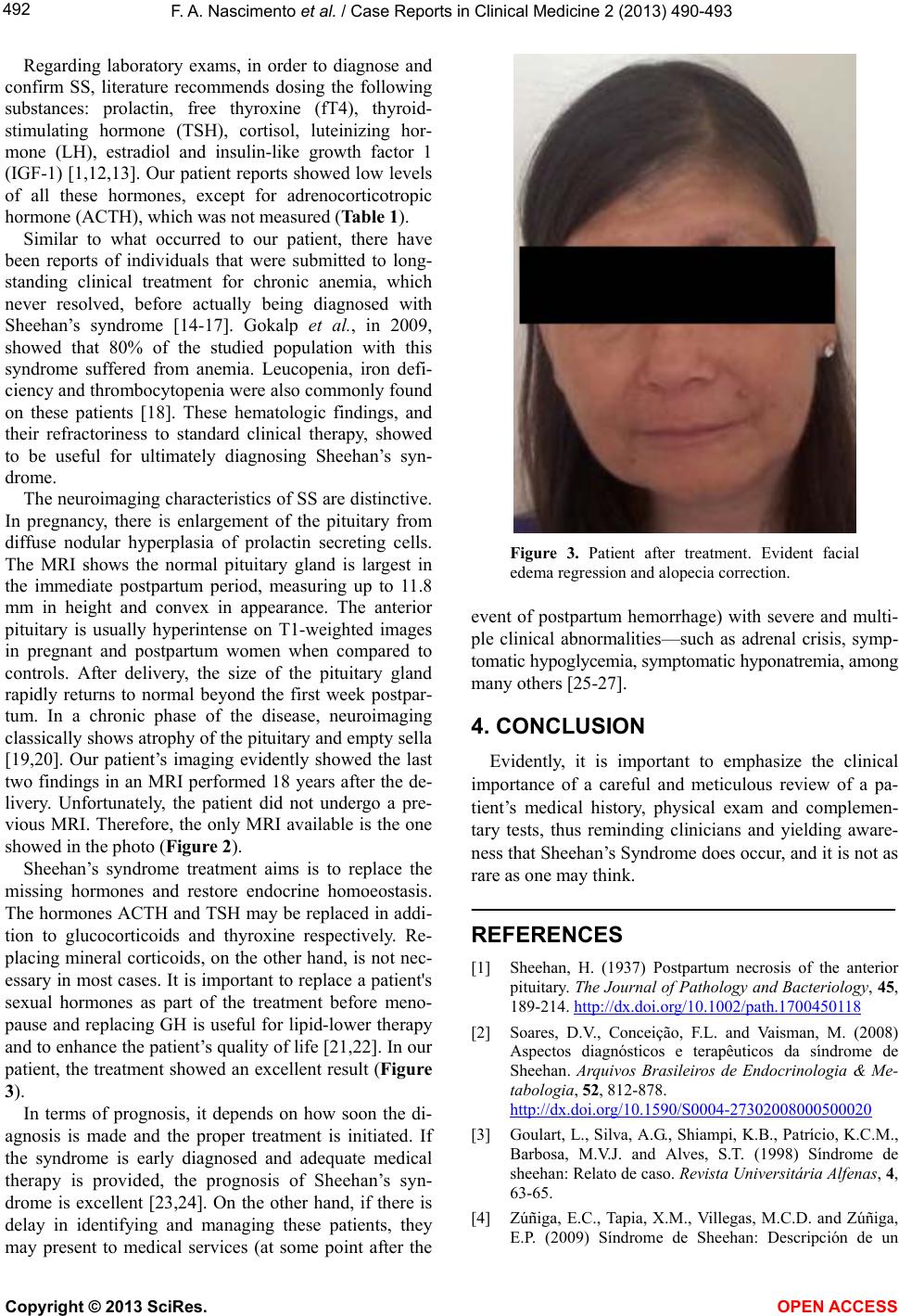 F. A. Nascimento et al. / Case Rep orts in Clinical Medicine 2 (2013) 490-493 492 Regarding laboratory exams, in order to diagnose and confirm SS, literature recommends dosing the following substances: prolactin, free thyroxine (fT4), thyroid- stimulating hormone (TSH), cortisol, luteinizing hor- mone (LH), estradiol and insulin-like growth factor 1 (IGF-1) [1,12,13]. Our patient reports showed low levels of all these hormones, except for adrenocorticotropic hormone (ACTH), which was not measured (Table 1). Similar to what occurred to our patient, there have been reports of individuals that were submitted to long- standing clinical treatment for chronic anemia, which never resolved, before actually being diagnosed with Sheehan’s syndrome [14-17]. Gokalp et al., in 2009, showed that 80% of the studied population with this syndrome suffered from anemia. Leucopenia, iron defi- ciency and thrombocytopenia were also commonly found on these patients [18]. These hematologic findings, and their refractoriness to standard clinical therapy, showed to be useful for ultimately diagnosing Sheehan’s syn- drome. The neuroimaging characteristics of SS are distinctive. In pregnancy, there is enlargement of the pituitary from diffuse nodular hyperplasia of prolactin secreting cells. The MRI shows the normal pituitary gland is largest in the immediate postpartum period, measuring up to 11.8 mm in height and convex in appearance. The anterior pituitary is usually hyperintense on T1-weighted images in pregnant and postpartum women when compared to controls. After delivery, the size of the pituitary gland rapidly returns to normal beyond the first week postpar- tum. In a chronic phase of the disease, neuroimaging classically shows atrophy of the pituitary and empty sella [19,20]. Our patient’s imaging evidently showed the last two findings in an MRI performed 18 years after the de- livery. Unfortunately, the patient did not undergo a pre- vious MRI. Therefore, the only MRI available is the one showed in the photo (Figure 2). Sheehan’s syndrome treatment aims is to replace the missing hormones and restore endocrine homoeostasis. The hormones ACTH and TSH may be replaced in addi- tion to glucocorticoids and thyroxine respectively. Re- placing mineral corticoids, on the other hand, is not nec- essary in most cases. It is important to replace a patient's sexual hormones as part of the treatment before meno- pause and replacing GH is useful for lipid-lower therapy and to enhance th e patient’s quality of life [21,22]. In our patient, the treatment showed an excellen t result (Figure 3). In terms of prognosis, it depends on how soon the di- agnosis is made and the proper treatment is initiated. If the syndrome is early diagnosed and adequate medical therapy is provided, the prognosis of Sheehan’s syn- drome is excellent [23,24]. On the other hand, if there is delay in identifying and managing these patients, they may present to medical services (at some point after the Figure 3. Patient after treatment. Evident facial edema regression and alopecia correction. event of postpartum hemorrhage) with severe and multi- ple clinical abnormalities—such as adrenal crisis, symp- tomatic hypogly c emia, symptomatic hy ponatremia, among many others [25-27]. 4. CONCLUSION Evidently, it is important to emphasize the clinical importance of a careful and meticulous review of a pa- tient’s medical history, physical exam and complemen- tary tests, thus reminding clinicians and yielding aware- ness that Sheehan’s Syndrome does occu r, and it is not as rare as one may think. REFERENCES [1] Sheehan, H. (1937) Postpartum necrosis of the anterior pituitary. The Journal of Pathology and Bacteriology, 45, 189-214. http://dx.doi.org/10.1002/path.1700450118 [2] Soares, D.V., Conceição, F.L. and Vaisman, M. (2008) Aspectos diagnósticos e terapêuticos da síndrome de Sheehan. Arquivos Brasileiros de Endocrinologia & Me- tabologia, 52, 812-878. http://dx.doi.org/10.1590/S0004-27302008000500020 [3] Goulart, L., Silva, A.G., Shiampi, K.B., Patrício, K.C.M., Barbosa, M.V.J. and Alves, S.T. (1998) Síndrome de sheehan: Relato de caso. Revista Universitária Alfenas, 4, 63-65. [4] Zúñiga, E.C., Tapia, X.M., Villegas, M.C.D. and Zúñiga, E.P. (2009) Síndrome de Sheehan: Descripción de un Copyright © 2013 SciRes. OPEN ACCESS  F. A. Nascimento et al. / Case Rep orts in Clinical Medicine 2 (2013) 490-493 Copyright © 2013 SciRes. OPEN ACCESS 493 caso clínico y revisión de la literatura. Revista Colombi- ana de Obstetriia e Ginecología, 60, 377-381. [5] Tessnow, A.H. and Wilson, J.D. (2010) The changing face of Sheehan’s syndrome. The American Journal of Medi- cal Sciences, 340, 402-406. http://dx.doi.org/10.1097/MAJ.0b013e3181f8c6df [6] Sheehan, H. and Davis, J.C. (1983) Post-partum hypopi- tuitarism. Annals of Internal Medicine, 99, 430. [7] Kan, A.K.S. and Calligerous, D. (1998) A case report of Sheehan syndrome presenting with diabetes insipidus. Australian and New Zealand Journal of Obstetrics and Gynaecology, 38, 224-226. http://d x.doi.org/ 10.1111/ j.1479-828X.1998.tb03009.x [8] Chávez, C.M.C., Castillo, L.M., Gaitán, P.B.M., Domín- guez, A.R. and Mendoza, P.E.C. (2013) Síndrome de sheehan. Descripción de un caso clínico y revisión de la literatura. Archivos de Medicina de Urgencia de Mexico, 5, 38-41. [9] Kelestimur, F. (2003) Sheehan’s syndrome. Pituitary, 6, 181-188. http://dx.doi.org/10.1023/B:PITU.0000023425.20854.8e [10] Guardia, O.G., Herrera, E.S., Brealey, A.G. and Hao, C.K.C. (2011) Sheehan’s syndrome in Costa Rica: Clini- cal experience with 60 cases. Endocrine Practice, 17, 337-344. http://dx.doi.org/10.4158/EP10145.OR [11] Moszkowski, E.F. (1973) Postpartum pituitary insuffi- ciency: Report of five unsual cases with long-term fol- low-up. Southern Medical Journal, 66, 878-882. http://dx.doi.org/10.1097/00007611-197308000-00008 [12] Prager, D. and Braunstein, G.D. (1995) Pituitary disorders during pregnancy. Endocrinology Metabolism Clinics of North America, 24, 1-14. [13] Rivers, J.-A. (2006) Lymphocitic hypophysitis: Disease spectrum and approach to diagnosis and therapy. Pituitary, 9, 35-45. http://dx.doi.org/10.1007/s11102-006-6598-z [14] Atzingen, A.C.V., Nunes, C.V., Toniolo, J.V., Araujo, M.F.A., Teixeira, V.A. and Junior, W.S. (2001) Insufi- ciência adrenal aguda devido síndrome de sheehan. Re- vista Brasileira de Medicina do Espo rte , 58, 762-765. [15] Melchardt, T., Namberger, K., Weiss, L., Egles, A., Faber, V. and Greil, R. (2010) An uncommon cause of anaemia: Sheehan’s syndrome. Wiener Klinische Wochenschrift, 122, 717-719. http://dx.doi.org/10.1007/s00508-010-1480-4 [16] Fatma, M., Mouna, E., Nabila, R., Mouna, M., Nadia, C. and Mohamed, A. (2011) Sheehan’s syndrome with pancytopenia: A case report and review of the literature. Journal of Medical Case Reports, 5, 1-3. http://dx.doi.org/10.1186/1752-1947-5-490 [17] Laway, B.A., Mir, S.A., Bashir, M.I., Bhat, J.R., Samoon, J. and Zargar, A.H. (2011) Prevalence of hematological abnormalities in patients with Sheehan’s syndrome: Re- sponse to replacement of glucocorticoids and thyroxine. Pituitary, 14, 39-43. http://dx.doi.org/10.1007/s11102-010-0255-2 [18] Gokalp, D., Tuzcu, A., Bahceci, M., Arikan, S., Bahceci, S. and Pasa, S. (2009) Sheehan’a syndrome as a rare cause of anaemia secondary to hypopituitarism. Annals of Hematology, 88, 405-410. http://dx.doi.org/10.1007/s00277-008-0607-4 [19] Gutenberg, A., Hans, V., Puchner, M.J.A. and Kreutzer, J. (2006) Primary hypophysitis: Clinical-pathological cor- relations. European Journal of Endocrinology, 155, 101- 107. http://dx.doi.org/10.1530/eje.1.02183 [20] Ishihara, T., Hino, M., Kurahachi, H., et al. (1996) Long-term clinical course of two cases of lymphocytic adenohypophysitis. Endocrine Journal, 46, 433-440. http://dx.doi.org/10.1507/endocrj.43.433 [21] Kaplun, J. (2008) Sequential Pituitary MR imaging in sheehan syndrome: Report of 2 cases. American Journal of Neuroradiology, 29, 941-943. http://dx.doi.org/10.3174/ajnr.A1016 [22] Kelstimur, F. and Colak, A.C. (2003) The baseline char- acteristics and the effects of GH replacement therapy in patients with Sheehan’s syndrome as compared to non- functioning pituitary adenomas. The Endocrine Society’s 85th Annual Meeting. Philadelphia, June 2003. [23] Storck, S. (2012) Síndrome de sheehan. http://www.nlm.nih.gov./medlineplus/spanish/ency/article /00175.htm [24] Mancini, M. (2010) Sindrome di sheehan. http://www.medicinalive.com/malattia/sindrome-di-sheeh an/ [25] Jain, D. (2013) A ray of hope for a woman with Shee- han’s syndrome. BMJ Case Reports. [26] Wani, A.M., Hussain, W.M., Mejally, M.A.A., Banjar, A.A., Ali, K.S., Khoujah, A.M., Raja, S.H., Bafaraj, M, G, Miamini, W.A. and Akhtar, M. (2010) Practice of symp- tomatictreatment in the era of evidence-based medicine: Report of 2 cases of diagnosis of Sheehan’s syndrome delayed till eighth decade. BMJ Case Reports. [27] Anfuso, S., Patrelli, T.S., Soncini, E., Chiodera, P., Fadda, G.M. and Nardelli, G.M. (2009) A case report of Shee- han’s syndromewith acute onset, hyponatremia and se- vere anemia. Acta Bio Medica, 80, 73-76. |