

Paper Menu >>
Journal Menu >>
 Vol.2, No.8, 470-472 (2013) Case Reports in Clinical Medicine http://dx.doi.org/10.4236/crcm.2013.28124 Is angiography still the best method to stratify stroke risk in symptomatic atherosclerotic carotid plaque? Gustavo W. Kuster1*, A. J. Da Rocha2, A. S. F. Silva2, R. A. Valiente3, M. S. G. Rocha3, A. R. Massaro4 1Division of Neurology, São Paulo University, São Paulo, Brazil; *Corresponding Author: [email protected] 2Department of Radiology and Pathology, Fleury Institute, São Paulo, Brazil 3Division of Neurology, Santa Marcelina Hospital, São Paulo, Brazil 4Sao Paulo University, São Paulo, Brazil Received 20 June 2013; revised 18 July 2013; accepted 25 August 2013 Copyright © 2013 Gustavo W. Kuster et al. This is an open access article distributed under the Creative Commons Attribution Li- cense, which permits unrestricted use, distribution, and reproduction in any medium, provided the original work is properly cited. ABSTRACT The degree of vessel lumen narrowing is an independent predictor of ischemic stroke. New development s in carotid plaque m orphology imag- ing (MR, CT), may bring new insights to the re- lationship between carotid atherosclerotic di- sease and stroke risk. Our aim is to review the stroke risk in a symptomatic patient with moder- ate carotid stenosis by CT imaging and histo- pathology. A 72-year-old patient with low ABCD2 scores TIA and moderate left internal carotid stenosis (50% by carotid ultrasound), was dis- charged with an optimized medical therapy. Four months later, he presented an ischemic stroke in the left frontal area. Carotid angi- ography showed a 60% stenosis in the left- internal carotid artery with a regular surface. CT plaque imaging detected a thin fibrous cap with calcification and an intraplaque hemorrhage (high- risk plaque). These findings were confirmed in the histolopathological study of the atheroscle- rotic plaque performed after the endarterectomy. After 1 year of follow-up, the patient returned independently to his daily activities. We propose, in this stud y, the inclusio n of nonin vasive p laque imaging in the evaluation of acute TIA with moderate carotid stenosis to better select pa- tients with higher risk of stroke recurrence. Keywords: Carotid Atherosclerotic Plaque; CT; Plaque Classification 1. INTRODUCTION Carotid atherosclerotic plaque has been identified since our ancient ancestors and some in the recent modern era have been evaluated to the prevention of catastrophic stroke [1]. The report from North America Symptomatic Carotid Endarterectomy Trial (NASCET) in 1991 has stimulated new interest in carotid stenosis and confirmed angiography as a gold standard method to stratify symptomatic patients to endarterectomy [2]. The degree of vessel lumen narrowing is an indepen- dent predictor of ischemic stroke, particularly in sympto- matic patients with severe carotid stenosis (70%). Ca- rotid endarterectomy is also associated with a moderate stroke risk reduction in patients with symptomatic moderate carotid stenosis (50% - 69%). However, treat- ment decisions in these cases should be also considered as other important issues including an exceptional sur- gical skill [3]. In addition, opti mal medical treatment has been improved and becomes a topic of equal impor- tance for managing carotid disease, especially in those patients with asymptomatic atherosclerotic plaque [4]. New developments in carotid plaque morphology imaging, particularly MR or CT, may bring new insights to the relationship between carotid atherosclerotic di- sease and stroke risk [5,6]. Our aim is to review the stroke risk in a symptomatic patient with moderate carotid stenosis according to the plaque surface morphology and the degree of stenosis on carotid angiography and to compare the carotid plaque morphology classification obtained by CT imaging and histopathology. 2. CASE A 72-year-old previously hypertensive patient arrived at emergency department with a sudden onset of right sided weakness lasting 10 minutes. He was evaluated using the TIA assessment protocol and obtained a low Copyright © 2013 SciRes. OPEN ACCESS 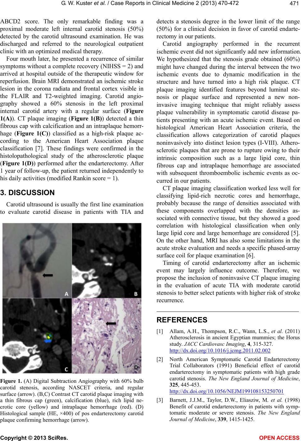 G. W. Kuster et al. / Case Reports in Clinical Medicine 2 (2013) 470-472 471 ABCD2 score. The only remarkable finding was a proximal moderate left internal carotid stenosis (50%) detected by the carotid ultrasound examination. He was discharged and referred to the neurological outpatient clinic with an optimized medical therapy. Four mouth later, he presented a recurrence of similar symptoms without a complete recovery (NIHSS = 2) and arrived at hospital outside of the therapeutic window for reperfusion. Brain MRI demonstrated an ischemic stroke lesion in the corona radiata and frontal cortex visible in the FLAIR and T2-weighted imaging. Carotid angio- graphy showed a 60% stenosis in the left proximal internal carotid artery with a regular surface (Figure 1(A)). CT plaque imaging (Figure 1(B)) detected a thin fibrous cap with calcification and an intraplaque hemorr- hage (Figure 1(C)) classified as a high-risk plaque ac- cording to the American Heart Association plaque classification [7]. These findings were confirmed in the histolopathological study of the atherosclerotic plaque (Figure 1(D)) performed after the endarterectomy. After 1 year of follow-up, the patien t returned indep enden tly to his daily activities (modified Rankin score = 1). 3. DISCUSSION Carotid ultrasound is usually the first line exa mination to evaluate carotid disease in patients with TIA and Figure 1. (A) Digital Subtraction Angiography with 60% bulb carotid stenosis, according NASCET criteria, and regular surface (arrow). (B,C) Contrast CT carotid plaque imaging with a thin fibrous cap (green), calcification (blue), rich lipid ne- crotic core (yellow) and intraplaque hemorrhage (red). (D) Histological sample (HE, ×400) of pos endarterectomy carotid plaque confirming hemorrhage (arrow). detects a stenosis degree in the lower limit of the range (50%) for a clinical decision in favor of carotid endarte- rectomy in our patients. Carotid angiography performed in the recurrent ischemic event did not significantly add new information. We hypothesized that the stenosis grade obtained (60%) might have changed during the interval between the two ischemic events due to dynamic modification in the structure and have turned into a high risk plaque. CT plaque imaging identified features beyond luminal ste- nosis or plaque surface and represented a new non- invasive imaging technique that might reliably assess plaque vulnerability in symptomatic carotid disease pa- tients presenting with an acute ischemic event. Based on histological American Heart Association criteria, the classification allows categorization of carotid plaques noninvasively into distinct lesion types (I-VIII). Athero- sclerotic plaques that are prone to rupture owing to their intrinsic composition such as a large lipid core, thin fibrous cap and intraplaque hemorrhage are associated with subsequent thromboembolic ischemic events as oc- curred in our patients. CT plaque imaging classification worked less well for classifying lipid-rich necrotic cores and hemorrhage, probably because the range of densities associated with these components overlapped with the densities as- sociated with connective tissue, but they showed a good correlation with histological classification when only large lipid core and large hemorrhage are considered [5]. On the other hand, MRI has also some limitations in the acute stroke evaluation and needs a specific phased-array surface coil for plaque examination [6]. Timing of carotid endarterectomy after an ischemic event may largely influence outcome. Therefore, we propose the inclusion of noninvasive CT plaque imaging in the evaluation of acute TIA with moderate carotid stenosis to better select patients with high er risk of strok e recurrence. REFERENCES [1] Allam, A.H., Thompson, R.C., Wann, L.S., et al. (2011) Atherosclerosis in ancient Egyptian mummies; the Horus study. JACC Cardiovasc Imaging, 4, 315-327. http://dx.doi.org/10.1016/j.jcmg.2011.02.002 [2] North American Symptomatic Carotid Endarterectomy Trial Collaborators (1991) Beneficial effect of carotid endarterectomy in symptomatic patients with high grade carotid stenosis. The New England Journal of Medicine, 325, 445-453. http://dx.doi.org/10.1056/NEJM199108153250701 [3] Barnett, J.J.M., Taylor, D.W., Eliasziw, M. et al. (1998) Benefit of carotid endarterectomy in patients with symp- tomatic moderate or severe stenosis. The New England Journal of Medicine, 339, 1415-1425. Copyright © 2013 SciRes. OPEN ACCESS  G. W. Kuster et al. / Case Reports in Clinical Medicine 2 (2013) 470-472 Copyright © 2013 SciRes. OPEN ACCESS 472 http://dx.doi.org/10.1056/NEJM199811123392002 [4] Marquadt, L., Geraghty, O.C., Mehta, Z. and Rothwell, P.M. (2010) Low risk of ipsilateral stroke in patients with asymptomatic carotid stenosis on best medical treatment: A prospective, population-based study. Stroke, 41, e11- e17. http://dx.doi.org/10.1161/STROKEAHA.109.561837 [5] Wintermark, M., Jawadi, S.S., Rapp, J.H., et al. (2008) High-resolution CT imagin of carotid artery atheroscle- rotic plaques. American Journal of Neuroradiology, 29, 875-882. http://dx.doi.org/10.3174/ajnr.A0950 [6] Saam, T., Cai, J., Ma, L., Cai, Y.Q., et al. (2006) Com- parison of symptomatic and asymptomatic atherosclerotic plaques features with in vivo MR imaging. Radiology, 20, 464-472. http://dx.doi.org/10.1148/radiol.2402050390 [7] Stary, H.C., Chandler, A.B. and Dinsmore, R.E., Ameri- can Heart Association (1995) A definition types of athero- sclerotic lesions and a histological classification of athero- sclerosis: A report from the Committee on vascular le- sions of the council on atherosclerosis. Circulation, 92, 1355-1374. http://dx.doi.org/10.1161/01.CIR.92.5.1355 |