

Paper Menu >>
Journal Menu >>
 Journal of Cancer Therapy, 2013, 4, 28-33 Published Online November 2013 (http://www.scirp.org/journal/jct) http://dx.doi.org/10.4236/jct.2013.410A004 Open Access JCT Pancreatic Neuroendocrine Tumors Are Characterized by Loss of Noxa Expression that Can be Recovered by the Proteasome Inhibitor Bortezomib E. P. Slater, J. Waldmann, C. López, E. Matthäi, V. Fendrich, D. K. Bartsch Department of Surgery, Philipps University, Marburg, Germany. Email: [email protected] Received August 2nd, 2013; revised September 2nd, 2013; accepted September 10th, 2013 Copyright © 2013 E. P. Slater et al. This is an open access article distributed under the Creative Commons Attribution License, which permits unrestricted use, distribution, and reproduction in any medium, provided the original work is properly cited. ABSTRACT Prognosis in well-differentiated neuroendocrine carcinomas varies considerably and therapeutic targets for metastatic diseas e are urgently need ed. cDNA microar ray studies in our laboratory revealed a significantly lower expression level of the Noxa-gene in human pancreatic neuroendocrine neoplasms (PNENs) as compared to normal islet cells. To deter- mine the validity of the downregulation of Noxa in PNENs, benign and malignant tumors from both sporadic and MEN1 patients were selected for expression analysis. To further verify the findings, neuroendocrine BON1 and QGP cell lines were tested for Noxa expression and its recovery by treatment with the proteasome inhibitor bortezomib. The expression of Noxa was significantly downregulated in 20 PNENs (p = 0.0036). There was no significant difference between MEN1 and sp oradic tumors. However, the malignant tumors showed a more significant decrease as compared to benign tumors (p = 0.0385 ) and the decrease in expression in tumor s greater than 20 mm was very highly sign ificant (p < 0.0001). Neither the BON1 nor QGP1 cell lines displayed expression of Noxa protein in the absence of the protea- some inhibitor, Bortezo mib. After stimulation with the drug for 16 h, the expression was induced in both cell lines that are correlated with an increase in the level of c-MYC expression, cleaved caspase 3 and cell death. The low expression level of Noxa in PNENs contributes to the inability o f these tumor entities to undergo apoptos is. The recovery of Noxa expression following treatment with the proteasome inhibitor, bortezomib, leading to caspase activation and cell death supports the use of such drugs for the treatment of these tumor entities. Keywords: Pancreatic Neuro end ocrine Neoplasms; Noxa, Bortezomib 1. Introduction Pancreatic neuroendocrine neoplasms (PNENs) represent 5% - 10% of pancreatic tumors and include gastrinoma, insulinoma and non-functioning endocrine tumors. They occur sporadically or in the autosomal dominant inher- ited syndromes such as multiple endocrine neoplasia type 1 (MEN1) and von-Hippel-Lindau. Although plasma chromogranin A and 24-h urine for 5-hydroxyindole ace- tic acid are known prognostic markers for survival in patients with such tumors, the role of other tumor mark- ers is not well described [1,2]. Altho ugh these tumors are surgically manageable, metastatic disease is present in ~50% of patients at the time of diagnosis, and the overall 5-year surviv al for patien ts with d istant metastasis is on ly 22%. Somatostatin analogues, IFN-α or hepatic artery chemoembolization prov ide good palliation of the symp- toms of carcinoid syndrome associated with such tumors. However, few systemic therapies have been shown to consistently elicit tumor responses and prolong survival. Recently, the mTor-inhibitor, everolimus, and the multi- targeting sunitinib have been shown to increase overall survival in randomized controlled trials [3,4]. The inef- fectiveness of most systemic chemotherapy in metastatic neuroendocrine tumors may relate to their unique bio- logical features such as slow growth pattern or hyper- vascularity [5]. The p53 tumor-suppressor protein functions as a tran- scriptional activator and several p53-indu cible genes that play a role in the induction of apoptosis in response to p53 have been described [6]. The pro-apoptotic gene, Noxa encodes a Bcl-2 homology 3 (BH3)-only member of the Bcl-2 family of proteins that inactivates the pro-  Pancreatic Neuroendocrine Tumors Are Characterized by Loss of Noxa Expression that Can be Recovered by the Proteasome Inhibitor Bortezomib 29 tective function of the pro-surv ival members of the Bcl-2 family and unleashes the mitochondrial-based apoptotic cascade by activating the Bax/Bak-like pro-apoptotic family members. In fact treatment of metastatic mela- noma or multiple myeloma with proteasome inhibitors has been reported to result in selective tumor cell killing by triggering the induction of Noxa expression [7,8]. A cDNA microarray analysis with 11,500 genes was performed using RNA from 10 gastrinoma tumor sam- ples, both sporadic and MEN1, and compared to RNA from normal islet cells obtained from healthy donors as reference. The comparative analysis of gene expression patterns revealed overexpression of 15 and underexpres- sion of 123 genes greater than 3-fold [9]. Included in this list was the gene Noxa, a proapoptotic member of the Bcl-2 family [10]. We hypothesized that this loss could contribute to tumor progression and proceeded to analyze the expression in a wider range of PNENs to verify these results and correlate them with clinical findings. 2. Materials and Methods 2.1. Patients We randomly selected 10 patients with benign and 10 patients with malignant neuroendocrine pancreatic tu- mors from the tumor bank at our institute. Five of each group were genetically confirmed MEN1 patients. Clini- cal data were prospectively recorded and analyzed with special regard to symptoms. The diagnosis of a func- tional tumor was established if hormone excess was pre- sent, excluding an isolated elevation of pancreatic poly- peptide (PP). Zollinger-Ellison Syndrome (ZES) was diagnosed in the presence of clinical symptoms, an ele- vated fasting serum gastrin level (ref < 125 pg/ml), a positive secretin stimulation test (increase > 200 pg/ml) and a low pH of the stomach. If symptomatic hypogly- cemia (<40 mg/ml) was combined with hyperinsulinism (>20 µU/ml) insulinoma was confirmed by a supervised fasting test. A Vipoma was defined in case of watery diarrhea (>6l/day) and elevated VIP serum levels (>130 pg/ml). Pancreatic tumors were considered non-func- tional if neither hormone levels were elevated nor symp- toms associated with hormone excess were evident ex- cept PP. Tumors were classified as malignant if they demonstrated infiltrating growth, angio invasion or lymph node or distant metastases. The pathological classifica- tion was performed according to Rindi and Klöppel [11]. The twenty fresh frozen tissue samples were obtained from the tumor bank of Philipps-University of Marburg. Clinical follow-up was conducted by the patients’ per- sonal physicians or at outpatient attendance. Survival was calculated as the time from surgical resection to ei- ther death or last follow up. Informed consent was ob- tained from all patients. The Ethics Committee of the University approved this study and all patients partici- pating in the study consented to sampling. Before proceeding with RNA isolation from these 20 tumor samples, an initial histochemical staining was performed to confirm a neoplastic cellularity of 85%. Again, the reference contained pooled RNA from normal human islet cells. 2.2. RNA Isolation and Quantitative Real-Time RT-PCR (qRT-PCR) Frozen control and tumor tissue samples were homoge- nized in the presence of guanidinium thiocyante-phe- nol-chloroform (Trizol Reagent; Invitrogen, Paisley, Scotland, UK) following the manufacturer’s protocol. Total RNA was then further purified by digestion with DNaseI and recovery of RNA using an RNeasy kit (Qiagen, Hilden, Germany) according to the supplier’s protocol. The relative expression of Noxa mRNA in tu- mors and normal islet cells was determined by Real Time PCR on a Roche Light Cycler using the following prim- ers: NOXA_for GTC CGA GGT GCT CCA GTT; NOXA_rev AAA CGT GCA CCT CCT GAG A (Ampli- con 298 bp; Accession number NM 21127; 6) and GAPDH_for CGT CTT CAC CAC CAT GGA GA; GAPDH_rev CGG CCA TCA CGC CAC AGT TT as previously described [12]. Negative controls contained no cDNA. Expression levels were normalized to GAPDH. 2.3. Cell Culture and Biological Reagents The BON-1 cell line was derived from a peripancreatic lymph node metastasis of a serotonin-producing PNEN [13]. The cells were cultured in a 1:1 mixture of Hams F12 Nutrient Mixture and 1x Dulbecco’s Modification of Eagle’s Medum (DMEM) (Life Technologies). The QGP-1 cell line was derived from a somatostatinoma and was grown in RPMI 1640. Both cell lines were supple- mented with 10% fetal calf serum, 1% L-glutamine (Biochrom KG, Berlin, Germany) and 1% penicillin- streptomycin (Life technologies) and grown in an incu- bator maintaining an atmosphere of 95% humidity and 5% CO2 at 37˚C. Each cell line was plated on day minus 1 and incu- bated on day 1 with 150 nM Bortezomib (LC Laborato- ries, Boston) or not for 16 h. The control cells received vehicle, DMSO. Both floating and adherent cells were harvested and whole cell extracts were obtained using RIPA buffer. 2.4. Western Blot For the Western blot, 30 µg of total protein from cell Open Access JCT  Pancreatic Neuroendocrine Tumors Are Characterized by Loss of Noxa Expression that Can be Recovered by the Proteasome Inhibitor Bortezomib Open Access JCT 30 lysates were analyzed by 17% SDS-PAGE and blotted onto nitrocellulose membrane (Schleicher & Schuell, Wh atman GmbH, Dassel, Germany). Filters were blocked with 5% nonfat dry milk in PBS and incubated overnight at 4˚C with specific antibodies (Noxa: Imgenex, San Diego, California, 1:100; Cleaved Caspase 3, Cell Sig- naling Technology, 1:1000; Actin, Sigma-Aldrich, 1: 2500; c-Myc, Santa Cruz sc-42, 1:200). After washing, blots were incubated with horseradish peroxidase-con- jugated secondary antibodies (Vectastain) and then re- vealed by enhanced chemiluminescence (Amersham Bio- sciences). 2.5. Statistics Data were analyzed using SPSS, version 14.0 for Micro- soft Windows. Means were compared using paired t-tests. A p-value < 0.05 was considered significant. 3. Results Based on previous results from our cDNA microarray analysis of 10 gastrinomas in comparison to pooled RNA from normal islet cells we proceeded to verify the finding of a downregulation of the Noxa gene in PNENs. For this purpose we selected 20 additional PNEN tumor samples. 3.1. Patients The median age of patients (12 male and 8 female) was 44 years (29 - 61) and these were followed for a median period of 61 months (13 - 193). The median tumor di- ameter was 15 mm (5 - 2 50). A summary appear s in Ta- ble 1. 3.2. PNENs vs. Normal Islet Cells Real time PCR analysis of 20 samples, 10 malignant and Table 1. Summary of clinical data. Pat ID Spordic/MEN 1 Tumor Stage WHO Age at Dx (yr) Tumor Size (mm)LNM Liver Met Ki 67Follow up (mos) Other therapies Ct Ct-Ref 1 MEN1 M GAS III 46 6 y N 1% NED (136)none 33.017.58 2 MEN1 M GAS III 49 5 n Y ND DOC (84) none 32.97.47 3 MEN1 M GAS III 35 8 y N 2-3%DOD (136)DOTATOC 31.095.66 4 MEN1 M VIP III 32 250 y N ND DOD (240)somatostatin, Interferon 34.38.87 5 MEN1 NECA III 33 10 n N 6% AWD (120)none 32.817.38 6 sporadic M GAS III 58 15 n Y ND DOD (78) none 30.515.08 7 sporadic M GAS III 29 15 y N 2% NED (146)none 35.129.69 8 sporadic M GAS III 50 20 n N ND NED (235)none 34.819.38 9 sporadic NECA III 45 25 y Y 30% AWD (25)somatosttin-LAR 35.8110.38 10 sporadic NECA III 44 15 y N 2% AWD (18)none 33.17.67 11 MEN1 INS II 48 40 n N ND NED (107)none 31.986.55 12 MEN1 INS I 32 20 n N 3% NED (79) none 31.245.81 13 MEN1 NFPET I 30 10 n N 1% AWD (72)none 29.794.36 14 MEN1 NFPET II 54 50 n N 2% AWD (108)none 35.6510.22 15 MEN1 NFPET II 37 40 n N 1% NED (60) none 34.969.53 16 sporadic GAS II 61 15 n N ND NED (125)none 30.735.3 17 sporadic GAS I 45 9 n N ND DOD (124)none 31.25.77 18 sporadic INS I 61 10 n N 1% NED (13) none 28.573.14 19 sporadic INS II 32 13 n N 5% NED (15) none 33.598.16 20 sporadic INS I 32 8 n N 2% NED (52) none 27.442.01 LNM, lym ph node m etasta sis; Me t, meta stasis; M , m aligna nt; Gas, gastri nom a; VIP, vipom a; NE CA, ne uroend ocrine carc inom a; IN S, i nsulin oma ; NFPE T, non- functional pancreatic endocrine tumor; NED, no evidence of disease; DOC, dead of unrelated causes; DOD, dead of disease; AWD, alive with disease; ND, not determin ed; y, yes; n, no; Ref, reference.  Pancreatic Neuroendocrine Tumors Are Characterized by Loss of Noxa Expression that Can be Recovered by the Proteasome Inhibitor Bortezomib 31 10 benign, was performed to determine the expression of Noxa using normal human islet cells pooled from three patients as a reference. All values for Noxa were normal- ized according to the results with GAPDH. All samples tested demonstrated a decrease in the level of expression of Noxa. Figure 1 shows the results of this analysis where the average threshold cycle (Ct) is presented for the group of 20 PNENs (Ct = 32.1) compared to that of normal islet cells (Ct = 25.3). A paired t test of these re- sults suggests that this result is very statistically signifi- cant (p = 0.0036). Of the 20 PNENs, 10 were from MEN1 patients: 5 malignant and 5 benign. Interestingly, there was no sig- nificant difference in the downregulation of Noxa be- tween these two groups: MEN1 and non-MEN1 (data not shown). 3.3. Malignant vs. Benign The results of the real time PCR analysis were graphed according to the designation malignant (Ct = 33.05) or benign (Ct = 31.21). Interestingly, h ere there is a statisti- cally significant difference between the two groups as determined by a two tailed test (p = 0.0385). The loss of Noxa expression is greater in malignant tumors as com- pared to benign tumors (Figure 1). 3.4. PNENs Larger Than 20 mm A further analysis compared the tumors >20 mm in size to the expression in normal islet cells. Here the decrease in expression of the tumor suppressor, Noxa is most evi- dent with an average Ct of 33.5. The statistical analysis results in a two-tailed p value of less than 0.0001 which is extremely statistically significant (Figure 1). Thus, the results of the initial microarray analysis were confirmed by real time PCR on 20 additio nal tumor sam- ples. 3.5. Noxa Expression in Tumor Cell Lines, QGP1 and BON1 The neuroendocrine tumor cell lines, BON1 (serotonin- producing PNEN) and QGP1 (somatostatinoma) were analyzed for their expression of Noxa. For this purpose the cells were grown in culture and total protein extracts were prepared and applied to a 17% SDS PAGE. The results of this Western blot analysis are presented in Figure 2. Lanes 1 and 3 contain the extracts from BON1 and QGP1 cells, respectively. Although the presence of Actin (42 kDa) verifies the presence of protein in the lane, there is no band at 11 kDa where Noxa protein would be expected to run. This lack of or very low level of expres- sion mimics the real ti me PCR results with the PNEN patient samples. Figure 1. Results of the real time PCR Analyses for Noxa expression in PNENs. 3.6. Expression of Noxa in Cell Lines Inducible by Bortezomib Both cell lines were treated with or without 150 nM bo r- tezomib for 16 h. The cell cultures were analyzed micro scopically to confirm cell death in the treated cultures. Both floating and adherent cells were harvested, pelleted and RIPA buffer was used to isolate cellular protein. Following quantitation, 30 µg were applied to the SDS PAGE, blotted and the membrane was hybridized with antibodies. As shown in Figure 2 Actin as a loading control was not changed by treatment with the drug. However, the level of expression of Noxa, which was not visible in untreated cells (lanes 1 and 3) was clearly in- duced following treatment with bortezomib (lanes 2 and 4). Cleavage of Caspase 3 was observed following the treatment with the drug as evidence that the cell killing is apoptosis. The known function of Noxa as a pro-apop- totic member of the Bcl-2 family would suggest that the increased expression of this protein leads to the apoptotic death of the cells. The level of expression of c-MYC was visible before treatment with the drug and was induced by treatment of the cells with bortezomib. As MYC is known to tran- scriptionally activate the expression of Noxa [14,15], th is would be the proposed mechanism for the increased ex- pression. Thus, treatment with Bortezomib stabilizes MYC that re-induces Noxa expression in these cells, leading to apoptotic killing. 4. Discussion In this study we have validated the down regulation of the pro-apoptotic Noxa gene in PNENs. These tumors express Noxa at a lower level than that of normal islet cells. Malignant tumors display a more significant de- crease than benign and larger tumors (>20 mm) show the greatest decrease. This low level of expression is mir- rored in the tumor cell lines, BON1 and QGP1. Open Access JCT 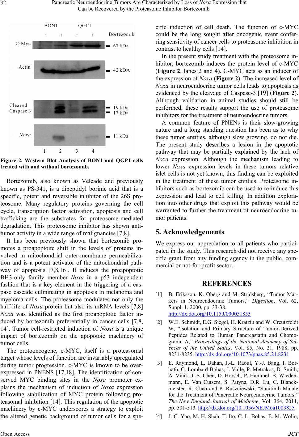 Pancreatic Neuroendocrine Tumors Are Characterized by Loss of Noxa Expression that Can be Recovered by the Proteasome Inhibitor Bortezomib 32 Figure 2. Western Blot Analysis of BON1 and QGP1 cells treated with and without bortezomib. Bortezomib, also known as Velcade and previously known as PS-341, is a dipeptidyl borinic acid that is a specific, potent and reversible inhibitor of the 26S pro- teosome. Many regulatory proteins governing the cell cycle, transcription factor activation, apoptosis and cell trafficking are the substrates for proteosome-mediated degradation. This proteosome inhibitor has shown anti- tumor activity in a wide range of malignancies [7,8]. It has been previously shown that bortezomib pro- motes a proapoptoitc shift in the levels of proteins in- volved in mitochondrial outer-membrane permeabiliza- tion and is a potent activator of the mitochondrial path- way of apoptosis [7,8,16]. It induces the proapoptotic BH3-only family member Noxa in a p53 independent fashion that is a key element in the triggering of a cas- pase cascade culminating in apoptosis in melanoma and myeloma cells. The proteasome modulates not only the half-life of Noxa protein but also its mRNA levels [7,8] Noxa was identified as the first proapoptotic factor in- duced by bortezomib preferentially in cancer cells [7,8, 14]. Tumor cell-restricted induction of Noxa is a unique impact of bortezomib on the apopotoic machinery of tumor cells. The protooncogene, c-MYC, itself is a proteasomal target whose levels of function are invariably upregulated during tumor progression. c-MYC is known to be over- expressed in PNENS [17,18]. The identification of con- served MYC binding sites in the Noxa promoter ex- plains the mechanism of induction of Noxa expression following stabilization of MYC protein following pro- teasomal inhibition [14]. This regulation of the apoptotic machinery by c-MYC underscores a strategy to exploit the altered genetic background of tumor cells for a spe- cific induction of cell death. The function of c-MYC could be the long sought after oncogenic event confer- ring sensitivity of cancer cells to proteaso me inh ibition in contrast to healthy cells [14]. In the present study treatment with the proteosome in- hibitor, bortezomib induces the protein level of c-MYC (Figure 2, lanes 2 and 4). C-MYC acts as an inducer of the expression of Noxa (Figure 2). The increased level of Noxa in neuroendocrine tumor cells leads to apoptosis as evidenced by the cleavage of Caspase-3 [19] (Figure 2). Although validation in animal studies should still be performed, these results support the use of proteasome inhibitors for the treatment of neuroendocrine tumors. A common feature of PNENs is their slow-growing nature and a long standing question has been as to why these tumor entities, although slow growing, do not die. The present study describes a lesion in the apoptotic pathway that may be partially explained by the lack of Noxa expression. Although the mechanism leading to lower Noxa expression levels in these tumors relative islet cells is not yet known, this finding can be exploited in the treatment of these tumor entities. Proteasome in- hibitors such as bortezomib can be used to re-induce this expression and lead to cell killing. In addition explora- tion into other drugs that exploit this pathway would be warranted to further the treatment of neuroendocrine tu- mor patients. 5. Acknowledgements We express our appreciation to all patients who partici- pated in the study. This research did not receive any spe- cific grant from any funding agency in the public, com- mercial or not-for-profit sector. REFERENCES [1] B. Eriksson, K. Oberg and M. Stridsberg, “Tumor Mar- kers in Neuroendocrine Tumors,” Digestion, Vol. 62, Suppl. 1, 2000, pp. 33-38. http://dx.doi.org/10.1159/000051853 [2] W.E. Schmidt, E.G. Siegel, H. Kratzin and W. Creutzfeldt W, “Isolation and Primary Structure of Tumor-Derived Peptides Related to Human Pancreastatin and Chomo- granin A,” Proceedings of the National Academy of Sci- ences of the United States, Vol. 85, No. 21, 1988, pp. 8231-8235. http://dx.doi.org/10.1073/pnas.85.21.8231 [3] E. Raymond, L. Dahan, J.-L. Raoul, Y.-J. Bang, I. Bor- bath, C. Lombard-Bohas, J. Valle, P. Metrakos, D. Smith, A. Vinik, J.-S. Chen, D. Hörsch, P. Hammel, B. Wi eden- mann, E. Van Cutsem, S. Patyna, D.R. Lu, C. Blanck- meister, R. Chao and P. Ruszniewski, “Sunitinib Malate for the Treatment of Pancreatic Neuroendocrine Tumors,” The New England Journal of Medicine, Vol. 364, 2011, pp. 501-513. http://dx.doi.org/10.1056/NEJMoa1003825 [4] J. C. Yao, M. H. Shah, T. Ito, C. L. Bohas, E. M. Wolin, Open Access JCT  Pancreatic Neuroendocrine Tumors Are Characterized by Loss of Noxa Expression that Can be Recovered by the Proteasome Inhibitor Bortezomib Open Access JCT 33 E. Van Cutsem, T.J. Hobday, T. Okusaka, J. Capdevila, E. G. E. de Vries, P. Tomassetti, M. E. Pa vel, S. Hoosen, T. Haas, J. Lincy, D. Lebwohl and K. Öberg, “Everolimus for Advanced Pancreatic Neuroendocrine Tumors,” The New England Journal of Medicine, Vol. 364, 2011, pp. 514-523. http://dx.doi.org/10.1056/NEJMoa1009290 [5] G. Klöppel, P. A. Intveld, B. Stamm and P. U. Heitz, “The Endocrine Pancreas,” In: K. Kovacs and S. L. Asa, Eds., Functional Endocrine Pathology, Blackwell Scien- tific Publications, Boston, 1990, pp. 396-421. [6] T. Shibue, K. Takeda, E. Oda, H. Tanaka, H. Murasawa, A. Takaoka, Y. Morishita, S. Akira, T. Taniguchi and N. Tanaka, “Integral Role of Noxa in p53-Mediated Apop- totic Response,” Genes & Development, Vol. 17, 2003, pp. 2233-2238. http://dx.doi.org/10.1101/gad.1103603 [7] J.-Z. Qin, J. Ziffra, L. Stennett, B. Bodner, B. K. Bonish, V. Chaturvedi, F. Bennett, P. M. Pollock, J. M. Trent, M. J. C. Hendrix, P. Rizzo, L. Miele and B. J. Nickoloff, “Proteasome Inhibitors Trigger NOXA-Mediated Apop- tosis in Melanoma and Myeloma Cells,” Cancer Re- search, Vol. 65, 2005, pp. 6282-6293. http://dx.doi.org/10.1158/0008-5472.CAN-05-0676 [8] Y. Fernandez, M. Verhaegen, T. P. Miller, J. L. Rush, P. Steiner, A. W. Opipari, S. W. Lowe and M. S. Soengas, “Differential Regulation of Noxa in Normal Melanocytes and Melanoma Cells by Proteasome Inhibition: Thera- peutic Implications,” Cancer Research, Vol. 65, 2005, pp. 6294-6304. http://dx.doi.org/10.1158/0008-5472.CAN-05-0686 [9] E. P. Slater, V. Fendrich, J. Waldmann, E. Nalbatow, B. Chaloupka and D. K. Bartsch, “Down-regulation of the Noxa Gene in Gastrinomas,” Experimental and Clinical Endocrinology & Diabetes, 2006. [10] A. K. Jansson, A. M. Emterling, G. Arbman and X.-F. Sun, “Noxa in Colorectal Cancer: A Study on DNA, mRNA and Protein Expression,” Oncogene, Vol. 22, 2003, pp. 4675-4678. http://dx.doi.org/10.1038/sj.onc.1206655 [11] G. Rindi and G. Klöppel, “Endocrine Tumors of the Gut and Pancreas Tumor Biology and Classification,” Neuro- endocrinology, Vol. 80, Suppl. 1, 2004, pp. 12-15. http://dx.doi.org/10.1159/000080733 [12] E. P. Slater, S. M. Diehl, P. Langer, B. Samans, A. Ramaswamy, A. Zielke and D. K. Bartsch, “Analysis by cDNA Microarrays of Gene Expression Patterns of Hu- man Adrenocortical Tumors,” European Journal of En- docrinology, Vol. 154, 2006, pp. 587-598. http://dx.doi.org/10.1530/eje.1.02116 [13] B. M. Evers, J. Ishizuka, C. M. Townsend and J. C. Thompson, “The Human Carcinoid Cell Line, BON,” Annals of the New York Academy of Sciences, Vol. 733, 1994, pp. 393-406. http://dx.doi.org/10.1111/j.1749-6632.1994.tb17289.x [14] M. A. Nikiforov, M. Riblett, W. H. Tang, V. Gratchouck, D. Zhuang, Y. Fernandez, M. Verhaegen, S. Varambally, A. M. Chinnaiyan, A. J. Jakubowiak and M. S. Soengas, “Tumor Cell-Selective Regulation of NOXA by c-MYC in Response to Proteasome Inhibition,” Proceedings of the National Academy of Sciences of the United States, Vol. 104, No. 9, 2007, pp. 19488-19493. http://dx.doi.org/10.1073/pnas.0708380104 [15] S. T. Nawrocki, J. S. Carew, K. H. Maclean, J. F. Cour- age, P. Huang, J. A. Houghton, J. L. Cleveland, F. J. Giles and D. J. McConkey, “Myc Regulates Aggresome Formation, the Induction of Noxa, and Apoptosis in Re- sponse to the Combination of Bortezomib and SAHA,” Blood, Vol. 112, No. 7, 2008, pp. 2917-2926. http://dx.doi.org/10.1182/blood-2007-12-130823 [16] J. Voortman, A. Checinska, G. Giaccone, J. A. Rodriguez and F. A. E. Kruyt, “Bortezomib, but not Cisplatin, In- duces Mitochondria-Dependent Apoptosis Accompanied by Up-Regulation of Noxa in the Non-Small Cell Lung Cancer Cell Line NCI-H460,” Molecular Cancer Thera- peutics, Vol. 6, No. 3, 2007, pp. 1046-1053. http://dx.doi.org/10.1158/1535-7163.MCT-06-0577 [17] D. Lindberg, O. Hessman, G. Akerström and G. Westin, “Cyclin-Dependent Kinase 4 (CDK4) Expression in Pan- creatic Endocrine Tumors,” Neuroendocrinology, Vol. 86, 2007, pp. 112-118. http://dx.doi.org/10.1159/000106762 [18] I. Arany, P. Rady, B. M. Evers, S. K. Tyring and C. M. Townsend, “Analysis of Multiple Molecular Changes in Human Endocrine Tumours,” Surgical Oncology, Vol. 3, No. 3, 1994, pp. 153-159. http://dx.doi.org/10.1016/0960-7404(94)90044-2 [19] D. E. Larsson, M. Wickström, S. Hassan, K. Oberg and D. Granberg, “The Cytotoxic Agents NSC-95397, Brefeldin A, Bortezomib and Sanguinarine Induce Apoptosis in Neuroendocrine Tumors in Vitro,” Anticancer Research, Vol. 30, No. 1, 2010, pp. 149-156. |