

Paper Menu >>
Journal Menu >>
 Vol.5, No.11A, 1-6 (2013) Health http://dx.doi.org/10.4236/health.2013.511A1001 Further studies on the effects of acamprosate on tolerance to the analgesic effects of morphine and NO synthesis in the brain Jacqueline Sepúlveda1*, Andrea Ortega1, Jorge Roa2, Enrique Contreras3 1Department of Pharmacology, School of Biological Sciences, University of Concepción, Concepción, Chile; *Corresponding Author: [email protected] 2Department of Physiology, School of Biological Sciences, University of Concepción, Concepción, Chile 3Department of Pre-Clinical and Clinical Sciences, School of Medicine, Catholic University of the Holy Conception, Concepción, Chile Received 18 July 2013; revised 19 August 2013; accepted 9 September 2013 Copyright © 2013 Jacqueline Sepúlveda et al. This is an open access article distributed under the Creative Commons Attribution License, which permits unrestricted use, distribution, and reproduction in any medium, provided the original work is properly cited. ABSTRACT The aim of this work was to investigate whether acamprosate modifies the expression of the en- zyme responsible for neuronal NO synthesis (nNOS) in the nucleus accumbens (NAc) of mice chronically treated with morphine and during the abstinence syndrome induced by naloxone. The enzyme was monitored by the NADPH dia- phorase method. The number of cells stained for NADPH diaphorase in the NAc of mice was counted in 40 µm thick coronal brain slices at 40X. The intensity of the histochemical reaction of stained cells from naive morphine plus saline and morphine plus acamprosate treated mice was analyzed by Image Pro Plus 4.5.1. Morphine administered in a slow release preparation in- creased the stain intensity of the positive neu- rons. The increase in the NADPH staining per- sisted after naloxone was given to mice chroni- cally treated with morphine. Acamprosate an- tagonized the effects induced by chronic mor- phine treatment in the NAc of mice. These re- sults indicate that up-regulation of nNOS in the NAc is a consequence of the sust a ined effect s of morphine stimulation, which, in turn, may result from an increased in glutamate release during the abstinence syndrome. Keywords: Morphine; Nitric Oxide; Nucleus Accumbens; Acamprosate 1. INTRODUCTION Acamprosate is a taurine analog (calcium acetylho- motaurinate) that has received considerable attention for its ability to prevent relapse in abstinent alcoholics [1-5]. The mechanisms by which acamprosate decreases re- lapse to alcohol users are still poorly understood. It has been demonstrated that the drug reduces Ca2+ fluxes through voltage-operated channels and that it interacts with NMDA receptor-mediated glutamatergic neuron- transmission in various brain regions with functional an- tagonistic properties [6-8]. The drug also inhibits the postsynaptic potential induced by glutamate in the neo- cortex and the hippocampus [9]. Further studies suggest that acamprosate may modify polyamine modulation of the NMDA receptor, but does not alter receptor function of the aminoacid [10]. The NAc is thought to be a determinant structure of the brain for the expression of rewarding effects of opi- ates [11,12]; additionally, neurochemical studies have demonstrated that the acute administration of morphine affects the concentrations of dopamine [13] acetylcholine [14], and that chronic morphine administration in rats augments glutamate release in the NAc, and in mor- phine-tolerant rats following naloxone administration [15]. There are now consisting evidences indicating that NO modulates several actions of opioids [16]. In mice and rats treated with morphine, the selective inhibitor of neu- ronal nitric oxide synthase (nNOS) 7-nitroindazole (7NI) has proven to suppress several actions induced by mor- phine, e.g. the activation of the extracellular signal-regu- lated kinase [17] and c-Fos expression in the striatum [18]. On the other hand, glutamate effects are associated with an increase in nitric oxide (NO) release [19,20]. Ac- cordingly, an increase of glutamate effects must be ex- Copyright © 2013 SciRes. OPEN A CCES S  J. Sepúlveda et al. / Health 5 (2013) 1-6 2 pected to induce a greater expression of nitric oxide syn- thase (NOS), the enzyme responsible for NO release in the central nervous system [21]. NADPH diaphorase is responsible for the calcium/ calmodulin-dependent synthesis of the guanylylcyclase activator nitric oxide from L-arginine. The enzyme selec- tively labels a number of discrete populations of neurons throughout the nervous system. The NADPH diaphorase histochemical technique is used for labeling nNO syn- thesis expression, considered to be a selective marker for distinct neural populations widely distributed throughout the CNS [22]. We have previously demonstrated that acamprosate re- duces tolerance to the antinociceptive effect of morphine and the intensity of the abstinence behavior in rats chro- nically treated with the opiate [23]. Besides, it reduces the high levels of the excitatory amino acid observed in the nucleus accumbens (NAc) of morphine dependent rats during the withdrawal period [24]. The present work extends these studies and investigates whether acampro- sate modifies the expression of NADPH diaphorase in the NAc of mice chronically treated with morphine and during the abstinence syndrome induced by naloxone, the opiate antagonist. 2. MATERIALS AND METHODS 2.1. Animals and Drugs Male adult albino Swiss Webster mice 12 - 15 weeks of age, weighing 25 - 32 g from the animal reproduction laboratory of the Department of Pharmacology of the University of Concepcion were used in all experiments. Mice were housed in groups of 10 and maintained on a 12/12-h light/dark cycles at a room temperature of 22˚C ± 2˚C with free access to food and water. All experiments were performed in accordance with institutional guide- lines for the use of experimental animals and with the National Institutes of Health Guide for the Care and Use of Laboratory Animals. The drugs used were morphine HCl (May and Baker, Dagenham, England), naloxone (Sigma, St. Louis, MO), and acamprosate (Lipha, Lyon, France). All other chemi- cal were purchased from Sigma, St. Louis, MO. 2.2. Induction of Morphine Dependence For the induction of chronic morphine effects, the opi- ate was administered in a suspension of the following composition: morphine, as the base form, 300 mg, 4.2 ml liquid paraffin, and 0.8 ml sorbital sesquioleate mixed with 5 ml saline. After 30 h the animals were sacrificed and destined for histological analyses. For the induction of the withdrawal syndrome, mor- phine (300 mg/kg) was administered 30 h before the i.p. administration of 4 mg/kg naloxone. To test the effect of acamprosate (100 mg/kg) on the expression of NADPH- diaphorase in morphine-dependent mice, this drug was i.p. administered during the chronic morphine treatment according to the following schedule: 30 min before and 12 and 24 h after the priming dose of morphine. Control groups were injected with the vehicle instead of acam- prosate. 2.3. Histological Preparations Mice destined for histological analyses were anesthe- tized with chloral hydrate (10 ml/kg solution 5% w/v) and after the induction of anesthesia; animals were in- jected with heparin (500 UI). Initially, isotonic saline so- lution (50 ml) for the intracardiac perfusion was used to flush out the blood, after which a mixture containing 4% (w/v) depolymerized paraformaldehyde in 0.1 M phos- phate buffer (PBS), pH 7.3, was perfused for 20 min. The brains were dissected out, cut into blocks, and post-fixed in the same solution for 4 hr at 4˚C. Tissue blocks were cryoprotected with 30% (w/v) sucrose in PBS until they sank, after which they were quickly fro- zen and stored at −80˚C. Tissue blocks were cut at 40 µm along coronal planes with a freezing sliding microtome (Leica SM 2000R, Germany) and collected in six series in cold PBS. One of these series was stained with the NADPH-diaphorase histochemical technique. 2.4. NADPH Histochemistry Free-floating sections were rinsed in PBS (3 × 10 min) and incubated at 37˚C in the darkness in a medium con- taining 0.08% (w/v) Triton X-100, 0.8 M nitrobluetetra- zolium, and 1 M -NADPH in 0.1 M Tris-HCl, pH 8.0, for 90 - 120 min. The reaction was controlled under the microscope to avoid undesirable formation of formozan crystals, and stopped with cold PBS. Stained sections were mounted onto gelatin-coated slides, dried overnight at 37˚C, dehydrated, cleared with xylene, and cover slip- ped with Entellan®. Controls for specificity of the NA- DPH histochemistry were incubated without NADPH or without chromogen. No reaction product was observed in the tissue when incubated without NADPH or without chromogen. 2.5. Evaluation of NADPH Histochemistry The boundaries of the region of the NAc were deter- mined, and NADPH stained cells of this nucleus were counted. These planimetric analyses were performed in four consecutive sections from the same series, corre- sponding to Bregma levels ranging from 0.60 to 1.70 mm [25]. Rectangular frames were captured with a 40X ob- jective and positive neurons were counted in each frame. Copyright © 2013 SciRes. OPEN A CCESS 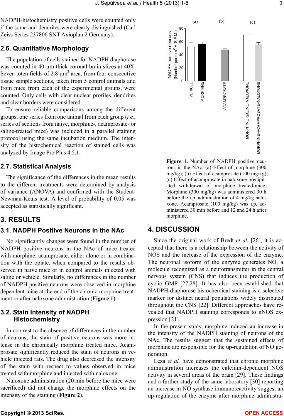 J. Sepúlveda et al. / Health 5 (2013) 1-6 3 NADPH-histochemistry positive cells were counted only if the soma and dendrites were clearly distinguished (Carl Zeiss Series 237806 SNT Axioplan 2 Germany). 2.6. Quantitative Morphology The population of cells stained for NADPH diaphorase was counted in 40 µm thick coronal brain slices at 40X. Seven toten fields of 2.8 µm2 area, from four consecutive tissue sample sections, taken from 5 control animals and from mice from each of the experimental groups, were counted. Only cells with clear nuclear profiles, dendrites and clear borders were considered. To ensure reliable comparisons among the different groups, one series from one animal from each group (i.e., series of sections from naive, morphine-, acamprosate- or saline-treated mice) was included in a parallel staining protocol using the same incubation medium. The inten- sity of the histochemical reaction of stained cells was analyzed by Image Pro Plus 4.5.1. 2.7. Statistical Analysis The significance of the differences in the mean results to the different treatments were determined by analysis of variance (ANOVA) and confirmed with the Student- Newman-Keuls test. A level of probability of 0.05 was accepted as statistically significant. 3. RESULTS 3.1. NADPH Positive Neurons in the NAc No significantly changes were found in the number of NADPH positive neurons in the NAc of mice treated with morphine, acamprosate, either alone or in combina- tion with the opiate, when compared to the results ob- served in naive mice or in control animals injected with saline or vehicle. Similarly, no differences in the number of NADPH positive neurons were observed in morphine dependent mice at the end of the chronic morphine treat- ment or after naloxone administration (Figure 1). 3.2. Stain Intensity of NADPH Histochemistry In contrast to the absence of differences in the number of neurons, the stain of positive neurons was more in- tense in the chronically morphine treated mice. Acam- prosate significantly reduced the stain of neurons in ve- hicle injected rats. The drug also decreased the intensity of the stain with respect to values observed in mice treated with morphine and injected with naloxone. Naloxone administration (20 min before the mice were sacrificed) did not change the morphine effects on the intensity of the staining (Figure 2). VEHICLE MORPHINE ACAMPROSATE MORPHINE+SALINE+NALOXONE MORPHINE+ACAMPROSATE+NALOXONE 0 20 40 60 80 AC B NADPH positi ve neur ons (Number per mm 2 ± S.E.M.) (a) (b) (c) Figure 1. Number of NADPH positive neu- rons in the NAc. (a) Effect of morphine (300 mg/kg); (b) Effect of acamprosate (100 mg/kg); (c) Effect of acamprosate in naloxone-precipit- ated withdrawal of morphine treated-mice. Morphine (300 mg/kg) was administered 30 h before the i.p. administration of 4 mg/kg nalo- xone. Acamprosate (100 mg/kg) was i.p. ad- ministered 30 min before and 12 and 24 h after morphine. 4. DISCUSSION Since the original work of Bredt et al. [26], it is ac- cepted that there is a relationship between the activity of NOS and the increase of the expression of the enzyme. The neuronal isoform of the enzyme generates NO, a molecule recognized as a neurotransmitter in the central nervous system (CNS) that induces the production of cyclic GMP [27,28]. It has also been established that NADPH-diaphorase histochemical staining is a selective marker for distinct neural populations widely distributed throughout the CNS [22]. Different approaches have re- vealed that NADPH staining corresponds to nNOS ex- pression [21]. In the present study, morphine induced an increase in the intensity of the NADPH staining of neurons of the NAc. The results suggest that the sustained effects of morphine are responsible for the up-regulation of NO ge- neration. Leza et al. have demonstrated that chronic morphine administration increases the calcium-dependent NOS activity in several areas of the brain [29]. These findings and a further study of the same laboratory [30] reporting an increase in NO synthase immunoreactivity suggest an up-regulation of the enzyme after morphine administra- Copyright © 2013 SciRes. OPEN A CCES S 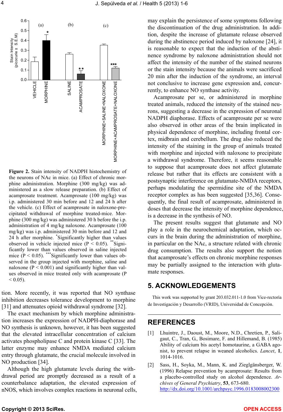 J. Sepúlveda et al. / Health 5 (2013) 1-6 4 VEHICLE MORPHINE SALINE ACAMPROSATE MORPHINE+SALINE+NALOXONE MORPHINE+ACAMPROSATE+NALOXONE 0.0 0.1 0.2 0.3 0.4 0.5 0.6 * **** ABC * S t ain Int ens i t y (picoc urie ± S. E . M ) (a) (b) (c) Figure 2. Stain intensity of NADPH histochemistry of the neurons of NAc in mice. (a) Effect of chronic mor- phine administration. Morphine (300 mg/kg) was ad- ministered as a slow release preparation. (b) Effect of acamprosate treatment. Acamprosate (100 mg/kg) was i.p. administered 30 min before and 12 and 24 h after the vehicle. (c) Effect of acamprosate in naloxone-pre- cipitated withdrawal of morphine treated-mice. Mor- phine (300 mg/kg) was administered 30 h before the i.p. administration of 4 mg/kg naloxone. Acamprosate (100 mg/kg) was i.p. administered 30 min before and 12 and 24 h after morphine. *Significantly higher than values observed in vehicle injected mice (P < 0.05). **Signi- ficantly lower than values observed in saline injected mice (P < 0.05). ***Significantly lower than values ob- served in the group injected with morphine, saline and naloxone (P < 0.001) and significantly higher than val- ues observed in mice treated only with acamprosate (P < 0.05). tion. More recently, it was reported that NO synthase inhibition decreases tolerance development to morphine [31] and attenuates opioid withdrawal syndrome [32]. The exact mechanism by which morphine administra- tion increases the expression of NADPH-diaphorase and NO synthesis is unknown, however, it has been suggested that the elevated intracellular concentration of calcium activates phospholipase C and protein kinase C [33]. The latter enzyme may enhance NMDA mediated calcium entry through glutamate, the crucial molecule involved in NO production [34]. Although the high glutamate levels during the with- drawal period are promptly decreased as a result of a counterbalance adaptation, the elevated expression of nNOS, which involves complex reactions in neuronal cells, may explain the persistence of some symptoms following the discontinuation of the drug administration. In addi- tion, despite the increase of glutamate release observed during the abstinence period induced by naloxone [24], it is reasonable to expect that the induction of the absti- nence syndrome by naloxone administration should not affect the intensity of the number of the stained neurons or the stain intensity because the animals were sacrificed 20 min after the induction of the syndrome, an interval not conclusive to increase gene expression and, concur- rently, to enhance NO synthase activity. Acamprosate per se, or administered in morphine treated animals, reduced the intensity of the stained neu- rons, suggesting a decrease in the expression of neuronal NADPH diaphorase. Effects of acamprosate per se were also observed in other areas of the brain implicated in physical dependence of morphine, including frontal cor- tex, midbrain and cerebellum. The drug also reduced the intensity of the staining in the group of animals treated with morphine and injected with naloxone to precipitate a withdrawal syndrome. Therefore, it seems reasonable to suppose that acamprosate does not affect glutamate release but rather that its effects are consistent with a postsynaptic interference on glutamate-NMDA receptors, perhaps modulating the spermidine site of the NMDA receptor complex as has been suggested [35,36]. Conse- quently, the final result of acamprosate, administered in doses that decrease the intensity of morphine dependence, is a decrease in the synthesis of NO. The present results suggest that glutamate and NO play a role in the neurochemical adaptation, which oc- curs in the brain during the administration of morphine, in particular on the NAc, a structure related with chronic drug consumption. The results also support the notion that acamprosate’s effects on chronic morphine responses may be partially assigned to the interaction with gluta- mate responses. 5. ACKNOWLEDGEMENTS This work was supported by grant 203.032.011-1.0 from Vice-rectoría de Investigación y Desarrollo (VRID), Universidad de Concepción. REFERENCES [1] Lhuintre, J., Daoust, M., Moore, N.D., Chretien, P., Sali- gaut, C., Tran, G., Bosimare, F. and Hillemand, B. (1985) Ability of calcium bis acetyl homotaurine, a GABA ago- nist, to prevent relapse in weaned alcoholics. Lancet, 1, 1014-1016. [2] Sass, H., Soyka, M., Mann, K. and Zieglgänsberger, W. (1996) Relapse prevention by acamprosate: Results from a placebo-controlled study on alcohol dependence. Ar- chives of General Psychiatry, 53, 673-680. http://dx.doi.org/10.1001/archpsyc.1996.0183008002300 Copyright © 2013 SciRes. OPEN A CCESS  J. Sepúlveda et al. / Health 5 (2013) 1-6 5 6 [3] Wilde, M.I. and Wagstaff, A.J. (1997) Acamprosate: A review of its pharmacology and clinical potential in the management of alcoholic dependence after detoxification. Drugs, 53, 1038-1053. http://dx.doi.org/10.2165/00003495-199753060-00008 [4] Chick, J., Howlett, H., Morgan, M.Y. and Ritson, B. (2000) United Kingdom Multicentre Acamprosate Study (UKMAS): A 6-month prospective study of acamprosate versus placebo in preventing relapse after withdrawal from alcohol. Alcohol and Alcoholism, 35, 176-187. http://dx.doi.org/10.1093/alcalc/35.2.176 [5] Han, D.H., Lyool, I.K., Sung, Y.H., Lee, S.H. and Ren- shaw, P.F. (2008) The effect of acamprosate on alcohol and food craving in patients with alcohol dependence. Drug and Alcohol Dependence, 93, 279-283. http://dx.doi.org/10.1016/j.drugalcdep.2007.09.014 [6] Spanagel, R. and Zieglgänsberger, W. (1997) Anti-craving compounds for ethanol: New pharmacological tools to study addictive processes. Trends in Pharmacological Sci- ences, 18, 54-59. http://dx.doi.org/10.1016/S0165-6147(97)89800-8 [7] Allgaier, C., Franke, H., Sobottka, H. and Scheibler, P. (2000) Acamprosate inhibits Ca2+ influx mediated by NMDA receptors and voltage-sensitive Ca2+ channels in cultured rat mesencephalicneurones. Naunyn-Schmiede- berg’s Archives of Pharmacology, 362, 440-443. http://dx.doi.org/10.1007/s002100000285 [8] Bachteler, D., Economidou, D., Danysz, W., Ciccocioppo, R. and Spanagel, R. (2005) The effects of acamprosate and neramexane on cue-induced reinstatement of etha- nol-seeking behavior in rat. Neuropsychopharmacology, 30, 1104-1110. http://dx.doi.org/10.1038/sj.npp.1300657 [9] Zeise, M.L., Kasparov, S., Capogna, M. and Zieglgäns- berger, W. (1993) Acamprosate (calcium acetyl homotau- rinate) decreases postsynaptic potentials in the rat neo- cortex: Possible involvement of excitatory amino acid re- ceptors. European Journal of Pharmacology, 231, 47-52. http://dx.doi.org/10.1016/0014-2999(93)90682-8 [10] Popp, R.L. and Lovinger, D.M. (2000) Interaction of acamprosate with ethanol and spermine on NMDA re- ceptors in primary cultured neurons. European Journal of Pharmacology, 394, 221-231. http://dx.doi.org/10.1016/S0014-2999(00)00195-3 [11] Koob, G.F., Wall, T.L. and Bloom, F.E. (1989) Nucleus accumbens as a substrate for the aversive stimulus effects of opiate withdrawal. Psychopharmacology, 98, 530-534. http://dx.doi.org/10.1007/BF00441954 [12] Stinus, L., Le Moal, M. and Koob, G.F. (1990) Nucleus accumbens and amygdale are possible substrate for the aversive stimulus effects of opiate withdrawal. Neurosci- ence, 37, 767-773. http://dx.doi.org/10.1016/0306-4522(90)90106-E [13] Pothos, E., Rada, P., Mark, G. and Hoebel, B.G. (1991) Dopamine microdialysis in the nucleus accumbens during acute and chronic morphine, naloxone precipitated with- drawal and clonidine treatment. Brain Research, 566, 348- 350. http://dx.doi.org/10.1016/0006-8993(91)91724-F [14] Rada, P.V., Mark, G.P., Taylor, K.M. and Hoebel, B.G. (1996) Morphine and naloxone ip or locally affect ex- tracellular acetylcholine in accumbens and prefrontal cor- tex. Pharmacology Biochemistry and Behavior, 53, 809- 816. http://dx.doi.org/10.1016/0091-3057(95)02078-0 [15] Sepúlveda, M.J., Hernandez, L., Rada, P., Tucci, S. and Contreras, E. (1998) Effect of precipitated withdrawal on extracellular glutamate and aspartate in the nucleus ac- cumbens of chronically morphine-treated rats: An in vivo microdialysis study. Pharmacology Biochemistry and Be- havior, 60, 255-262. http://dx.doi.org/10.1016/S0091-3057(97)00550-9 [16] Kielstein, A., Tsikas, D., Galloway, G.P., Mendelson, J.E. (2007) Asymmetric dimethylarginine (ADMA)—A mo- dulator of nociception in opiate tolerance and addiction? Nitric Oxide, 17, 55-59. http://dx.doi.org/10.1016/j.niox.2007.05.005 [17] Komatsu, T., Sakurada, C., Sasaki, M., Sanai, K., Tsuzuki, M., Bagetta, G., Sakurada, S., Sakurada, T. (2007) Ex- tracellular signal-regulated kinase (ERK) and nitric oxide synthase mediate intrathecal morphine-induced nocicep- tive behavior. Neuropharmacology, 52, 1237-1243. http://dx.doi.org/10.1016/j.neuropharm.2007.01.003 [18] Harlan, R.E., Webber, D.S. and Garcia, M.M. (2001) In- volvement of nitric oxide in morphine induced c-Fos ex- pression in the rat striatum. Brain Research Bulletin, 54, 207-212. http://dx.doi.org/10.1016/S0361-9230(00)00451-2 [19] Garthwaite, J. (1991) Glutamate, nitric oxide and cell-cell signaling in the nervous system. Trends in Neuroscience, 14, 60-67. http://dx.doi.org/10.1016/0166-2236(91)90022-M [20] Nestler, E., Alreja, M. and Aghajanian, M. (1994) Mo- lecular and cellular mechanisms of opiate action: Studies in the rat locus coeruleus. Brain Research Bulletin, 35, 521-528. http://dx.doi.org/10.1016/0361-9230(94)90166-X [21] Hope, B.T., Michael, G.J., Knigge, K.M. and Vincent, S.R. (1991) Neuronal NADPH diaphorase is a nitric oxide synthase. Proceedings of the National Academy of Sci- ences of the United States of America, 88, 2811-2814. http://dx.doi.org/10.1073/pnas.88.7.2811 [22] Vincent, S.R. and Kimura, H. (1992) Histochemical map- ping of nitric oxide synthase in the rat brain. Neurosci- ence, 46, 755-784. http://dx.doi.org/10.1016/0306-4522(92)90184-4 [23] Sepúlveda, M.J., Ortega, A., Zapata, G. and Contreras, E. (2002) Acamprosate decreases the induction of tolerance and physical dependence in morphine-treated mice. Euro- pean Journal of Pharmacology, 445, 87-91. http://dx.doi.org/10.1016/S0014-2999(02)01767-3 [24] Sepúlveda, J., Oliva, P. and Contreras, E. (2004) Neuro- chemical changes of the extracellular concentrations of glutamate and aspartate in the nucleus accumbens of rats after chronic administration of morphine. European Jour- nal of Pharmacology, 483, 249-258. http://dx.doi.org/10.1016/j.ejphar.2003.10.037 [25] Paxinos, G. and Franklin, K.B.J. (2001) The mouse brain in stereotaxic coordinates. 2nd Edition, Elsevier, New York. Copyright © 2013 SciRes. OPEN A CCES S  J. Sepúlveda et al. / Health 5 (2013) 1-6 Copyright © 2013 SciRes. OPEN A CCES S 6 [26] Bredt, D.S., Hwang, P.M., Glatt, C.E., Lowenstein, C., Reed, R.R. and Snyder, S.H. (1991) Cloned and ex- pressed nitric oxide synthase structurally resembles cyto- chrome P450 reductase. Nature, 351, 714-718. http://dx.doi.org/10.1038/351714a0 [27] Garthwaite, J., Charles, S.L. and Chess-Williams, R. (1988) Endothelium-derived relaxing factor release on activation of NMDA receptors suggests role as intercellular mes- senger in the brain. Nature, 33, 385. http://dx.doi.org/10.1038/336385a0 [28] Bredt, D.S. and Snyder, S.H. (1992) Nitric oxide, a novel neuronal messenger. Neuron, 8, 3-11. http://dx.doi.org/10.1016/0896-6273(92)90104-L [29] Leza, J.C., Lizasoain, I., San Martin-Clark, O. and Lor- enzo, P. (1995) Morphine-induced changes on cerebral and cerebellar nitric oxide synthase activity. European Journal of Pharmacology, 285, 95-98. http://dx.doi.org/10.1016/0014-2999(95)00474-Y [30] Cuellar, B., Fernandez, A.P., Lizasoain, I., Moro, M.A., Lorenzo, P., Bentura, M.L., Rodrigo, J. and Leza, J.C. (2000) Up-regulation of neuronal NO synthase immuno- reactivity in opiate dependence and withdrawal. Psycho- pharmacology, 148, 66-73. http://dx.doi.org/10.1007/s002130050026 [31] Santamarta, M.T., Ulibarri, I. and Pineda, J. (2005) Inhi- bition of neuronal nitric oxide synthase attenuates the development of morphine tolerance in rats. Synapse, 57, 38-46. http://dx.doi.org/10.1002/syn.20151 [32] Mori, T., Ito, S., Matsubayashi, K. and Sawaguchi, T. (2007) Comparison of nitric oxidesynthase inhibitors, phospholipase A2 inhibitor and free radical scavengers as attenuators of opioid withdrawal syndrome. Behavioural Pharmacology, 18, 725-729. http://dx.doi.org/10.1097/FBP.0b013e3282f18da6 [33] Smart, D. and Lambert, D.G. (1996) The stimulatory ef- fects of opioids and their possible role in the development of tolerance. Trends in Pharmacological Sciences, 7, 264- 269. http://dx.doi.org/10.1016/0165-6147(96)10023-7 [34] Chen, L. and Huang, L.Y. (1991) Sustained potentiation of NMDA receptor-mediated glutamate responses through activation of protein kinase C by a mu opioid. Neuron, 7, 319-326. http://dx.doi.org/10.1016/0896-6273(91)90270-A [35] Naassila, M., Hammoumi, S., Legrand, E., Durbin, P. and Daoust, M. (1998) Mechanism of action of acamprosate. Part I Characterization of spermidine-sensitive acampro- sate binding site in rat brain. Alcoholism: Clinical and Experimental Research, 22, 802-809. http://dx.doi.org/10.1111/j.1530-0277.1998.tb03871.x [36] Mayer, S., Harris, B., Gibson, D.A., Blanchard, J., Pren- dergast, M.A., Holley, R.C. and Littleton, J. (2002) Acam- prosate has no effect on NMDA-induced toxicity but re- duces toxicity induced by spermidine or by changing the medium in organotypic hippocampal slice cultures from rat. Alcoholism: Clinical and Experimental Research, 26, 655-662. http://dx.doi.org/10.1111/j.1530-0277.2002.tb02587.x |