

Paper Menu >>
Journal Menu >>
 Vol.3, No.4, 221-226 (2013) Journal of Diabetes Mellitus http://dx.doi.org/10.4236/jdm.2013.34034 Anti-diabetic and antioxidant effects of virgin coconut oil in alloxan induced diabetic male Sprague Dawley rats Bolanle Iranloye1*, Gabriel Oludare1, Makinde Olubiyi1,2 1Department of Physiology, College of Medicine, University of Lagos, Lagos, Nigeria; *Corresponding Author: [email protected] 2Department of Physiology, Kogi State University, Ayangba, Nigeria Received 25 September 2013; revised 20 October 2013; accepted 28 October 2013 Copyright © 2013 Bolanle Iranloye et al. This is an open access article distributed under the Creative Commons Attribution License, which permits unrestricted use, distribution, and reproduction in any medium, provided the original work is properly cited. ABSTRACT Oxidative stress has been discovered to be in- volved in the progression of diabetes mellitus. The antioxidant properties of virgin coconut oil (VCO) among other functions might have a be- neficial effect in ameliorating the disease. This study w as aimed to determine the glycemic and antioxidant effects of VCO in alloxan induced diabetic rats. 24 male Sprague-Dawley rats w ere divided into 4 group s as follows: control (C), dia- betes untreated (DUT), diabetes treated with 7.5 ml/kg VCO (DT7.5) and diabetes treated with 10 ml/kg VCO (DT10). Alloxan (100 mg/kg b.w I.P) was used to induce diabetes and VCO was ad- ministered orally once daily for 4 w eeks. Fasting blood glucose level w as measured on Day 0 (72 hours post alloxan injection) and after 4 weeks. Glucose tolerance test was conducted on the 4th week as well as the determination of serum insulin and liver antioxidant parameters using standard biochemical methods. Values are means ± S.E.M., compared by ANOVA and Tukey’s post hoc test. The result s show that VCO significantly reduced the fasting blood glucose level in DT7.5 rats (132.4 ± 6.911) and DT10 rats (131.6 ± 12.2) are compared with DUT rats (320.4 ± 22.99) and improved the oral glucose tolerance. Serum in- sulin was increased in DT10 rats. GSH activities significantly increased p < 0.05 in DT10 rats (0.39 ± 0.022) when compared to DUT rats (0.032 ± 0.004). CAT activities also significantly increased p < 0.05 in DT7.5 (17.63 ± 0.61) and DT10 rats (30.88 ± 0.97) w hen compared to DUT rats (10.98 ± 0.6). SOD activities significantly increased p < 0.05 in DT7.5 (2.634 ± 0.04) and DT10 rats (2.258 ± 0.32) when compared to DUT rats (1.366 ± 0.05) while MDA significantly reduced p < 0.05 in DT7.5 (49.16 ± 0.51) and DT10 (33.64 ± 0.42) rats when compared to DUT rats (99.93 ± 4.79). This study revealed that VCO has a hypoglycemic action, enhances insulin secretion and also ameliorates oxidative stress induced in type I (alloxan-in- duced diabetic) male rats. Keywords: Virgin Coconut Oil; Oxidative Stress; Blood Glucose; Glucose Tolerance 1. INTRODUCTION Diabetes mellitus characterized by hyperglycaemia, is due to the deficiency of insulin secretion or its action. It has been associated with a syndrome of disturbance in the homeostasis of carbohydrate, fat and protein metabo- lism [1]. Diabetes mellitus has been categorized into type 1 and type 2 diabetes. Type 1 diabetes refers to defi- ciency of endogenous insulin which is caused by a cellu- lar-mediated auto immune destruction of the beta cells in the pancreas which produces insulin. And type 2 diabetes is as a result of a decreased response to insulin by its receptors, which is also referred to as insulin resistance [1,2]. Oxidative stress contributes significantly to the patho- physiology of several diseases which include diabetes [3]. Alloxan, a chemical used in inducing diabetes acts mainly by the generation of reactive oxygen species (ROS) [3]. It preferentially accumulates in the GLUT2 glucose transporter in the pancreatic beta cells and sub- sequently leads to the death of the cells. Therefore al- loxan is a model compound when studying diabetes as a Copyright © 2013 SciRes. OPEN ACCESS  B. Iranloye et al. / Journal of Diabetes Mellitus 3 (2013) 221-226 222 result of ROS mediated beta cell toxicity. Historically, coconut oil has been renowned for its medicinal and nutritional value. Studies on the biological effects of coconut oil have proven that it ameliorates oxidative stress by boosting the antioxidant defense sys- tem, mopping up free radicals and reducing lipid peroxi- dation [4,5]. It has also been reported to suppress micro- bial and viral activities [6], promote weight loss and en- hance thyroid function [7]. Other researches have also re- ported that coconut oil possesses anti-inflammatory and anti-ulcerogenic effect [8], while also having the ability to increase the level of high density lipoprotein (HDL) cholesterol and to reduce the level of low density lipo- protein (LDL) in serum and tissues [4]. Copra oil and virgin coconut oil (VCO) are the two main types of coconut oil. Copra oil is extracted from the dried endosperm of the coconut fruit while VCO is pro- duced by a “wet” extraction process from the fresh en- dosperm of the coconut fruit [9]. The mode of extraction of VCO makes it more beneficial than copra oil. This is because no chemicals are used and there is little or no application of heat during its extraction. Therefore it re- tains more of the natural active components which in- clude polyphenols which have been proven to boost the antioxidant defense system [4]. The present study therefore, determined the possible role of the antioxidant effect of VCO on oxidation/per- oxidation linked with diabetes mellitus in alloxan in- duced diabetic rats as well as its possible effect on glu- cose homeostasis. 2. MATERIALS AND METHODS 2.1. Animals Male Sprague-Dawley rats weighing 120 - 150 g were obtained from the Laboratory Animal House of the Col- lege of Medicine of the University of Lagos. The rats were allowed to acclimatize for two weeks before the commencement of the experiment and were fed with standard rat chow and water ad libitum at 20˚C - 25˚C under a 12 h light/dark cycle. All animal handling and experiment protocols complied with the international guidelines for laboratory animals as supported by the College of Medicine of the University of Lagos ethical committee. 2.2. Experimental Groups Rats were randomly divided into 4 groups (n = 6): Group 1, control (C) received 0.5 ml distilled water; Group 2, diabetic untreated (DUT); Group 3, diabetic treated with 7.5 ml/kg body weight of VCO (DT7.5) and Group 4, diabetic treated with 10 ml/kg body weight of VCO (DT10). Seventy two hours following the induction of diabetes, VCO was administered orally for 4 weeks daily at the appropriate dose for Groups 3 and 4 animals. 2.3. Induction of Diabetes Following 2 weeks acclimatization of the rats, Alloxan monohydrate (manufactured by Denixco Private limited, India) was used to induce type 1 diabetes in Groups 2, 3 and 4. A dose of 100 mg/kg body weight of Alloxan monohydrate was administered only once intraperito- neally. A mild pressure was applied at the spot of injec- tion to enhance absorption. After 3 days of administra- tion the fasting blood glucose level of these rats were measured. Rats with fasting blood glucose level above 200 mg/dl were considered diabetic. 2.4. Measurement of Blood Glucose Blood glucose level was measured using One Touch Ultra test strips (Lifescan Inc. Milpitas, USA). Blood was obtained from the rats at the tip of rat’s tail. The blood was dropped on the test strips already inserted in a One Touch Ultra Easy Glucometer (Lifescan Inc. Milpitas, USA). The glucose levels of the animal were displayed on the glucometer in about 5 seconds. Blood glucose level was measured at the beginning of the ex- periment and after 4 weeks. 2.5. Preparation of Virgin Coconut Oil Mature coconuts were bought from Mushin Market, Lagos, Nigeria. VCO was extracted using the wet extrac- tion method [4]. The solid endosperm of mature coconut was crushed and made into thick slurry. About 500 ml of water was added to the slurry obtained and squeezed through a fine sieve to obtain coconut milk. The resultant coconut milk was left for about 24 hours to facilitate the gravitational separation of the emulsion. Demulsification produced layers of an aqueous phase (water) on the bot- tom, an emulsion phase (cream) in the middle layer and an oil phase on top. The oil on top was scooped and warmed for about 3 minutes to remove moisture. The obtained oil was then filtered and stored at room tem- perature. 2.6. Oral Glucose Tolerance Test (OGTT) On the 4th week of the experiment, all groups were subjected to oral glucose tolerance test (OGTT). The rats were fasted overnight for sixteen-hour (16-h) and subse- quently challenged with a glucose load of 2 ug/kg body weight. Blood glucose levels were determined at 0 h (pre-glucose treatment) and at 30, 60, 90, 120 and 180 min (post glucose treatment). The glucose levels were measured using a complete blood glucose monitoring system (One-Touch Ultra Easy Glucose Meter, Lifescan Copyright © 2013 SciRes. OPEN ACCES S 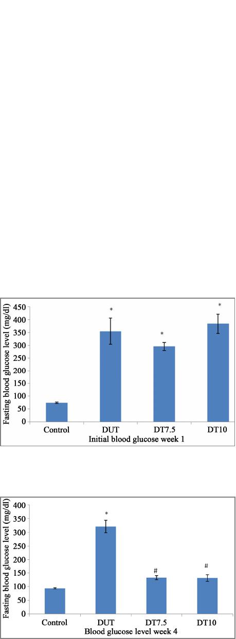 B. Iranloye et al. / Journal of Diabetes Mellitus 3 (2013) 221-226 223 Inc. Milpitas, USA). 2.7. Sample Collection The rats were anesthetized by intramuscular injection of 50 mg/kg of ketamine. The liver was removed and homogenized in phosphate buffer, pH 7.4 and stored at −20˚C. Blood samples were also collected from the ven- tricle of the heart, allowed to clot and spun at 3000 rmp to obtain serum sample for insulin assay. 2.8. MDA L evel As a marker of lipid peroxidation, the level of malon- dialdehyde (MDA) in the liver homogenate was meas- ured [10]. 1 ml of the tissue homogenate was thoroughly mixed with 2 ml of TCA-TBA-HCl solution and heated for 15 minutes in a water bath. After cooling, the pre- cipitate is removed by centrifugation and the absorbance measured at 535 nm is taken as an index of lipid peroxi- dation. 2.9. SOD, CAT and GSH Activities At the end of the 4 week period of the experiment, the activity of the superoxide dismutase (SOD) enzyme in the liver homogenate was determined [11]. The reaction was carried out in 0.5m sodium carbonate buffer pH 10.2 and was initiated by the addition of 3 × 10⁻⁴ epinephrine in 0.005 N HCl. The absorbance was read at 320 nm. Catalase (CAT) activity was determined by measuring the exponential disappearance of H2O2 at 240 nm and expressed in units/mg of protein [12]. Reduced glu- tathione (GSH) content of the liver homogenate was de- termined [13], based on the reaction of Ellman’s reagent 5,5’dithiobis-2-nitrobenzoic acid (DNTB) with the thiol group of GSH at pH 8.0 to produce 5-thiol-2-nitroben- zoate which is yellow at 412 nm. Absorbance was re- corded using UV-Visible Spectrophotometer in all meas- urement. The protein concentrations of the samples were measured using the method of Bradford [14]. 2.10. Serum Insulin Level Enzyme-Linked Immunosorbent Assay (ELISA) was used to measure the level of insulin in the serum sample obtained from the animal. The protocol used was as de- scribed by the manufacturer of the assay kit (Enzo-Life Science). 2.11. Statistical Analysis Data were presented as mean and Standard Error of Mean (SEM). One-way ANOVA and Tukey’s post hoc test was used to determine the specific pairs of groups that were statistically different at p < 0.05. Analysis was performed with GraphPad software. 3. RESULTS 3.1. Fasting Blood Glucose Fasting blood glucose was measured at 72 hours post alloxan injection. Hyperglycemia was observed in DUT, DT7.5, and DT10 rats. After four weeks of coconut oil treatment, DT7.5 and DT10 rats showed a significant re- duction (p < 0.05) in fasting blood glucose level com- pared with DUT rats (Figures 1 and 2). 3.2. Oral Glucose Tolerance Test (OGTT) After 4 weeks administration of coconut oil, glucose concentration (pre glucose challenge) in both DT7.5 and DT10 rats showed a significantly reduced glucose con- centration when compared with DUT rats. As expected, there was an initial increase in blood glucose 30 minutes post glucose challenge which reduced over time as pre- sented in Figure 2. Three hours post glucose challenge showed that DT7.5 and DT10 significantly reduced blood glucose level when compared with DUT rats. The effect Figure 1. Fasting blood glucose level (mg/dl) of diabetic rats induced with alloxan. Values are expressed as mean ± S.E.M. *p < 0.05 is significant compared with Group 1 (control). Figure 2. Effect of 4 weeks VCO supplementation on fasting blood glucose level (mg/dl) in alloxan induced diabetic rats. Values are expressed as mean ± S.E.M. *p < 0.05 is significant compared with Group 1 (control). #p < 0.05 is significant com- pared with Group 2 (diabetes untreated). Copyright © 2013 SciRes. OPEN ACCES S 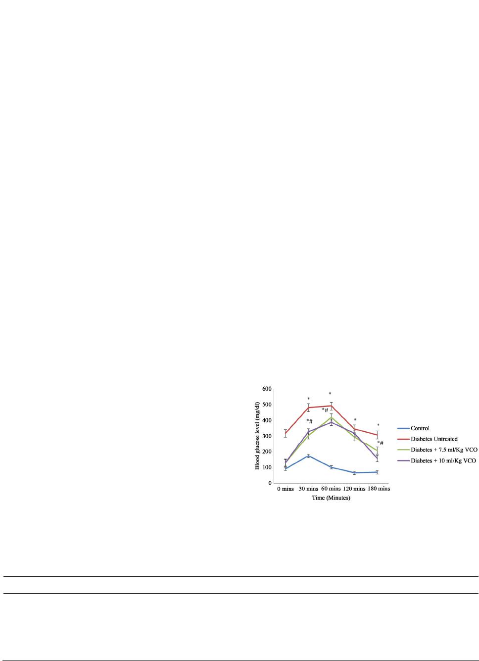 B. Iranloye et al. / Journal of Diabetes Mellitus 3 (2013) 221-226 Copyright © 2013 SciRes. 224 of DT10 was more effective than that of DT7.5 (Figure 3). antioxidative defence capacity, thus the generation of ROS by alloxan leads to the death of these cells. This is possible due to the reduction product of the reaction, dialuric acid, which generates hydrogen peroxide, su- peroxide radicals and hydroxyl radicals. These radicals are responsible for the death of the beta cells and the ensuing state of insulin-dependent alloxan diabetes [3]. 3.3. Antioxidant Enzymes Activities and MDA Levels MDA level was significantly increased in DUT rats when compared to control (p < 0.05), however, MDA levels were significantly reduced in DT7.5 and DT10 rats compared with DUT rats. Though a significant reduction in DT7.5 rats was observed when compared with DUT rats, the value observed compared with the control still shows lipid peroxidation (Table 1). In the antioxidant enzymes, SOD activity was significantly reduced in DUT rats when compared with control. DT7.5 and DT10 sig- nificantly increased the activity of SOD when compared with DUT rats. The enhancement in SOD activity how- ever was still significantly lower compared with the con- trol values. The activity of GSH was significantly re- duced in DUT rats when compared with control. How- ever, DT10 rats significantly increased the activity of GSH while DT7.5 had no effect on the activity of GSH when compared with DUT rats. Lastly, CAT activity was reduced in DUT treated rats when compared with control rats. DT7.5 and DT10 significantly increased the activity of CAT when compared with DUT rats. DT10 enhanced this activity more than the control rats while this activity was still decreased in DT7.5 rats compared to control rats (Table 1). This study reports marked hyperglycemia 72 hours post alloxan injection (100 mg/kg body weight). This is supported by other previous studies and reports [15-17]. Four weeks of treatment with VCO decreased the fasting blood glucose level in DT7.5 and DT10 rats when com- pared with DUT rats. Supporting the report that coconut oil has a hypoglycemic effect [18,19]. Since, alloxan generates ROS to impair the beta cell function; it is pos- sible that VCO alleviates blood glucose level due to its antioxidant property. It is possible that the beta cells re- sponse to oxidative stress might have been enhanced thus enabling the cells to carry out their function of insulin production. Consequently, this increase in insulin pro- duction will lead to reduced blood glucose. Oral glucose tolerance test is used to measure insulin function or the degree of peripheral utilization of glucose [20]. In this study, following glucose administration, there was a minimal rise in the blood glucose level which fell below the control value after 2 hours in the control rats. In DUT, DT7.5 and DT10 rats there was a marked rise in blood glucose level after the glucose challenge and the blood glucose level failed to return to the control value 3.4. Serum Insulin Level Table 2 shows the serum insulin level of male rats treated with VCO. DUT rats shows a significantly re- duced insulin level when compared control. DT10 alone significantly increased the level of insulin when com- pared with DUT rats. Though the values of DT7.5 were increased it was however not significant and the values obtained was still significantly lower than those of the control rats. 4. DISCUSSION Figure 3. Effect of virgin coconut oil (VCO) on oral glucose tolerance test (OGTT). Values are expressed as mean ± S.E.M. *p < 0.05 is significant compared with Group 1 (control). #p < 0.05 is significant compared with Group 2 (diabetes untreated). Alloxan, used in inducing diabetes is a toxic glucose analogue that generates ROS in the presence of intracel- lular thiols [3]. The beta cells of the pancreas have a low Table 1. Effect of VCO on the activity of superoxide dismutase, glutathione, catalase and malondialdehyde levels. Control (C)Diabetes untreated (DUT)Diabetes + 7.5 ml/kg VCO (DT7.5) Diabetes + 10 ml/kg VCO (DT10) MDA (U/mg protein) 31.78 ± 2.1899.93 ± 4.79* 49.16 ± 0.51*# 33.64 ± 0.42# SOD (U/mg protein) 3.91 ± 0.14 1.37 ± 0.05* 2.63 ± 0.04*# 2.26 ± 0.32*# GSH (U/mg protein) 0.11 ± 0.0070.03 ± 0.004* 0.04 ± 0.008* 0.39 ± 0.022*# CAT (U/mg protein) 25.87 ± 0.9610.98 ± 0.60* 17.63 ± 0.61*# 30.88 ± 0.97*# V alues are expressed as mean ± S.E.M. *p < 0.05 is significant compared with control. #p < 0.05 is significant compared with diabetes untreated group. OPEN ACCESS  B. Iranloye et al. / Journal of Diabetes Mellitus 3 (2013) 221-226 225 Table 2. Effect of virgin coconut oil (VCO) on serum insulin level. Insulin level (μiu/ml) Control (C) 3.05 ± 0.25 Diabetes untreated (DUT) 1.19 ± 0.04* Diabetes + 7.5 ml/kg VCO (DT7.5) 1.90 ± 0.40* Diabetes + 10 ml/kg VCO (DT10) 2.50 ± 0.03# Values are expressed as mean ± S.E.M. *p < 0.05 is significant compared with Control. #p < 0.05 is significant compared with Diabetes untreated group. even after 3 hours indicating impairment in glucose tol- erance which is an indication of diabetes. In DT7.5 and DT10 rats there was a significant improvement in glucose tolerance compared with the DUT rats, supporting the view that ingestion of VCO improves glucose tolerance in diabetic rats [21]. In addition, the 10 ml/kg dosage of VCO proved to have a greater effect as the blood glucose level in DT10 rats after 3 hours of glucose challenge was closer to the control value than in DT7.5 rats. It has been reported that the lauric oil in VCO pos- sesses insulino-tropic properties [18]. Serum insulin was increased in DT10 rats with a non significant increase in DT7.5 rats compared with DUT rats. Since the dosage of 7.5 ml/kg body weight of VCO could not elevate serum insulin, it implies that the 10 ml/kg body weight dose is more effective in the control of glucose homeostasis than the 7.5 ml/kg body weight. This is evidenced by the re- duction in blood glucose level and the improvement in glucose tolerance compared to DUT rats as discussed earlier. Antioxidant enzymes are critical part of cellular pro- tection against reactive oxygen species and ultimately oxidative stress. Oxidative stress is determined by the balance between the generation of ROS such as super- oxide anion (2 O) and the antioxidant defense systems such as superoxide dismutase (SOD). Antioxidants en- zymes involved in the elimination of ROS include SOD, CAT and GSH, respectively. The present study showed a decrease in the activity of all measured antioxidants en- zymes in DUT rats. This indicates a decrease in the anti- oxidant defense system. However treatment with VCO in DT7.5 and DT10 rats increased the activities of the anti- oxidant enzymes. Since oxidative stress contributes sig- nificantly to the pathophysiology of diabetes [22], sub- stances that suppress oxidative stress might be therapeu- tically beneficial. Studies have shown that exogenously administered antioxidants have protective effects on dia- betes, thus providing insight into the relationship be- tween free radicals and diabetes [20,22-24]. The reduc- tion in fasting blood glucose of rats treated with VCO after 4 weeks and a decrease in the OGTT of the rats compared with the diabetic untreated rats can be associ- ated to the antioxidant effect of VCO. 5. CONCLUSION VCO alleviates hyperglycemia and improves glucose tolerance probably by its antioxidant effect which con- sequently leads to improvement of insulin secretion as examined in this study. The study also shows that a dos- age of 10 ml/kg body weight of VCO is quite beneficial and more effective than that of 7.5 ml/kg body weight. This was evident because the 7.5 ml/kg body weight did not increase insulin secretion and possibly because of the higher OGTT values. REFERENCES [1] American Diabetes Association (2010) Diagnosis and classification of diabetes mellitus. Diabetic Care, 33. [2] World Health Organization (2011) Diabetes fact sheet. Sheet number 312 August. [3] Lenzen, S. (2008) The mechanisms of alloxan- and strep- tozotocin-induced Diabetes. Diabetologia, 51, 216-226. http://dx.doi.org/10.1007/s00125-007-0886-7 [4] Nevin, K.G. and Rajamohan, T. (2006) Virgin coconut oil supplemented diet increases the antioxidant status in rats. Food Chemistry, 99, 260-266. http://dx.doi.org/10.1016/j.foodchem.2005.06.056 [5] Dosumu, O.O., Duru, F.I.O., Osinubi, A.A., Oremosu, A.A. and Noronha, C.C. (2010) Influence of virgin coco- nut oil (VCNO) on oxidative stress, serum testosterone and gonadotropic hormones (FSH, LH) in chronic ethanol ingestion. Agriculture and Biology Journal of North Ame- rica, 1, 1126-1132. [6] Van Immerseel, F., De Buck, J. and Boyen, F. (2004) Medium-chain fatty acids decrease colonization and in- vasion through hilA suppression shortly after infection of chickens with Salmonella enterica serovar enteritidis. Ap- plied and Environmental Microbiology, 70, 3582-3587. http://dx.doi.org/10.1128/AEM.70.6.3582-3587.2004 [7] Takeuchi, H., Sekine, S., Kojima, K. and Aoyama, T. (2008) The application of medium-chain fatty acids: Edi- ble oil with a suppressing effect on body fat accumulation. Asia Pacific Journal of Clinical Nutrition, 17, 320-324. [8] Anosike, C.A. and Obidoa, O. (2010) Anti-inflammatory and anti-ulcerogenic effect of ethanol extract of coconut (Cocos nucifera) on experimental rats. African Journal of Food, Agriculture, Nutrition and Development, 10, 10-16. [9] Cretney, J. and Tafunai, A. (2004) Tradition, trade, and technology: Virgin coconut oil in samoa. In Chains of Fortune: Linking Women Producers and Workers with Global Markets, 45-74. [10] Uchiyama, M. and Mihara, M. (1978) Determination of malonaldehyde precursor in tissues by thiobarbituris acid test. Analytical Biochemistry, 86, 271-278. http://dx.doi.org/10.1016/0003-2697(78)90342-1 [11] Sun, M. and Zigmam S. (1978) An improved spectro- Copyright © 2013 SciRes. OPEN ACCES S  B. Iranloye et al. / Journal of Diabetes Mellitus 3 (2013) 221-226 226 photomeric assay for superoxide dismutase based on epi- nephrine autooxidation. Analytical Biochemistry, 90, 81- 89. http://dx.doi.org/10.1016/0003-2697(78)90010-6 [12] Aebi, H. (1984) Catalase in vitro. Methods in Enzymology, 8, 121-126. http://dx.doi.org/10.1016/S0076-6879(84)05016-3 [13] Van Dooran, R., Liejdekker, C.M. and Handerson, P.T. (1978) Synergistic effects of phorone on the hepatotoxic- ity of bromobenzene and paracetamol in mice. Toxicology, 11, 225-233. http://dx.doi.org/10.1016/S0300-483X(78)91389-6 [14] Bradford, M.M. (1976) A rapid and sensitive method for the quantification of microgram quantities of protein util- izing the principle of protein-dye binding. Analytical Bio- chemistry, 72, 248-254. http://dx.doi.org/10.1016/0003-2697(76)90527-3 [15] Szkudelski, T. (2001) The mechanism of alloxan and streptozotocin action in B cells of the rat pancreas. Phy- siological Research, 50, 536-546. [16] Lenzen, S., Tiedge, M., Jorns, A. and Munday, R. (1996) Alloxan derivatives as a tool for the elucidation of the mechanism of the diabetogenic action of alloxan. In: Shafrir, E., Ed., Lessons from Animal Diabetes. Birk- häuser, Boston, 113-122. http://dx.doi.org/10.1007/978-1-4612-4112-6_8 [17] Jorns, A., Munday, R., Tiedge, M. and Lenzen, S. (1997) Comparative toxicity of alloxan, N-alkylalloxans and nin- hydrin to isolated pancreatic islets in vitro. Journal of Endocrinology, 155, 283-293. http://dx.doi.org/10.1677/joe.0.1550283 [18] Garfinkel, M., Lee, S., Opara, E.C. and Akwari, O.E. (1992) Insulinotropic potency of lauric acid. A metabolic rationale for medium chain fatty acids (MCF) in TPN formulation. Journal of Surgical Research, 52, 328-333. http://dx.doi.org/10.1016/0022-4804(92)90111-C [19] Sadikot, S.M. (2005) Coconut for health nutrition. Jakarta, APCC, 6. [20] Kim, S.S., Gallaher, D.D. and Csallany, A.S. (2000) Vi- tamin E and probucol reduce urinary lipophilic aldehydes and renal enlargement in streptozotocin induced diabetic rats. Lipids, 35, 1225-1237. http://dx.doi.org/10.1007/s11745-000-0639-2 [21] Siddalingaswamy, M., Rayaorth, A. and Khanum, F. (2011) Anti-diabetic effects of cold and hot extracted vir- gin coconut oil. Journal of Diabetes Mellitus, 1, 118-123. http://dx.doi.org/10.4236/jdm.2011.14016 [22] Maritim, A.C., Sanders, R.A. and Watkins, J.B. (2003) Diabetes, oxidative stress and antioxidants: A review. Jour- nal of Biochemical and Molecular Toxicology, 17, 24-38. http://dx.doi.org/10.1002/jbt.10058 [23] Mekinova, D., Chorvathova, V., Volkovova, K., Staru- chova, M., Grancicova, F., Klvanova, J., Nevin, K.G. and Rajamohan, T. (2004) Beneficial effects of virgin coconut oil on lipid parameters and in vitro LDL oxidation. Clini- cal Biochemistry, 37, 830-835. http://dx.doi.org/10.1016/j.clinbiochem.2004.04.010 [24] Borenshtein, D.R., Ofri, M., Werman, A., Stark, H.J., Tritschler, W. and Moeller Madar Z. (2001) Cataract de- velopment in diabetic sand rats treated with alpha-lipoic acid and its gamma-linolenic acid conjugate. Diabetes/ Metabolism Research and Reviews, 17, 44-50. http://dx.doi.org/10.1002/1520-7560(0000)9999:9999<:: AID-DMRR153>3.0.CO;2-S Copyright © 2013 SciRes. OPEN ACCES S |