

Paper Menu >>
Journal Menu >>
 Surgical Science, 2013, 4, 1-7 Published Online December 2013 (http://www.scirp.org/journal/ss) http://dx.doi.org/10.4236/ss.2013.412A001 Open Access SS Surgical Technique Affects the Incidence of Marginal Ulceration after Roux-en-Y in Gastric Bypass Yong Kwon Lee, Corrigan McBride, Valerie Shostrom, Jon Thompson Department of Surgery, College of Public Health, University of Nebraska Medical Center, Omaha, USA Email: jthom[email protected] Received September 18, 2013; revised October 15, 2013; accepted October 23, 2013 Copyright © 2013 Yong Kwon Lee et al . This is an open access article distributed under the Creative Commons Attribution License, which permits unrestricted use, distribution, and reproduction in any medium, provided the original work is properly cited. ABSTRACT Background: Marginal ulceration (MU) is a recognized complication of Roux-en-Y gastric bypass (RYGB) surgery. While several possible risk factors have been reported, the mechanism of MU remains incompletely understood. The aim of this study was to compare the effect of surgical technique on the incidence of MU. Methods and Material: This was a retrospective study of 749 patients undergoing RYGB over a ten-year period with at least one year of follow-up. The diagnosis of MU was made based on clinical symptoms and confirmed by postoperative endoscopy (POE). We assessed four different gastric bypass (GBP) techniques: T1—Open, non-divided stomach, circular stapler, non-vago- tomy (n = 332); T2—Open, divided stomach, circular stapler, vagotomy (n = 91); T3—Laparoscopic, divided, circular stapler, vagotomy (n = 152); T4—Laparoscopic, divided, linear stapler, vagotomy (n = 174). Results: The four groups were similar with respect to age and mean BMI. The frequency of POE was 16%, 25%, 28% and 27% in groups T1-T4 respectively (NS). The incidence of MU was significantly lower in T1 (2.1%) compared to T2 (5.5%), T3 (15.1%) and T4 (12.6%), p < 0.05. MU occurred significantly more frequently with an antecolic Roux limb versus retrocolic (14.5% vs 5.6%, p < 0.05). Conclusion: The incidence of MU after RYGB surgery is influenced by surgical technique. The lowest incidence of MU was with a non-divided stomach, no vagotomy, transverse staple line, and circular stapled an- astomosis. A retrocolic Roux limb was protective. There was no difference in the incidence of MU using laparoscopic versus open bypass if a similar technique was employed or using linear versus circular stapler for the gastrojeju- nostomy. Keywords: Gastric Bypass; Marginal Ulceration 1. Introduction Marginal ulceration (MU) is a well-known complication after Roux-en-Y gastric bypass (RYGB). The symptoms can range from minimal abdominal discomfort to life threatening bleeding or perforation [1]. The reported in- cidence of MU has varied widely in the literature from 1% to 16% [1-10]. These differences may be related to differences in surgical technique and patient factors, but also vary depending on definition, method of detection and follow-up. The mechanism of MU formation after RYGB is in- completely understood. Several technical factors have been implicated, including pouch size and orientation, an association with foreign material, staple line disruption and gastrogastric fistula, and anastomotic technique [5,9, 11,12]. A variety of patient-related factors such as non- steroidal antiinflammatory medication (NSAID), helico- bacter pylori infection and smoking may also be impor- tant [13]. The aim of the present study was to evaluate the influence of different surgical techniques of RYGB on MU. 2. Methods Patients This was a retrospective study of 749 adult patients (>19 years of age), who underwent primary open or laparo- scopic RYGB procedures. Inclusion criteria were preop- erative BMI ≥ 35, patient with unsuccessful non-surgical weight loss treatment and one of the four RYGB tech- niques received (Table 1). We had excluded techniques that had less than 50 procedures performed (n = 18) and revisional procedures (n = 90). All operations were per- formed by four surgeons at a single institution from Oc- tober 1995 to November 2005. Overall, there were 653 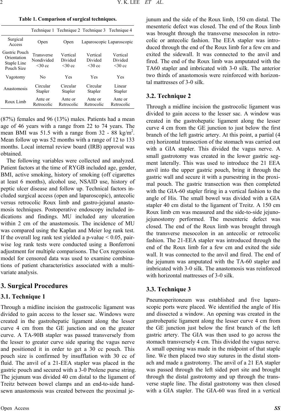 Y. K. LEE ET AL. 2 Table 1. Comparison of surgical techniques. Technique 1 Technique 2 Technique 3 Technique 4 Surgical Access Open Open Laparoscopic Laparoscopic Gastric Pouch Orientation Staple Line Pouch Size Transverse Nondivided <30 cc Vertical Divided <30 cc Vertical Divided <30 cc Vertical Divided <30 cc Vagotomy No Yes Yes Yes Anastomosis Circular Stapler Circular Stapler Circular Stapler Linear Stapler Roux Limb Ante or Retrocolic Ante or Retrocolic Ante or Retrocolic Ante or Retrocolic (87%) females and 96 (13%) males. Patients had a mean age of 46 years with a range from 22 to 74 years. The mean BMI was 51.5 with a range from 32 - 88 kg/m2. Mean follow up was 52 months with a range of 12 to 133 months. Local internal review board (IRB) approval was obtained. The following variables were collected and analyzed. Patient factors at the time of RYGB included age, gender, BMI, active smoking, history of smoking (off cigarettes at least 6 months), alcohol use, NSAID use, history of peptic ulcer disease and follow up. Technical factors in- cluded surgical access (open and laparoscopic), antecolic versus retrocolic Roux limb and gastro-jejunal anasto- mosis techniques. Postoperative endoscopy included in- dications and findings. MU included any ulceration within 2 cm of the anastomosis. The incidence of MU was compared using the Kaplan and Meier log rank test. If the overall log rank test yielded a p-value < 0.05, pair- wise log rank tests were conducted using a Bonferroni adjustment for multiple comparisons. The Cox regression model for censored data was used to examine combina- tions of patient characteristics associated with a multi- variate analysis. 3. Surgical Procedures 3.1. Technique 1 Through a midline incision the gastrocolic ligament was divided to gain access to the lesser sac. Windows were created in the gastrohepatic ligament along the lesser curve 4 cm from the GE junction and on the greater curve. A TA-90B stapler was passed transversely from the lesser to greater curve side sparing the vagus nerve and positioned it in order to get a 30 cc pouch. This pouch size is confirmed by insufflation with 30 cc of fluid. The anvil of a 21-EEA stapler was placed in the gastric pouch and secured with a 3-0 Prolene purse string. The jejunum was divided 40 cm distal to the ligament of Treitz between bowel clamps and an end-to-side hand- sewn anastomosis was created between the proximal je- junum and the side of the Roux limb, 150 cm distal. The mesenteric defect was closed. The end of the Roux limb was brought through the transverse mesocolon in retro- colic or antecolic fashion. The EEA stapler was intro- duced through the end of the Roux limb for a few cm and exited the sidewall. It was connected to the anvil and fired. The end of the Roux limb was amputated with the TA60 stapler and imbricated with 3-0 silk. The anterior two thirds of anastomosis were reinforced with horizon- tal mattresses of 3-0 silk. 3.2. Technique 2 Through a midline incision the gastrocolic ligament was divided to gain access to the lesser sac. A window was created in the gastrohepatic ligament along the lesser curve 4 cm from the GE junction to just below the first branch of the left gastric artery. At this point, a partial (4 cm) horizontal transection of the stomach was carried out with a GIA stapler. This divided the vagus nerve. A small gastrotomy was created in the lower gastric seg- ment laterally. This was used to introduce the 21 EEA anvil into the upper gastric pouch, bring it through the gastric wall and secure it with a pursestring in the proxi- mal pouch. The gastric transection was then completed with the GIA-60 stapler firing in a vertical fashion to the angle of His. The small bowel was divided with a GIA stapler 40 cm distal to the ligament of Treitz. A 150 cm Roux limb cm was measured and the side-to-side jejuno- jejunostomy performed. The mesenteric defect was closed. The end of the Roux limb was brought through the transverse mesocolon in an antecolic or retrocolic fashion. The 21-EEA stapler was introduced through the end of the Roux limb for a few cm and exited the side wall. It was connected to the anvil and fired. The end of the jejunum was amputated with the TA-60 stapler and imbricated with 3-0 silk. The anastomosis was reinforced with horizontal mattresses of 3-0 silk. 3.3. Technique 3 Pneumoperitoneum was established and five laparo- scopic ports were placed. We identified the angle of His and dissected a window. An opening was created in the gastrohepatic ligament along the lesser curve 4 cm from the GE junction just below the first branch of the left gastric artery. The GIA was then used to go across the stomach transversely 4 cm. This divided the vagus nerve. A small opening was made in the midpoint of that staple line. We then placed two stay sutures in the distal stom- ach and made a gastrotomy. The anvil of a 21 EA stapler was passed through the left sided port site and brought through the distal gastrotomy and up through the trans- verse staple line. The distal gastrotomy was then closed with a GIA stapler. The GIA-60 was fired in a vertical Open Access SS 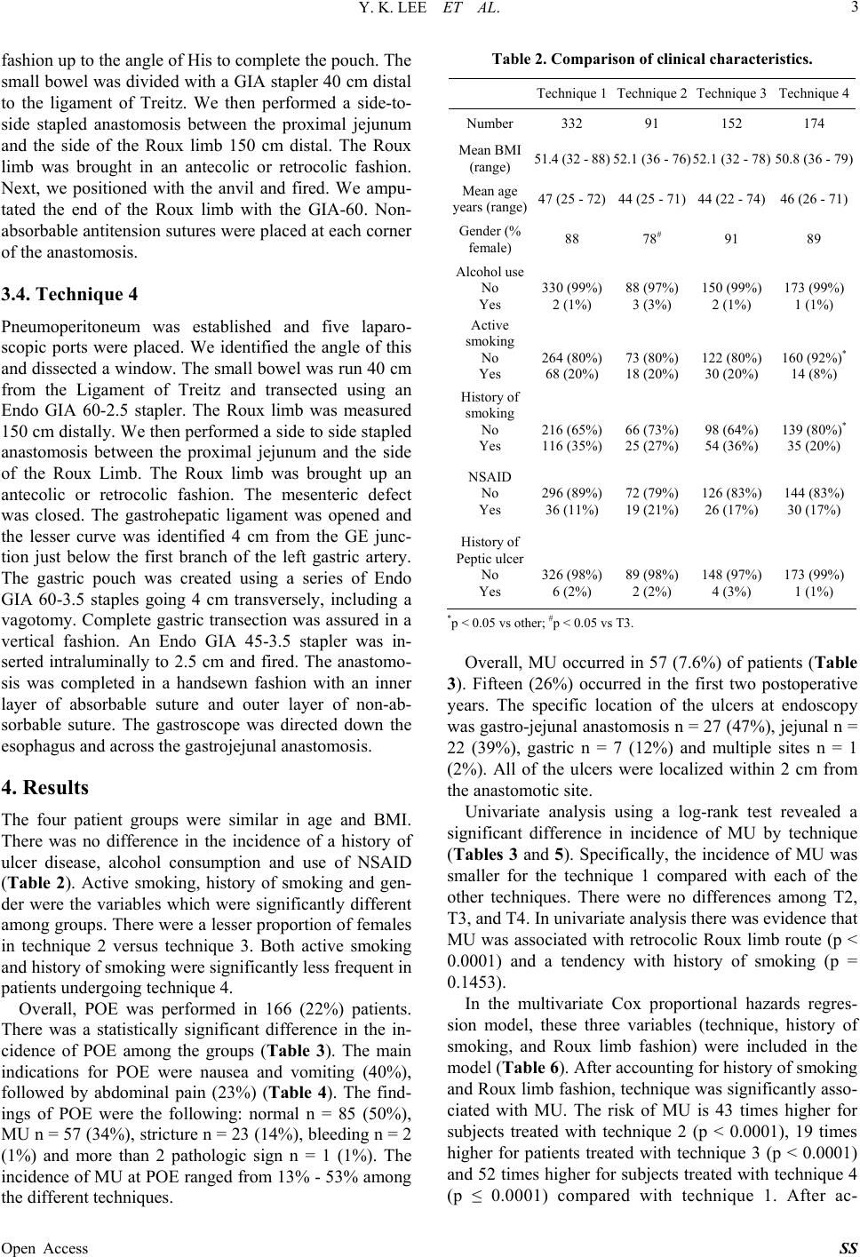 Y. K. LEE ET AL. 3 fashion up to the angle of His to complete the pouch. The small bowel was divided with a GIA stapler 40 cm distal to the ligament of Treitz. We then performed a side-to- side stapled anastomosis between the proximal jejunum and the side of the Roux limb 150 cm distal. The Roux limb was brought in an antecolic or retrocolic fashion. Next, we positioned with the anvil and fired. We ampu- tated the end of the Roux limb with the GIA-60. Non- absorbable antitension sutures were placed at each corner of the anastomosis. 3.4. Technique 4 Pneumoperitoneum was established and five laparo- scopic ports were placed. We identified the angle of this and dissected a window. The small bowel was run 40 cm from the Ligament of Treitz and transected using an Endo GIA 60-2.5 stapler. The Roux limb was measured 150 cm distally. We then performed a side to side stapled anastomosis between the proximal jejunum and the side of the Roux Limb. The Roux limb was brought up an antecolic or retrocolic fashion. The mesenteric defect was closed. The gastrohepatic ligament was opened and the lesser curve was identified 4 cm from the GE junc- tion just below the first branch of the left gastric artery. The gastric pouch was created using a series of Endo GIA 60-3.5 staples going 4 cm transversely, including a vagotomy. Complete gastric transection was assured in a vertical fashion. An Endo GIA 45-3.5 stapler was in- serted intraluminally to 2.5 cm and fired. The anastomo- sis was completed in a handsewn fashion with an inner layer of absorbable suture and outer layer of non-ab- sorbable suture. The gastroscope was directed down the esophagus and across the gastrojejunal anastomosis. 4. Results The four patient groups were similar in age and BMI. There was no difference in the incidence of a history of ulcer disease, alcohol consumption and use of NSAID (Table 2). Active smoking, history of smoking and gen- der were the variables which were significantly different among groups. There were a lesser proportion of females in technique 2 versus technique 3. Both active smoking and history of smoking were significantly less frequent in patients undergoing technique 4. Overall, POE was performed in 166 (22%) patients. There was a statistically significant difference in the in- cidence of POE among the groups (Table 3). The main indications for POE were nausea and vomiting (40%), followed by abdominal pain (23%) (Table 4). The find- ings of POE were the following: normal n = 85 (50%), MU n = 57 (34%), stricture n = 23 (14%), bleeding n = 2 (1%) and more than 2 pathologic sign n = 1 (1%). The incidence of MU at POE ranged from 13% - 53% among the different techniques. Table 2. Comparison of clinical characteristi cs. Technique 1Technique 2 Technique 3 Technique 4 Number 332 91 152 174 Mean BMI (range) 51.4 (32 - 88)52.1 (36 - 76) 52.1 (32 - 78) 50.8 (36 - 79) Mean age years (range)47 (25 - 72)44 (25 - 71) 44 (22 - 74) 46 (26 - 71) Gender (% female) 88 78# 91 89 Alcohol use No Yes 330 (99%) 2 (1%) 88 (97%) 3 (3%) 150 (99%) 2 (1%) 173 (99%) 1 (1%) Active smoking No Yes 264 (80%) 68 (20%) 73 (80%) 18 (20%) 122 (80%) 30 (20%) 160 (92%)* 14 (8%) History of smoking No Yes 216 (65%) 116 (35%) 66 (73%) 25 (27%) 98 (64%) 54 (36%) 139 (80%)* 35 (20%) NSAID No Yes 296 (89%) 36 (11%) 72 (79%) 19 (21%) 126 (83%) 26 (17%) 144 (83%) 30 (17%) History of Peptic ulcer No Yes 326 (98%) 6 (2%) 89 (98%) 2 (2%) 148 (97%) 4 (3%) 173 (99%) 1 (1%) *p < 0.05 vs other; #p < 0.05 vs T3. Overall, MU occurred in 57 (7.6%) of patients (Table 3). Fifteen (26%) occurred in the first two postoperative years. The specific location of the ulcers at endoscopy was gastro-jejunal anastomosis n = 27 (47%), jejunal n = 22 (39%), gastric n = 7 (12%) and multiple sites n = 1 (2%). All of the ulcers were localized within 2 cm from the anastomotic site. Univariate analysis using a log-rank test revealed a significant difference in incidence of MU by technique (Tables 3 and 5). Specifically, the incidence of MU was smaller for the technique 1 compared with each of the other techniques. There were no differences among T2, T3, and T4. In univariate analysis there was evidence that MU was associated with retrocolic Roux limb route (p < 0.0001) and a tendency with history of smoking (p = 0.1453). In the multivariate Cox proportional hazards regres- sion model, these three variables (technique, history of smoking, and Roux limb fashion) were included in the model (Table 6). After accounting for history of smoking and Roux limb fashion, technique was significantly asso- ciated with MU. The risk of MU is 43 times higher for subjects treated with technique 2 (p < 0.0001), 19 times higher for patients treated with technique 3 (p < 0.0001) and 52 times higher for subjects treated with technique 4 p ≤ 0.0001) compared with technique 1. After ac- ( Open Access SS 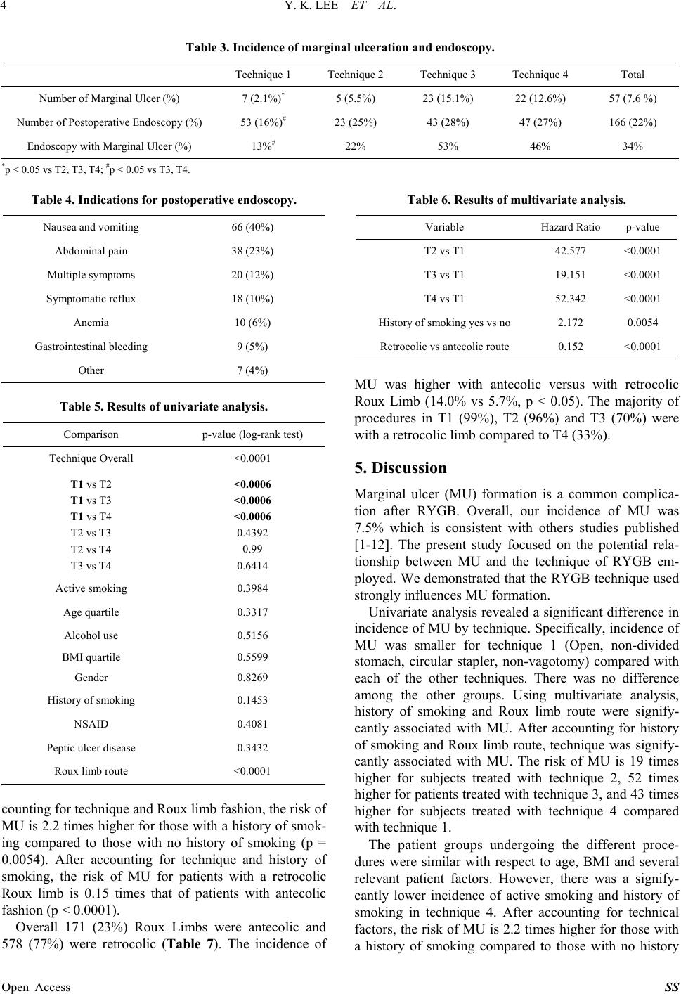 Y. K. LEE ET AL. Open Access SS 4 Table 3. Incidence of marginal ulceration and endoscopy. Technique 1 Technique 2 Technique 3 Technique 4 Total Number of Marginal Ulcer (%) 7 (2.1%)* 5 (5.5%) 23 (15.1%) 22 (12.6%) 57 (7.6 %) Number of Postoperative Endoscopy (%) 53 (16%)# 23 (25%) 43 (28%) 47 (27%) 166 (22%) Endoscopy with Marginal Ulcer (%) 13%# 22% 53% 46% 34% *p < 0.05 vs T2, T3, T4; #p < 0.05 vs T3, T4. Table 4. Indications for postoperative endoscopy. Nausea and vomiting 66 (40%) Abdominal pain 38 (23%) Multiple symptoms 20 (12%) Symptomatic reflux 18 (10%) Anemia 10 (6%) Gastrointestinal bleeding 9 (5%) Other 7 (4%) Table 5. Results of univariate analysis. Comparison p-value (log-rank test) Technique Overall <0.0001 T1 vs T2 T1 vs T3 T1 vs T4 T2 vs T3 T2 vs T4 T3 vs T4 <0.0006 <0.0006 <0.0006 0.4392 0.99 0.6414 Active smoking 0.3984 Age quartile 0.3317 Alcohol use 0.5156 BMI quartile 0.5599 Gender 0.8269 History of smoking 0.1453 NSAID 0.4081 Peptic ulcer disease 0.3432 Roux limb route <0.0001 counting for technique and Roux limb fashion, the risk of MU is 2.2 times higher for those with a history of smok- ing compared to those with no history of smoking (p = 0.0054). After accounting for technique and history of smoking, the risk of MU for patients with a retrocolic Roux limb is 0.15 times that of patients with antecolic fashion (p < 0.0001). Overall 171 (23%) Roux Limbs were antecolic and 578 (77%) were retrocolic (Table 7). The incidence of Table 6. Results of multivariate analysis. Variable Hazard Ratio p-value T2 vs T1 42.577 <0.0001 T3 vs T1 19.151 <0.0001 T4 vs T1 52.342 <0.0001 History of smoking yes vs no 2.172 0.0054 Retrocolic vs antecolic route 0.152 <0.0001 MU was higher with antecolic versus with retrocolic Roux Limb (14.0% vs 5.7%, p < 0.05). The majority of procedures in T1 (99%), T2 (96%) and T3 (70%) were with a retrocolic limb compared to T4 (33%). 5. Discussion Marginal ulcer (MU) formation is a common complica- tion after RYGB. Overall, our incidence of MU was 7.5% which is consistent with others studies published [1-12]. The present study focused on the potential rela- tionship between MU and the technique of RYGB em- ployed. We demonstrated that the RYGB technique used strongly influences MU formation. Univariate analysis revealed a significant difference in incidence of MU by technique. Specifically, incidence of MU was smaller for technique 1 (Open, non-divided stomach, circular stapler, non-vagotomy) compared with each of the other techniques. There was no difference among the other groups. Using multivariate analysis, history of smoking and Roux limb route were signify- cantly associated with MU. After accounting for history of smoking and Roux limb route, technique was signify- cantly associated with MU. The risk of MU is 19 times higher for subjects treated with technique 2, 52 times higher for patients treated with technique 3, and 43 times higher for subjects treated with technique 4 compared with technique 1. The patient groups undergoing the different proce- dures were similar with respect to age, BMI and several relevant patient factors. However, there was a signify- cantly lower incidence of active smoking and history of smoking in technique 4. After accounting for technical factors, the risk of MU is 2.2 times higher for those with a history of smoking compared to those with no history 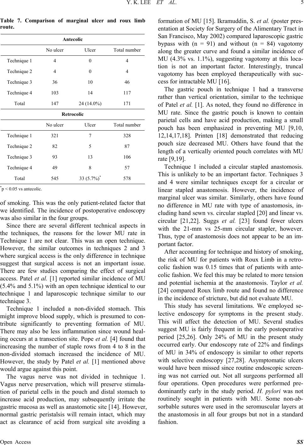 Y. K. LEE ET AL. 5 Table 7. Comparison of marginal ulcer and roux limb route. Antecolic No ulcer Ulcer Total number Technique 1 4 0 4 Technique 2 4 0 4 Technique 3 36 10 46 Technique 4 103 14 117 Total 147 24 (14.0%) 171 Retrocolic No ulcer Ulcer Total number Technique 1 321 7 328 Technique 2 82 5 87 Technique 3 93 13 106 Technique 4 49 8 57 Total 545 33 (5.7%)* 578 *p < 0.05 vs antecolic. of smoking. This was the only patient-related factor that we identified. The incidence of postoperative endoscopy was also similar in the four groups. Since there are several different technical aspects in the techniques, the reasons for the lower MU rate in Technique 1 are not clear. This was an open technique. However, the similar outcomes in techniques 2 and 3 where surgical access is the only difference in technique suggest that surgical access is not an important issue. There are few studies comparing the effect of surgical access. Patel et al. [1] reported similar incidence of MU (5.4% and 5.1%) with an open technique identical to our technique 1 and laparoscopic technique similar to our technique 3. Technique 1 included a non-divided stomach. This might improve blood supply, which is presumed to con- tribute significantly to preventing formation of MU. There may also be less inflammation since wound heal- ing occurs at a transection site. Pope et al. [4] found that increasing the number of staple rows from 4 to 8 in the non-divided stomach increased the incidence of MU. However, the study by Patel et al. [1] mentioned above would argue against this point. The vagus nerve was not divided in technique 1. Vagus nerve preservation, which will preserve stimula- tion of parietal cells in the pouch and distal stomach to increase acid production, may subsequently irritate the gastric mucosa as well as anastomotic site [14]. However, normal gastric peristalsis will remain intact, which may act as clearance of acid from surgical site avoiding a formation of MU [15]. Ikramuddin, S. et al. (poster pres- entation at Society for Surgery of the Alimentary Tract in San Francisco, May 2002) compared laparoscopic gastric bypass with (n = 91) and without (n = 84) vagotomy along the greater curve and found a similar incidence of MU (4.3% vs. 1.1%), suggesting vagotomy at this loca- tion is not an important factor. Interestingly, truncal vagotomy has been employed therapeutically with suc- cess for intractable MU [16]. The gastric pouch in technique 1 had a transverse rather than vertical orientation, similar to the technique of Patel et al. [1]. As noted, they found no difference in MU rate. Since the gastric pouch is known to contain parietal cells and have acid production, making a small pouch has been emphasized in preventing MU [9,10, 12,14,17,18]. Printen [18] demonstrated that reducing pouch size decreased MU. Others have found that the length of a vertically oriented pouch correlates with MU rate [9,19]. Technique 1 included a circular stapled anastomosis. This is unlikely to be an important factor. Techniques 3 and 4 were similar techniques except for a circular or linear stapled anastomosis. However, the incidence of marginal ulcer was similar. Similarly, others have found no difference in MU rate with type of anastomosis, in- cluding hand sewn vs. circular stapled [20] and linear vs. circular [21,22]. Suggs et al. [23] found fewer ulcers with the 21-mm vs 25-mm circular stapler, however. Thus, type of anastomosis does not appear to be an im- portant factor. After accounting for technique and history of smoking, the risk of MU for patients with Roux Limb in a retro- colic fashion was 0.15 times that of patients with ante- colic fashion. We feel this may be related to more tension and potential ischemia at the anastomosis. Taylor et al. [24] compared Roux limb route and found no difference in the incidence of stricture, but did not evaluate MU. This study has several limitations. We employed se- lective endoscopy for symptoms in the present study. This will affect the detection of MU. Several studies suggest MU is fairly frequent in the early postoperative period [25,26]. Only 24% of MU in the present study occurred early. Our endoscopy rate of 22% and findings of MU in 34% of endoscopy is similar to other reports with selective endoscopy [27,28]. Asymptomatic ulcers would have been missed since routine endoscopic screen- ing was not carried out. Not all surgeons performed all four operations. Open procedures were performed pre- dominantly early in the study period. H. pylori was not routinely sought in patients with MU. Some non-ab- sorbable sutures were used in the seromuscular layers at the anastomosis in all four groups but not in a standard fashion. Open Access SS  Y. K. LEE ET AL. 6 6. Conclusion In conclusion, the incidence of MU after RGBP surgery is influenced by surgical technique. The overall 7.5% incidence of MU is consistent with other studies. The lowest incidence of MU was the technique with a non- divided stomach, no vagotomy, and a circular anastomo- sis. A retrocolic Roux limb was protective. There was no difference of MU using linear or circular stapler for the gastrojejunostomy and no difference in laparoscopic ver- sus open bypass if a similar technique was employed. REFERENCES [1] R. A. Patel, R. E. Brolin and A. Gandhi, “Revisional Ope- rations for Marginal Ulcer after Roux-en-Y Gastric By- pass,” Surgery for Obesity and Related Diseases, Vol. 5, No. 3, 2009, pp. 317-322. http://dx.doi.org/10.1016/j.soard.2008.10.011 [2] L. D. MacLean, B. M. Rhode, C. Nohr, S. Katz and A. P. McLean, “Stomal Ulcer after Gastric Bypass,” Journal of the American College of Surgeons, Vol. 185, No. 1, 1997, pp. 1-7. http://dx.doi.org/10.1016/S1072-7515(01)00873-0 [3] J. F. Capella and R. F. Capella, “Gastro-Gastric Fistulas and Marginal Ulcers in Gastric Bypass Procedures for Weight Reduction,” Obesity Surgery, Vol. 9, No. 1, 1999, pp. 22-27. http://dx.doi.org/10.1381/096089299765553674 [4] G. D. Pope, P. P. Goodney, K. W. Burchard, R. R. Proia, A. Olafsson, B. E. Lacy and L. J. Burrows, “Peptic Ul- cer/Stricture after Gastric Bypass: A Comparison of Tech- nique and Acid Suppression Variables,” Obesity Surgery, Vol. 12, No. 1, 2002, pp. 30-33. http://dx.doi.org/10.1381/096089202321144540 [5] B. C. Sacks, S.G. Mattar, F.G. Qureshi, G.M. Eid, J.L. Collins, E.J. Barinas-Mitchell, P.R. Schauer and R.C. Ramanathan, “Incidence of Marginal Ulcers and the Use of Absorbable Anastomotic Sutures in Laparoscopic Roux- en-Y Gastric Bypass,” Surgery for Obesity and Related Diseases, Vol. 2, No. 1, 2006, pp. 11-16. http://dx.doi.org/10.1016/j.soard.2005.10.013 [6] R. M. Dallal and L. A. Bailey, “Ulcer Disease after Gas- tric Bypass Surgery,” Surgery for Obesity and Related Diseases, Vol. 2, No. 4, 2006, pp. 455-459. http://dx.doi.org/10.1016/j.soard.2006.03.004 [7] A. A. Gumbs, A. J. Duffy and R. L. Bell, “Incidence and Management of Marginal Ulceration after Laparoscopic Roux-Y Gastric Bypass,” Surgery for Obesity and Re- lated Diseases, Vol. 2, No. 4, 2006, pp. 460-463. http://dx.doi.org/10.1016/j.soard.2006.04.233 [8] J. A. Wilson, J. Romagnuolo, T. K. Byrne, K. Morgan and F. A. Wilson, “Predictors of Endoscopic Findings af- ter Roux-en-Y Gastric Bypass,” The American Journal of Gastroenterology, Vol. 101, No. 10, 2006, pp. 2194- 2199. http://dx.doi.org/10.1111/j.1572-0241.2006.00770.x [9] J. H. Jordan, M. P. Hocking, W. R. Rout and E. R. Wood- ward, “Marginal Ulcer Following Gastric Bypass for Mor- bid Obesity,” The American Surgeon, Vol. 57, No. 5, 1991, pp. 286-288. [10] J. A. Sapala, M. H. Wood, M. A. Sapala and T. M. Flake Jr., “Marginal Ulcer after Gastric Bypass: A Prospective 3-Year Study of 173 Patients,” Obesity Surgery, Vol. 8, No. 5, 1998, pp. 505-516. http://dx.doi.org/10.1381/096089298765554061 [11] E. E. Frezza, H. Herbert, R. Ford and M. S. Wachtel, “En- doscopic Suture Removal at Gastrojejunal Anastomosis after Roux-en-Y Gastric Bypass to Prevent Marginal Ul- ceration,” Surgery for Obesity and Related Diseases, Vol. 3, No. 6, 2007, pp. 619-622. http://dx.doi.org/10.1016/j.soard.2007.08.019 [12] J. Hedberg, H. Hedenstrom, S. Nilsson, M. Sundbom and S. Gustavsson, “Role of Gastric Acid in Stomal Ulcer af- ter Gastric Bypass,” Obesity Surgery, Vol. 15, No. 10, 2005, pp. 1375-1378. http://dx.doi.org/10.1381/096089205774859380 [13] C. S. Yang, W. J. Lee, H. H. Wang, S. P. Huang, J. T. Lin and M. S. Wu, “The Influence of Helicobacter Pylori In- fection on the Development of Gastric Ulcer in Sympto- matic Patients after Bariatric Surgery,” Obesity Surgery, Vol. 16, No. 6, 2006, pp. 735-739. http://dx.doi.org/10.1381/096089206777346754 [14] H. Siilin, A. Wanders, S. Gustavsson and M. Sundbom, “The Proximal Gastric Pouch Invariably Contains Acid- Producing Parietal Cells in Roux-en-Y Gastric Bypass,” Obesity Surgery, Vol. 15, No. 6, 2005, pp. 771-777. http://dx.doi.org/10.1381/0960892054222849 [15] J. A. Sapala, M. H. Wood and M. P. Schuhknecht, “Va- gotomy at the Time of Gastric Bypass: Can It Be Harm- ful?” Obesity Surgery, Vol. 14, No. 5, 2004, pp. 575-576. http://dx.doi.org/10.1381/096089204323093327 [16] J. Hunter, R. D. Stahl, M. Kakade, I. Breitman, J. Grams and R. H. Clements, “Effectiveness of Thoracoscopic Truncal Vagotomy in the Treatment of Marginal Ulcers after Laparoscopic Roux-en-Y Gastric Bypass,” The Ame- rican Surgeon, Vol. 78, No. 6, 2012, pp. 663-668. [17] E. E. Mason, J. R. Munns, G. P. Kealey, R. Wangler, W. R. Clarke, H. F. Cheng and K. J. Printen, “Effect of Gas- tric Bypass on Gastric Secretion,” American Journal of Surgery, Vol. 131, No. 2, 1976, pp. 162-168. http://dx.doi.org/10.1016/0002-9610(76)90090-8 [18] K. J. Printen, D. Scott and E. E. Mason, “Stomal Ulcers after Gastric Bypass,” Archives of Surgery, Vol. 115, No. 4, 1980, pp. 525-527. http://dx.doi.org/10.1001/archsurg.1980.01380040147026 [19] D. E. Azagury, B. K. Abu Dayyeh, I. T. Greenwalt and C. C. Thompson, “Marginal Ulceration after Roux-en-Y Gas- tric Bypass Surgery: Characteristics, Risk Factors, Treat- ment and Outcomes,” Endoscopy, Vol. 43, No. 11, 2011, pp. 950-954. http://dx.doi.org/10.1055/s-0030-1256951 [20] R. Gonzalez, E. Lin, K. R. Venkatesh, S. P. Bowers and C. D. Smith, “Gastrojejunostomy during Laparoscopic Gastric Bypass: Analysis of 3 Techniques,” Archives of Surgery, Vol. 138, No. 2, 2003, pp. 181-184. [21] F. P. Bendewald, J. N. Choi, L. S. Blythe, D. J. Selzer, J. H. Ditslear and S. G. Mattar, “Comparison of Hand-Sewn, Linear-Stapled, and Circular-Stapled Gastrojejunostomy Open Access SS  Y. K. LEE ET AL. Open Access SS 7 in Laparoscopic Roux-en-Y Gastric Bypass,” Obesity Surgery, Vol. 21, No. 11, 2011, pp. 1671-1675. http://dx.doi.org/10.1007/s11695-011-0470-6 [22] S. Giordano, P. Salminen, F. Biancari and M. Victorzon, “Linear Stapler Technique May Be Safer than Circular in Gastrojejunal Anastomosis for Laparoscopic Roux-en-Y Gastric Bypass: A Meta-analysis of Comparative Stud- ies,” Obesity Surgery, Vol. 21, No. 12, 2011, pp. 1958- 1964. http://dx.doi.org/10.1007/s11695-011-0520-0 [23] W. J. Suggs, W. Kouli, M. Lupovici, W. Y. Chau and R. E. Brolin, “Complications at Gastrojejunostomy after La- paroscopic Roux-en-Y Gastric Bypass: Comparison Be- tween 21-and 25-mm Circular Staplers,” Surgery for Obe- sity and Related Diseases, Vol. 3, No. 5, 2007, pp. 508- 514. http://dx.doi.org/10.1016/j.soard.2007.05.003 [24] J. D. Taylor, I. M. Leitman, J. B. Rosser, B. Davis and E. Goodman, “Does the Position of the Alimentary Limb in Roux-en-Y Gastric Bypass Surgery Make a Difference?” Journal of Gastrointestinal Surge ry, Vol. 10, No. 10, 2006, pp. 1397-1399. http://dx.doi.org/10.1016/j.gassur.2006.09.007 [25] K. El-Hayek, P. Timratana, H. Shimizu and B. Chand, “Marginal Ulcer after Roux-en-Y Gastric Bypass: What Have We Really Learned?” Surgical Endoscopy, Vol. 26, No. 10, 2012, pp. 2789-2796. http://dx.doi.org/10.1007/s00464-012-2280-x [26] A. B. Garrido Jr., M. Rossi, S. E. Lima Jr., A. S. Brenner and C. A. Gomes, “Early Marginal Ulcer Following Roux- en-Y Gastric Bypass Under Proton Pump Inhibitor Treat- ment: Prospective Multicentric Study,” Arquivos de Gas- troenterologia, Vol. 47, No. 2, 2010, pp. 130-134. http://dx.doi.org/10.1590/S0004-28032010000200003 [27] J. A. Wilson, J. Romagnuolo, T. K. Byrne, K. Morgan and F. A. Wilson, “Predictors of Endoscopic Findings af- ter Roux-en-Y Gastric Bypass,” The American Journal of Gastroenterolo g y, Vol. 101, No. 10, 2006, pp. 2194-2199. http://dx.doi.org/10.1111/j.1572-0241.2006.00770.x [28] F. Obeid, A. Falvo, H. Dabideen, J. Stocks, M. Moore and M. Wright, “Open Roux-en-Y Gastric Bypass in 925 Patients without Mortality,” American Journal of Surgery, Vol. 189, No. 3, 2005, pp. 352-356. http://dx.doi.org/10.1016/j.amjsurg.2004.11.023 |