

Paper Menu >>
Journal Menu >>
 International Journal of Otolaryngology and Head & Neck Surgery, 2013, 2, 271-275 Published Online November 2013 (http://www.scirp.org/journal/ijohns) http://dx.doi.org/10.4236/ijohns.2013.26056 Open Access IJOHNS Giant R etropha ryngeal Abscess in a Nigerian Adult Following Fish Bone Throat Injury Stanley B. Amutta1*, Daniel Aliyu1, Mohammed Abdullahi1, Mohammed Danfulani2, Yunusa A. Mufutau3 1ENT Department, Usmanu Danfodiyo University Teaching Hospital, Sokoto, Nigeria 2Radiology Department, Usmanu Danfodiyo University Teaching Hospital, Sokoto, Nigeria 3Psychiatry Department, Usmanu Danfodiyo University Teaching Hospital, Sokoto, Nigeria Email: *[email protected] Received August 26, 2013; revised September 25, 2013; accepted October 6, 2013 Copyright © 2013 Stanley B. Amutta et al. This is an open access article distributed under the Creative Commons Attribution Li- cense, which permits unrestricted use, distribution, and reproduction in any medium, provided the original work is properly cited. ABSTRACT Retropharyngeal abscess (RPA) is an uncommon disease entity that typically occurs in children under 5 years of age, which has the potential to cause upper airway obstruction. Upper respiratory tract infection is the most common predis- posing factor to RPA in children while cervical spine tuberculosis and trauma are the major aetiological factors in the adult. We report a case of a 21-year-old Nigerian farmer with giant retropharyngeal abscess following fish bone throat injury causing life-threatening upper airway obstruction. The patient responded well to emergency tracheostomy, intra- oral surgical drainage and empirical antibiotic therapy. We highlight the diagnostic and therapeutic challenges of man- aging such a patient. Keywords: Giant Retropharyngeal Abscess; Fish Bone; Tracheostomy; Antibiotic; Intra-Oral Drainage 1. Introduction Retropharyngeal abscess (RPA) though an uncommon disease entity occurs usually in children under the age of 5 years [1-3]. However, recent findings indicate that adults could also be involved [4,5]. The involvement of the airway results in upper airway obstruction which could lead to life threatening condition thus, it is a surgi- cal emergency. The pathological basis for the higher prevalence in children is due to the loose aggregate of lymph nodes in the prevertebral space which may be- come infected with resultant suppuration. These lymph nodes are not usually present in adults because they at- rophy while the child grows; hence retropharyngeal ab- scess is not a common disorder among adults. Upper respiratory tract infection is the most common predis- posing factor to RPA in children [3-6] while cervical spine tuberculosis and trauma are the major aetiological factors in adults [3-5,7]. Other factors reported among adults include diabetes mellitus [4] and human immuno- deficiency virus (HIV) infection [7,8]. In a retrospective study in Ibadan, Nigeria [9], thirty cases of retropharyngeal abscess were reported compris- ing of 25 children and 5 adults. In that study, adult pa- tients presented early while the children presented late. A recent study from north central Nigeria reported RPA due to fish bone but in a child [5]. We report a case of giant retropharyngeal abscess in a 21-year-old subsis- tence adult Nigerian farmer following fish bone throat injury while highlighting the diagnostic and therapeutic challenges of managing this type of patient. 2. Case Report A 21-year-old subsistence farmer, presented at the Emer- gency department of our hospital with 10-day history of progressive sore throat, persistent low grade fever, neck pain, dyspnoea, odynophagia and voice changes. There was an antecedent history of fish bone impaction in the throat two weeks prior to the onset of symptoms however; the patient did not seek medical intervention. He was unable to take both fluid and solid diet at presentation. There was associated hoarseness and inability to sit or stand without support. There was no history suggestive of recurrent tonsillitis, dental infection, chronic cough, night sweats or contact with a patient with pulmonary tuberculosis and was not a known diabetic. *Corresponding author. 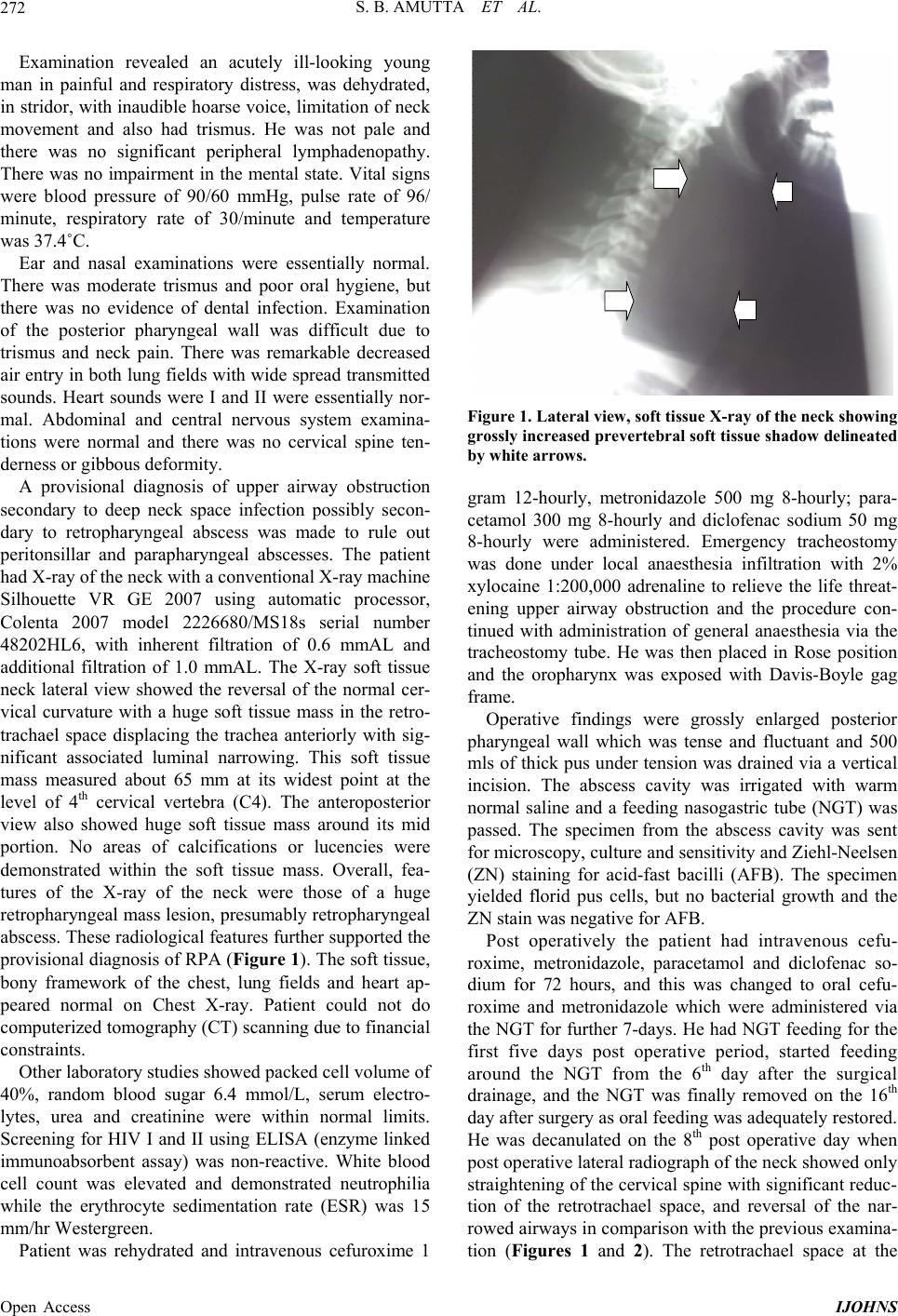 S. B. AMUTTA ET AL. 272 Examination revealed an acutely ill-looking young man in painful and respiratory distress, was dehydrated, in stridor, with inaudible hoarse voice, limitation of neck movement and also had trismus. He was not pale and there was no significant peripheral lymphadenopathy. There was no impairment in the mental state. Vital signs were blood pressure of 90/60 mmHg, pulse rate of 96/ minute, respiratory rate of 30/minute and temperature was 37.4˚C. Ear and nasal examinations were essentially normal. There was moderate trismus and poor oral hygiene, but there was no evidence of dental infection. Examination of the posterior pharyngeal wall was difficult due to trismus and neck pain. There was remarkable decreased air entry in both lung fields with wide spread transmitted sounds. Heart sounds were I and II were essentially nor- mal. Abdominal and central nervous system examina- tions were normal and there was no cervical spine ten- derness or gibbous deformity. A provisional diagnosis of upper airway obstruction secondary to deep neck space infection possibly secon- dary to retropharyngeal abscess was made to rule out peritonsillar and parapharyngeal abscesses. The patient had X-ray of the neck with a conventional X-ray machine Silhouette VR GE 2007 using automatic processor, Colenta 2007 model 2226680/MS18s serial number 48202HL6, with inherent filtration of 0.6 mmAL and additional filtration of 1.0 mmAL. The X-ray soft tissue neck lateral view showed the reversal of the normal cer- vical curvature with a huge soft tissue mass in the retro- trachael space displacing the trachea anteriorly with sig- nificant associated luminal narrowing. This soft tissue mass measured about 65 mm at its widest point at the level of 4th cervical vertebra (C4). The anteroposterior view also showed huge soft tissue mass around its mid portion. No areas of calcifications or lucencies were demonstrated within the soft tissue mass. Overall, fea- tures of the X-ray of the neck were those of a huge retropharyngeal mass lesion, presumably retropharyngeal abscess. These radiological features further supported the provisional diagnosis of RPA (Figure 1). The soft tissue, bony framework of the chest, lung fields and heart ap- peared normal on Chest X-ray. Patient could not do computerized tomography (CT) scanning due to financial constraints. Other laboratory studies showed packed cell volume of 40%, random blood sugar 6.4 mmol/L, serum electro- lytes, urea and creatinine were within normal limits. Screening for HIV I and II using ELISA (enzyme linked immunoabsorbent assay) was non-reactive. White blood cell count was elevated and demonstrated neutrophilia while the erythrocyte sedimentation rate (ESR) was 15 mm/hr Westergreen. Patient was rehydrated and intravenous cefuroxime 1 Figure 1. Lateral view, soft tissue X-ray of the neck showing grossly increased prevertebral soft tissue shadow delineated by white arrows. gram 12-hourly, metronidazole 500 mg 8-hourly; para- cetamol 300 mg 8-hourly and diclofenac sodium 50 mg 8-hourly were administered. Emergency tracheostomy was done under local anaesthesia infiltration with 2% xylocaine 1:200,000 adrenaline to relieve the life threat- ening upper airway obstruction and the procedure con- tinued with administration of general anaesthesia via the tracheostomy tube. He was then placed in Rose position and the oropharynx was exposed with Davis-Boyle gag frame. Operative findings were grossly enlarged posterior pharyngeal wall which was tense and fluctuant and 500 mls of thick pus under tension was drained via a vertical incision. The abscess cavity was irrigated with warm normal saline and a feeding nasogastric tube (NGT) was passed. The specimen from the abscess cavity was sent for microscopy, culture and sensitivity and Ziehl-Neelsen (ZN) staining for acid-fast bacilli (AFB). The specimen yielded florid pus cells, but no bacterial growth and the ZN stain was negative for AFB. Post operatively the patient had intravenous cefu- roxime, metronidazole, paracetamol and diclofenac so- dium for 72 hours, and this was changed to oral cefu- roxime and metronidazole which were administered via the NGT for further 7-days. He had NGT feeding for the first five days post operative period, started feeding around the NGT from the 6th day after the surgical drainage, and the NGT was finally removed on the 16th day after surgery as oral feeding was adequately restored. He was decanulated on the 8th post operative day when post operative lateral radiograph of the neck showed only straightening of the cervical spine with significant reduc- tion of the retrotrachael space, and reversal of the nar- rowed airways in comparison with the previous examina- tion (Figures 1 and 2). The retrotrachael space at the Open Access IJOHNS 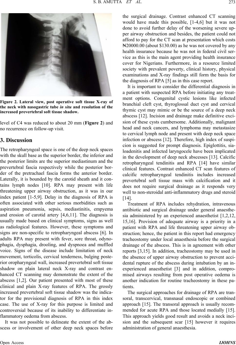 S. B. AMUTTA ET AL. 273 Figure 2. Lateral view, post operative soft tissue X-ray of the neck with nasogastric tube in situ and resolution of the increased prevertebral soft tissue shadow. level of C4 was reduced to about 20 mm (Figure 2) and no recurrence on follow-up visit. 3. Discussion The retropharyngeal space is one of the deep neck spaces with the skull base as the superior border, the inferior and the posterior limits are the superior mediastinum and the prevertebral fascia respectively while the posterior bor- der of the pretrachael fascia forms the anterior border. Laterally, it is bounded by the carotid sheath and it con- tains lymph nodes [10]. RPA may present with life threatening upper airway obstruction, as it was in our index patient [1-5,9]. Delay in the diagnosis of RPA is often associated with other serious morbidities such as aspiration pneumonia, sepsis, mediastinitis, empyema and erosion of carotid artery [4,6,11]. The diagnosis is usually made based on clinical symptoms, signs as well as radiological features. However, these symptoms and signs are non-specific to retropharyngeal abscess [6]. In adults RPA may present with fever, sore throat, odyno- phagia, dysphagia, drooling, and dyspnoea and muffled voice. Signs of the disease include limitation of neck movement, torticolis, cervical tenderness, bulging poste- rior oropharyngeal wall, increased prevertebral soft tissue shadow on plain lateral neck X-ray and contrast en- hanced CT scanning may demonstrate the extent of the abscess [1,2]. Our patient presented with most of these clinical and plain X-ray features of RPA. The grossly increased prevertebral soft tissue shadow was the indica- tor for the provisional diagnosis of RPA in this index case. The use of X-ray for this purpose is limited and controversial because of its inability to differentiate in- flammatory oedema from abscess. It was not possible to delineate the extent of the ab- scess or involvement of other deep neck spaces before the surgical drainage. Contrast enhanced CT scanning would have made this possible, [1-4,6] but it was not done to avoid further delay of the worsening severe up- per airway obstruction and besides, the patient could not afford to pay for the CT scan at presentation which costs ₦20000.00 (about $130.00) as he was not covered by any health insurance because he was not in federal civil ser- vice as this is the main agent providing health insurance cover for Nigerians. Furthermore, in a resource limited society with prevalent poverty, clinical history, physical examinations and X-ray findings still form the basis for the diagnosis of RPA [5] as in this case report. It is important to consider the differential diagnosis in a patient with suspected RPA before initiating any treat- ment options. Congenital cystic lesions like infected branchial cleft cyst, thyroglossal duct cyst and cervical thymic cyst may mimic or be the source of a deep neck abscess [12]. Incision and drainage make definitive exci- sion of these cysts cumbersome. Additionally, malignant head and neck cancers, and lymphoma may metastasize to cervical lymph node and present with deep neck space infection or abscess [12]. Therefore, high index of suspi- cion is suggested for prompt diagnosis. Epiglottitis, sia- loadenitis and infected laryngocele have been implicated in the development of deep neck abscesses [13]. Calcific retropharyngeal tendinitis and RPA [14] have similar clinical features. Contrast enhanced CT scan features of calcific retropharyngeal tendinitis includes increased prevertebral soft tissue mass with calcification and it does not require surgical drainage as it responds very well to non-steroidal anti-inflammatory drugs and steroid [14]. Treatment of RPA includes rehydration, intravenous antibiotic and surgical drainage under general anaesthe- sia administered by an experienced anaesthetist [1,2,12, 15,16]. Provision of adequate airway is a priority in a patient with RPA and life threatening upper airway ob- struction; hence, the patient in this report had emergency tracheostomy under local anaesthesia before the surgical drainage of the abscess. This is in agreement with other reports [3,15]. In addition, tracheostomy may be used in the absence of upper airway obstruction to prevent acci- dental rupture of the abscess during intubation by an in- experienced anaesthetist [3] and in addition, compro- mised airways resulting from post operative oedema is another indication for routine tracheostomy in these pa- tients. The surgical approaches for drainage of RPA are tran- soral, transcervical, transnasal endoscopic or combined approach [15]. The transoral approach is usually recom- mended for acute RPA and those located medially [15]. This approach yields good result and avoids a neck inci- sion and the subsequent scar [15] however it requires administration of general anaesthesia. Open Access IJOHNS  S. B. AMUTTA ET AL. 274 Trans-nasal endoscopic guided needle aspiration is suitable for very small and unilocular abscesses [15-17]. It has the advantage of avoiding general anaesthesia and probable injury to local structures such as blood vessels and nerves [17], although repeated aspirations may be required to achieve complete resolution. Additionally, it is not appropriate for large abscesses with potential for airways obstruction [17]. Cold RPA from tuberculosis is drained by external transcervical approach to prevent dissemination of the infection into the gastrointestinal tract. Besides, large or recurrent acute RPA that extend behind carotid artery can be safely approach by the trans- cervical route [17] to limit bleeding. Disadvantages are neck scar and injury to surrounding structures. The main aetiological factors of RPA in adult are trauma induced by foreign bodies, iatrogenic, orodental infection and tuberculosis [3,4,6-8]. Fish bone injury was the responsible agent in our index patient. It is important to note that the patient did not seek medical help until complications set in thus compelling him to come to the hospital. Ignorance coupled with poverty and poor access to specialist otolaryngologic service was some of the reasons that hindered the patient from seeking medical care early. In contrast to this, adults with RPA in another report from Western Nigeria presented early [9]. The high level of awareness, better economic environment and exposure to western education in western region of Nigeria may be the explanation for this difference. Clinical conditions associated with impaired immunity such as tuberculosis, diabetes mellitus and infection with HIV which are relatively common in developing coun- tries are sometimes associated with RPA [18-21]. The patient in this report did not have destructive lesion of the cervical vertebra and Ziehl-Neelsen staining of the specimen smear for acid-fast bacilli was negative while random blood sugar value was within normal range and he was non-reactive to HIV I and II viruses. The organisms frequently encountered in RPA are po- lymicrobial including aerobes (group A β-haemolytic streptococcus, staphylococcus aureus and haemophilus influenza), anaerobes (gram negative cocci and bacter- oides) [11,22]. The specimen from our patient yielded no bacterial growth. This could be due to prior antibiotic therapy from rural patent medicine shops which is a common practice by our patients though inappropriate culture technique and media for the isolation of anaer- obes could be responsible. The retropharyngeal abscess was described as giant because it measured 65 mm in diameter and this so far the biggest reported from our sub-region. However, we could not verify if larger size has been reported else- where. In an earlier report Ozbek et al., [3] reported a giant RPA which was 61 mm in transverse diameter but this is smaller than that reported in the present case. The prognosis for patients with RPA is very good es- pecially with early diagnosis and appropriate intervention. However, with late presentation, delayed diagnosis and in the presence of significant co-morbidities such as im- munosuppression, HIV, tuberculosis, diabetes mellitus, head and neck malignancies the prognosis may be poor. Ridder et al. [12] reported 6 (2.6%) mortality among 234 cases over an 8-year period and the deaths were due to sepsis and multiple organ failure. Overall, the patient in this report responded very well to the tracheostomy, in- tra-oral drainage and the broad spectrum antibiotics. He was decanulated on the 8th day post surgical drainage and was discharged home after achieving full recovery and no recurrence on follow-up visit. 4. Conclusion The prognosis of giant retropharyngeal abscess is good with tracheostomy, surgical drainage and medical inter- vention as seen in this case report. REFERENCES [1] B. Hartley, “Cervicofacial Infection in Children,” In: G. Michael, Ed., Scott’s Browns Otorhinolaryngology, Head and Neck Surgery, 7th Edition, Hodder Arnold, London, 2008, pp. 1213-1214. http://dx.doi.org/10.1201/b15118-107 [2] L. W. Tom and I. N. Jacobs, “Diseases of the Oral Cavity, Oropharynx, and Nasopharynx,” In: B. James, Ed., Ballenger’s Otorhinolaryngology, Head and Neck Sur- gery, 16th Edition, BC Decker Inc., Hamilton, 2003, pp. 1039-1040. [3] C. Ozbek, S. Dagli, E. E. U. Tuna, O. Ciffci and C. Oz- dem, “Giant Retropharyngeal Abscess in an Adult as a Complication of Acute Tonsillitis: Case Report,” Ear Nose Throat Journal, Vol. 88, No. 11, 2009, p. E20. [4] S. B. Sharma and P. Hong, “Ingestion and Pharyngeal Trauma Causing Secondary Retropharyngeal Abscess in Five Adult Patients,” Case Report in Emergency Medi- cine, Vol. 2012, 2012, Article ID: 943090. [5] O. A. Afolabi, J. O. Fadare, E. O. Oyewole and S. A. Ogah, “Fish Bone Foreign Body Presenting with an Acute Fulminating Retropharyngeal Abscess in a Resource- Challenge Centre: A Case Report,” Journal of Medical Case Reports, Vol. 5, 2011, p. 165. http://dx.doi.org/10.1186/1752-1947-5-165 [6] A. Harkani, R. Hassani, T. Ziad, L. Aderdour, H. Nouri, Y. Rochdi and A. Raji, “Retropharyngeal Abscess in Adults: Five Case Reports And Review of the Literature,” The Scientific World Journal, Vol. 11, 2011, pp. 1623- 1629. http://dx.doi.org/10.1100/2011/915163 [7] E. Mevio, P. Calabro, F. De Paoli, A. Maccabruni and G. Michelone, “Unusual Extracranial Complications of Otitis Media in a Young HIV Patient: Retropharyngeal and Mouret’s Abscess,” Revue de Laryngologie-Otologie- Rhinologie, Vol. 119, No. 3, 1998, pp. 199-201. [8] R. Meyer, S. Argarwal and I. Singh, “Tuberculous Retro- Open Access IJOHNS  S. B. AMUTTA ET AL. Open Access IJOHNS 275 pharyngeal Abscess in an HIV Patient,” Hong Kong Medi- cal Journal, Vol. 12, No.6, 2006, pp. 483-485. [9] O. G. Nwaorgu, P. A. Onakoya, J. A. Fasunla and T. S. Ibekwe, “Retropharyngeal Abscess: A Clinical Experi- ence at the University College Hospital Ibadan,” Nigeria Journal of Medicine, Vol. 14, No. 4, 2005, pp. 415-418. [10] C. R. Jennings, “Surgical Anatomy of the Neck,” In: G. Michael, Ed., Scott’s Browns Otorhinolaryngology, Head and Neck Surgery, 7th Edition, Vol. II, Hodder Arnold, London, 2008, pp. 1744-1745. http://dx.doi.org/10.1201/b15118-153 [11] I. Brook, “Microbiology and Management of Peritonsillar, Retropharyngeal Abscess and Pharyngeal Abscesses,” Journal of Oral & Maxillofacial Surgery, Vol. 62, No. 12, 2004, pp. 1545-1550. http://dx.doi.org/10.1016/j.joms.2003.12.043 [12] G. J. Ridder, K. Technau-Ihling, A. Sander and C. C. Boedeker, “Spectrum and Management of Deep Neck Space Infections: An 8-Year Experience of 234 Cases,” Otolaryngology—Head and Neck Surgery, Vol. 133, No. 5, 2005, pp. 709-714. http://dx.doi.org/10.1016/j.otohns.2005.07.001 [13] J. Stalfors, A. Adielsson, A. Ebenfelt, G. Nethander and T. Westin, “Deep Neck Space Infections Remain a Surgical Challenge. A Study of 72 Patients,” Acta Otolaryngology, Vol. 124, No. 10, 2004, pp. 119-1196. http://dx.doi.org/10.1080/00016480410017864 [14] K. Terao, T. Kusunoki, K. Mori. K. Murata and K. Doi, “A Case of Calcific Retropharyngeal Tendinitis: The Sig- nificance of Early Diagnosis,” Ear, Nose and Throat Journal, Vol. 92, No. 2, 2013, pp. 74-83. [15] P. J. Schuller, M. Cohen, J. Greve, C. Plettenberg, J. Chereath, M. Wagemann, et al., “Surgical Management of Retropharyngeal Abscess,” Acta Oto-Laryngologica, Vol. 129, No. 11, 2009, pp. 1274-1279. http://dx.doi.org/10.3109/00016480802642088 [16] D. K. C. Wong, C. Brown, N. Mills, P. Spielmann and M. Neeff, “To Drain or Not to Drain-Management of Pediat- ric Deep Neck Abscesses: A Case Conrol Study,” Inter- national Journal Pediatric Otorhinolaryngology, Vol. 76, No. 12, 2012, pp. 1810-1813. http://dx.doi.org/10.1016/j.ijporl.2012.09.006 [17] K. Chang, Y. Chen, S. Hao and S. Chen, “Ultrasound- Guided Closed Drainage for Abscesses of the Head and Neck,” Otolaryngology—Head and Neck Surgery, Vol. 132, No. 1, 2004, pp. 119-124. http://dx.doi.org/10.1016/j.otohns.2004.08.004 [18] M. P. Kamath, K. M. Bhojwani, S. U. Kamath, C. Maha- bala and S. Argawal, “Tuberculous Retropharyngeal Ab- scess,” Ear Nose Throat Journal, Vol. 86, No. 4, 2007, pp. 236-237. [19] H. A. Soub, “Retropharyngeal Abscess Associated with Tuberculosis of Cervical Spine,” Tubercle and Lung Dis- ease, Vol. 77, No. 6, 1996, pp. 563-565. http://dx.doi.org/10.1016/S0962-8479(96)90057-X [20] A. Christoforidou, S. Metallidis, P. Kollaras, A. Agathange- lidis, P. Nikolaidia, V. Vital and K. Markou, “Tubercu- lous Retropharyngeal Abscess as a Cause of Oropharyn- geal Dysphagia,” American Journal of Otolaryngology- Head and Neck Medicine and Surgery, Vol. 33, No. 2, 2012, pp. 272-274. [21] C. Patil, R. Kharat, P. Deshmukh, S. Singhal and B. D’Souza, “Tuberculous Retropharyngeal Abscess without Cervical Spine TB,” Asian Pacific Journal of Tropical Medicine, Vol. 4, No. 3, 2011, pp. 251-252. http://dx.doi.org/10.1016/S1995-7645(11)60080-1 [22] H. S. Sharma, D. S. Kurl and M. Hamza, “Retropharyn- geal Abscess: Recent Trend,” Auris Nasus Larynx, Vol. 25, No. 4, 1998, pp. 403-406. http://dx.doi.org/10.1016/S0385-8146(98)00007-8 |