

Paper Menu >>
Journal Menu >>
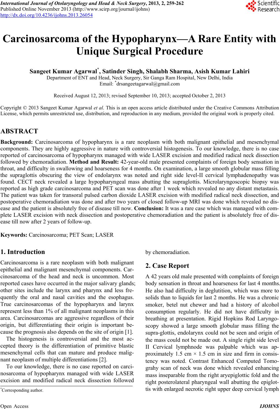 International Journal of Otolaryngology and Head & Neck Surgery, 2013, 2, 259-262 Published Online November 2013 (http://www.scirp.org/journal/ijohns) http://dx.doi.org/10.4236/ijohns.2013.26054 Open Access IJOHNS Carcinosarcoma of the Hypopharynx—A Rare Entity with Unique Surgical Procedure Sangeet Kumar Agarwal*, Satinder Singh, Shalabh Sharma, Asish Kumar Lahiri Department of ENT and Head, Neck Surgery, Sir Ganga Ram Hospital, New Delhi, India Email: *[email protected] Received August 12, 2013; revised September 10, 2013; accepted October 2, 2013 Copyright © 2013 Sangeet Kumar Agarwal et al. This is an open access article distributed under the Creative Commons Attribution License, which permits unrestricted use, distribution, and reproduction in any medium, provided the original work is properly cited. ABSTRACT Background: Carcinosarcoma of hypopharynx is a rare neoplasm with both malignant epithelial and mesenchymal components. They are highly aggressive in nature with controversial histogenesis. To our knowledge, there is no case reported of carcin osarcoma of hypopharynx managed w ith wide LASER excision and modified radical neck dissection followed by chemorad iation. Method and Result: 42-year-old male presented complaints of foreign body sensation in throat, and difficulty in swallo wing and hoarseness for 4 months. On examination, a large smooth globular mass filling the supraglottis obscuring the view of endolarynx was noted and right side level-II cervical lymphadenopathy was found. CECT neck revealed a large hypopharyngeal mass abutting the supraglottis. Microlaryngoscopic biopsy was reported as high grade carcinosarcoma and PET scan was done after 1 week which revealed no any distant metastasis. The patient was taken for transoral pulsed carbon dioxide LASER excision with modified radical neck dissection, and postoperative chemoradiation was done and after two years of closed follow-up MRI was done which revealed no dis- ease and the patient is absolu tely free of disease till now. Conclusion: It was a rare case which was managed with com- plete LASER excision with neck dissection and postoperative chemoradiation and the patient is absolutely free of dis- ease till now after 2 years of follow-up. Keywords: Carcinosarcoma; PET Scan; LASER 1. Introduction Carcinosarcoma is a rare neoplasm with both malignant epithelial and malignant mesenchymal components. Car- cinosarcoma of the head and neck is uncommon. Most reported cases have occurred in the major salivary glands; other sites include the larynx and pharynx and less fre- quently the oral and nasal cavities and the esophagus. True carcinosarcomas of the hypopharynx and larynx represent less than 1% of all malign ant neoplasms in this area. Carcinosarcomas are aggressive regardless of their origin, but differentiating their origin is important be- cause the prognosis also depends on the site of origin [1]. The histogenesis is controversial and the most ac- cepted theory is the differentiation of primitive blastic mesenchymal cells that can mature and produce malig- nant neoplasm of multiple differentiations [2]. To our knowledge, there is no case reported on carci- nosarcoma of hypopharynx managed with wide LASER excision and modified radical neck dissection followed by chemoradiation. 2. Case Report A 42 years old male presented with complain ts of foreign body sensation in throat and hoarseness for last 4 months. He also had difficulty in deglutition, which was more to solids than to liquids for last 2 months. He was a chronic smoker, betel nut chewer and had a history of alcohol consumption regularly. He did not have difficulty in breathing at presentation. Rigid Hopkins Rod Laryngo- scopy showed a large smooth globular mass filling the supra-glottis, endolarynx could not be seen and origin of the mass could not be made out. A single right side level II Cervical lymphnode was palpable which was ap- proximately 1.5 cm × 1.5 cm in size and firm in consis- tency was noted. Contrast Enhanced Computed Tomo- grahy scan of neck was done which revealed enhancing mass inseparable from the right aryepiglottic fold and the right posterolateral pharyngeal wall abutting the epiglot- tis with enlarged necrotic right upper deep cervical lymp h *Corresponding author. 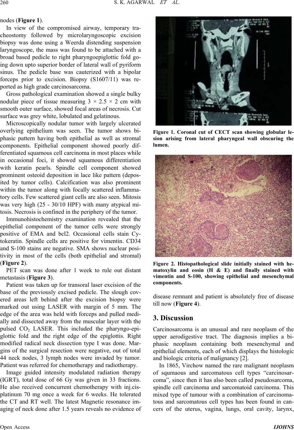 S. K. AGARWAL ET AL. 260 nodes (Figure 1). In view of the compromised airway, temporary tra- cheostomy followed by microlaryngoscopic excision biopsy was done using a Weerda distending suspension laryngoscope, the mass was found to be attached with a broad based pedicle to right pharyngoepiglottic fold go- ing down upto superior border of lateral wall of pyriform sinus. The pedicle base was cauterized with a bipolar forceps prior to excision. Biopsy (S1607/11) was re- ported as high grade carcinosarcoma. Gross pathological examination showed a single bulky nodular piece of tissue measuring 3 × 2.5 × 2 cm with smooth outer surface, showed focal areas of necrosis. Cut surface was grey white, lobulated and gelatinous. Microscopically nodular tumor with largely ulcerated overlying epithelium was seen. The tumor shows bi- phasic pattern having both epithelial as well as stromal components. Epithelial component showed poorly dif- ferentiated squamous cell carcinoma in most places while in occasional foci, it showed squamous differentiation with keratin pearls. Spindle cell component showed prominent osteoid deposition in lace like pattern (depos- ited by tumor cells). Calcification was also prominent within the tumor along with focally scattered inflamma- tory cells. Few scattered giant cells are also seen. Mito sis was very high (25 - 30/10 HPF) with many atypical mi- tosis. Necrosis is confined in the periphery of the tumor. Immunohistochemistry examination revealed that the epithelial component of the tumor cells were strongly positive of EMA and bcl2. Occasional cells stain Cy- tokeratin. Spindle cells are positive for vimentin. CD34 and S-100 stains are negative. SMA shows nuclear posi- tivity in most of the cells (both epithelial and stromal) (Figure 2). PET scan was done after 1 week to rule out distant metastasis (Figure 3). Patient was taken up for transoral laser excision of the base of the previously excised pedicle. The slough cov- ered areas left behind after the excision biopsy were marked out using LASER with margin of 5 mm. The edge of the area was held with forceps and pulled medi- ally and dissected away from the muscular layer with the pulsed CO2 LASER. This included the pharyngo-epi- glottic fold and the right edge of the epiglottis. Right modified radical neck dissection type I was done. Mar- gins of the surgical resection were negative, out of total 44 neck nodes, 3 lymph nodes were invaded by tumor. Patient was referred for chemotherapy and radiotherapy. Image guided intensity modulated radiation therapy (IGRT), total dose of 66 Gy was given in 33 fractions. He also received concurrent chemotherapy with inj.cis- platinum 70 mg once a week for 6 weeks. He tolerated the CT and RT well. The latest Magnetic resonance im- aging of neck done after 1.5 years reveals no evidence of Figure 1. Coronal cut of CECT scan showing globular le- sion arising from lateral pharyngeal wall obscuring the lumen. Figure 2. Histopathological slide initially stained with he- matoxylin and eosin (H & E) and finally stained with vimentin and S-100, showing epithelial and mesenchymal components. disease remnant and patient is absolutely free of disease till now (Figure 4). 3. Discussion Carcinosarcoma is an unusual and rare neoplasm of the upper aerodigestive tract. The diagnosis implies a bi- phasic neoplasm containing both mesenchymal and epithelial elements, each of wh ich displays the histologic and biologic criteria of malignancy [2]. In 1865, Virchow named the rare malignant neoplasm of squmaous and sarcomatous cell types “carcinosar- coma”, since then it has also been called pseudosarcoma, spindle cell carcinoma and sarcomatoid carcinoma. This mixed type of tumour with a combination of carcinoma- tous and sarcomatous cell types has been found in can- cers of the uterus, vagina, lungs, oral cavity, larynx, Open Access IJOHNS 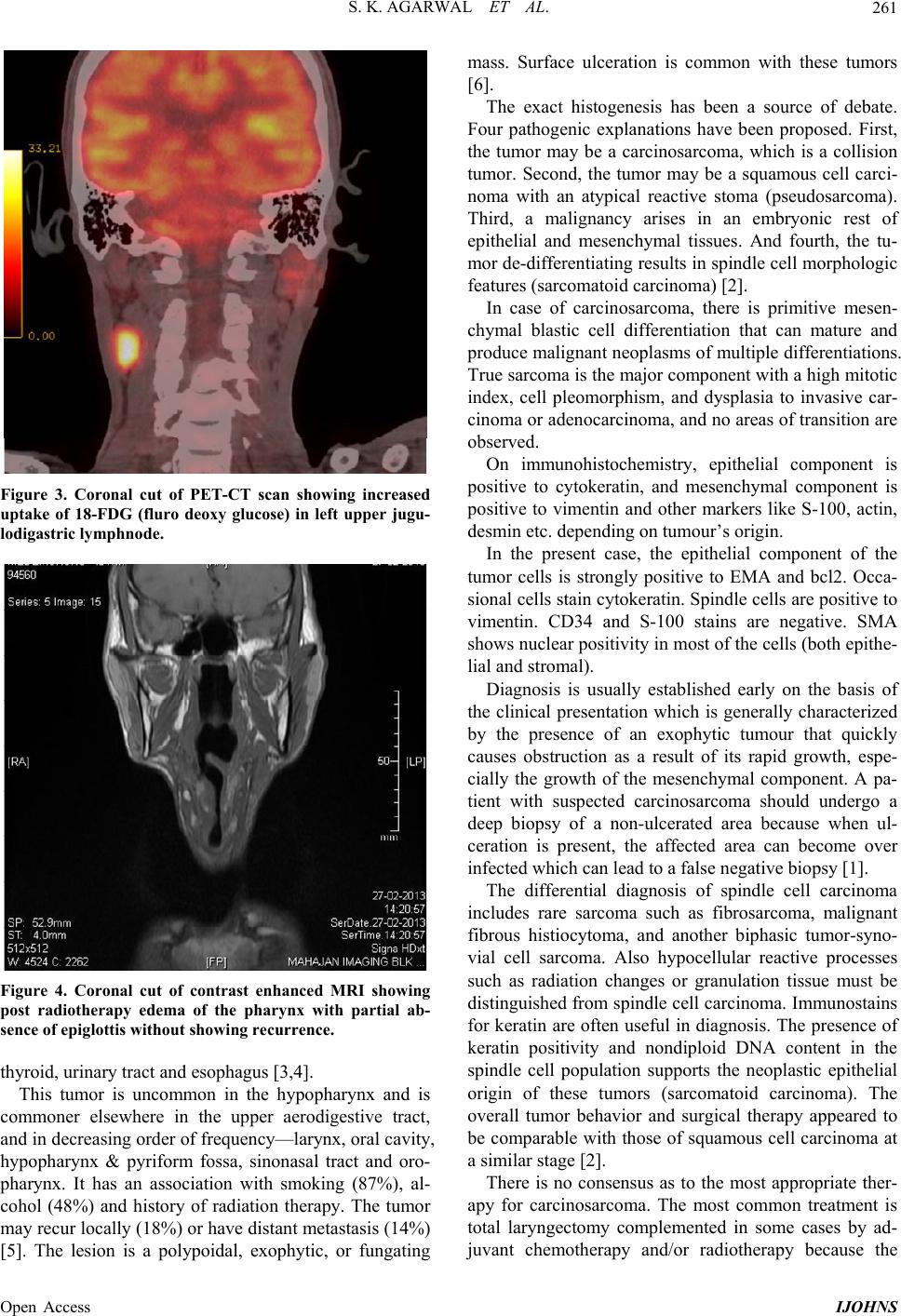 S. K. AGARWAL ET AL. 261 Figure 3. Coronal cut of PET-CT scan showing increased uptake of 18-FDG (fluro deoxy glucose) in left upper jugu- lodigastric lymphnode. Figure 4. Coronal cut of contrast enhanced MRI showing post radiotherapy edema of the pharynx with partial ab- sence of epiglottis without showing recurrence. thyroid, urinary tract and esophagus [3,4]. This tumor is uncommon in the hypopharynx and is commoner elsewhere in the upper aerodigestive tract, and in decreasing order of frequency—larynx, oral cavity, hypopharynx & pyriform fossa, sinonasal tract and oro- pharynx. It has an association with smoking (87%), al- cohol (48%) and history of radiation therapy. The tumor may recur locally (18%) or have distant metastasis (14%) [5]. The lesion is a polypoidal, exophytic, or fungating mass. Surface ulceration is common with these tumors [6]. The exact histogenesis has been a source of debate. Four pathogenic explanations have been proposed. First, the tumor may be a carcinosarcoma, which is a collision tumor. Second, the tumor may be a squamous cell carci- noma with an atypical reactive stoma (pseudosarcoma). Third, a malignancy arises in an embryonic rest of epithelial and mesenchymal tissues. And fourth, the tu- mor de-differentiating results in spindle cell morphologic features (sarcomatoid carcinoma) [2]. In case of carcinosarcoma, there is primitive mesen- chymal blastic cell differentiation that can mature and produce malignant neoplasms of multiple differen tiations. True sarcoma is the major component with a high mitotic index, cell pleomorphism, and dysplasia to invasive car- cinoma or adeno carcinoma, and no areas of transition are observed. On immunohistochemistry, epithelial component is positive to cytokeratin, and mesenchymal component is positive to vimentin and other markers like S-100, actin, desmin etc. depending on tumour’s origin. In the present case, the epithelial component of the tumor cells is strongly positive to EMA and bcl2. Occa- sional cells stain cytokeratin. Spindle cells are positive to vimentin. CD34 and S-100 stains are negative. SMA shows nuclear positivity in mo st of the cells (both epith e- lial and stromal). Diagnosis is usually established early on the basis of the clinical presentation which is generally characterized by the presence of an exophytic tumour that quickly causes obstruction as a result of its rapid growth, espe- cially the growth of the mesenchymal component. A pa- tient with suspected carcinosarcoma should undergo a deep biopsy of a non-ulcerated area because when ul- ceration is present, the affected area can become over infected which can lead to a f a lse negative biopsy [1] . The differential diagnosis of spindle cell carcinoma includes rare sarcoma such as fibrosarcoma, malignant fibrous histiocytoma, and another biphasic tumor-syno- vial cell sarcoma. Also hypocellular reactive processes such as radiation changes or granulation tissue must be distinguished from spindle cell carcinoma. Immunostains for keratin are often useful in diagnosis. The presence of keratin positivity and nondiploid DNA content in the spindle cell population supports the neoplastic epithelial origin of these tumors (sarcomatoid carcinoma). The overall tumor behavior and surgical therapy appeared to be comparable with those of squamous cell carcinoma at a similar stage [2]. There is no consensus as to the most appropriate ther- apy for carcinosarcoma. The most common treatment is total laryngectomy complemented in some cases by ad- juvant chemotherapy and/or radiotherapy because the Open Access IJOHNS 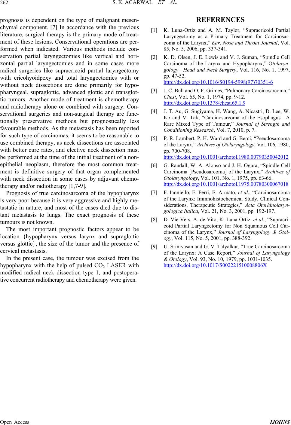 S. K. AGARWAL ET AL. Open Access IJOHNS 262 prognosis is dependent on the type of malignant mesen- chymal component. [7] In accordance with the previous literature, surgical therapy is the primary mode of treat- ment of these lesions. Conservational operations are per- formed when indicated. Various methods include con- servation partial laryngectomies like vertical and hori- zontal partial laryngectomies and in some cases more radical surgeries like supracricoid partial laryngectomy with cricohyoidpexy and total laryngectomies with or without neck dissections are done primarily for hypo- pharyngeal, supraglottic, advanced glottic and transglot- tic tumors. Another mode of treatment is chemotherapy and radiotherapy alone or combined with surgery. Con- servational surgeries and non-surgical therapy are func- tionally preservative methods but prognostically less favourable methods. As the metastasis has been reported for such type of carcinomas, it seems to be reasonable to use combined therapy, as neck dissections are associated with better cure rates, and elective neck dissection must be performed at the time of the initial treatment of a non- epithelial neoplasm, therefore the most common treat- ment is definitive surgery of that organ complemented with neck dissection in some cases by adjuvant chemo- therapy and/ or radiotherapy [1,7- 9]. Prognosis of true carcinosarcoma of the hypopharynx is very poor because it is very aggressive and highly me- tastatic in nature, and most of the cases died due to dis- tant metastasis to lungs. The exact prognosis of these tumours is not known. The most important prognostic factors appear to be location {hypopharynx versus larynx and supraglottic versus glottic}, the size of the tumor and the presence of cervical metastasis. In the present case, the tumour was excised from the hypopharynx with the help of pulsed CO2 LASER with modified radical neck dissection type 1, and postopera- tive co ncu rrent radi othe rapy a nd c hemot hera py we re gi ven. REFERENCES [1] K. Luna-Ortiz and A. M. Taylor, “Supracricoid Partial Laryngectomy as a Primary Treatment for Carcinosar- coma of the Larynx,” Ear, Nose and Throat Journal, Vol. 85, No. 5, 2006, pp. 337-341. [2] K. D. Olsen, J. E. Lewis and V. J. Suman, “Spindle Cell Carcinoma of the Larynx and Hypopharynx,” Otolaryn- gology—Head and Neck Surgery, Vol. 116, No. 1, 1997, pp. 47-52. http://dx.doi.org/10.1016/S0194-5998(97)70351-6 [3] J. C. Bull and O. F. Grimes, “Pul mo nary Carcinosarcom a ,” Chest, Vol. 65, No. 1, 1974, pp. 9-12. http://dx.doi.org/10.1378/chest.65.1.9 [4] J. T. Au, G. Sugiyama, H. Wang, A. Nicastri, D. Lee, W. Ko and V. Tak, “Carcinosarcoma of the Esophagus—A Rare Mixed Type of Tumour,” Journal of Strength and Conditioning Research, Vol. 7, 2010, p. 7. [5] P. R. Lambert, P. H. Ward and G. Berci, “Pseudosarcoma of the Larynx,” Archives of Otola ryn gology , Vol. 106, 1980, pp. 700-708. http://dx.doi.org/10.1001/archotol.1980.00790350042012 [6] G. Randall, W. A. Alonso and J. H. Ogura, “Spindle Cell Carcinoma [Pseudosarcoma] of the Larynx,” Archives of Otolaryngology, Vol. 101, No. 1, 1975, pp. 63-66. http://dx.doi.org/10.1001/archotol.1975.00780300067018 [7] F. Ianniello, E. Ferri, E. Armato, et al., “Carcinosarcoma of the Larynx: Immnohistochemical Study, Clinical Con- siderations, Therapeutic Strategies,” Acta Otorhinolaryn- gologica Italica, Vol. 21, No. 3, 2001, pp. 192-197. [8] D. Vie Vers, A. de Vito, K. Luna-Ortiz, et al., “Supracri- coid Partial Laryngectomy for Non Squamous Cell Car- cinoma of the Larynx,” Journal of Laryngology & Otol- ogy, Vol. 115, No. 5, 2001, pp. 388-392. [9] U. Srinivasan and G. V. Talyalkar, “True Carcinosarcoma of the Larynx: A Case Report,” Journal of Laryngology & Otology, Vol. 93, No. 10, 1979, pp. 1031-1035. http://dx.doi.org/10.1017/S002221510008806X |