

Paper Menu >>
Journal Menu >>
 Open Journal of Physical Chemistry, 2013, 3, 157-162 Published Online November 2013 (http://www.scirp.org/journal/ojpc) http://dx.doi.org/10.4236/ojpc.2013.34019 Open Access OJPC The Role of Eumelanin in Generating Reactive Oxygen and Reactive Nitrogen in Solution: Possible Relevance to Keloid Formation Julian M. Menter1, Comnuan Nokkaew2, Danita Eatman3, Aquilla Sprewell1, Natalia Silvestrov3, Abrienne M. Patta1, Sandra Harris-Hooker2 1Department of Microbiology, Biochemistry and Immunology, Morehouse School of Medicine, Atlanta, USA 2Office of Sponsored Research, Department of Pathology, Morehouse School of Medicine, Atlanta, USA 3Analytical Laboratory, Department of Pharmacology and Toxicology, Morehouse School of Medicine, Atlanta, USA Email: [email protected] Received March 26, 2013; revised April 25, 2013; accepted May 1, 2013 Copyright © 2013 Julian M. Menter et al. This is an open access article distributed under the Creative Commons Attribution License, which permits unrestricted use, distribution, and reproduction in any medium, provided the original work is properly cited. ABSTRACT Recently, nitric oxide (NO) has been implicated as an epigenetic factor in keloids, a scarring disease occurring primarily in dark skinned people who have relatively high amounts of pigment melanin. In this work, we tested whether a mela- nin-mediated redox reaction involving adsorbed NO and O2 can couple NO oxidation with O2 reduction to form reac- tive oxygen species (ROS) or reactive nitrogen species (RNS) in vitro at pH 7.4. We measured the formation of reactive species that oxidize dihydrorhodamine123 (DHR) to fluorescent rhodamine123 in the presence and absence of sepia melanin. In separate experiments, we monitored NO concentration with 4,5-diaminofluorescein (DAF) by measuring the highly fluorescent NO-adduct, DAF-2T. We attempted to detect peroxynitrite with 5 µM 3-methyl-1,2-cyclopenta- nedione (MCP), a selective scavenger of peroxynitrite (IC50 = 3.6 µM for ONOO vs. 63.8 µM and >> 100 µM for NO and respectively). However, MCP itself oxidized DHR. We found that in the absence of NO, melanin itself oxi- dizes DHR, with no loss of DAF-fluorescence (i.e. no net consumption of NO). In the presence of NO, there was a ~57% loss of DAF fluorescence, indicating that NOx is formed at the expense of NO. The data provided good fit (r2 = 0.94) to a Langmuir adsorption isotherm, with pseudo first order rate k' = 8.2 × 10⁷) s1 and adsorption coefficient Kad = 4.04 M1. Both of these parameters are consistent with a facile chemisorption reaction between NO and O2 on the mela- nin surface. Possible reactions are a) NO and 2 O 2 O ONOO and/or b) 2NO + O2 2NO2. The latter reaction is dis- favored in solution but is significantly accelerated on the melanin surface via an entropy effect. Keywords: Melanin; Nitric Oxide; NOx; Redox 1. Introduction Pigment eumelanin binds a wide variety of compounds [1] and can act as a redox reagent [2,3]. Depending on its chemical environment, melanin can either donate or accept electrons from ambient molecules and/or couple the oxidation of one compound with the reduction of another [4-6]. Melanin is well known to undergo auto- xidation as well as Fenton chemistry with bound or adventitious transition metals (usually iron or copper) to produce .OH radicals. Depending on the relative metal and pigment concentrations, melanin may act as either an antioxidant or a pro-oxidant [7,8]. People of color are particularly susceptible to keloids, a recalcitrant consequence of aberrant wound healing, characterized by excessive collagen deposition that ex- tends beyond the original wound [9-11]. The etiology of keloids is complex, and both genetic [12] and epigenetic [13] factors are involved. Recent work [14-16] has indi- cated that nitric oxide (NO) enhances the synthesis of type I collagen by dermal fibroblasts and that the keloid lesion may arise from the NO/cGMP signaling path- way. The particular susceptibility of darkly-pigmented persons to keloids is intriguing, and it leads one to ask whether pigment itself may play an active role in keloid pathogenesis, given its ability to bind and mediate redox 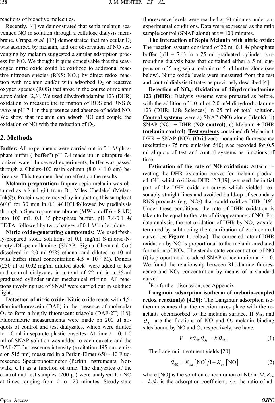 J. M. MENTER ET AL. 158 reactions of bioactive molecules. Recently, [4] we demonstrated that sepia melanin sca- venged NO in solution through a cellulose dialysis mem- brane. Crippa et al. [17] demonstrated that molecular O2 was adsorbed by melanin, and our observation of NO sca- venging by melanin suggested a similar adsorption proc- ess for NO. We thought it quite conceivable that the scav- enged nitric oxide could be oxidized to additional reac- tive nitrogen species (RNS; NOx) by direct redox reac- tion with melanin and/or with adsorbed O2 or reactive oxygen species (ROS) that arose in the course of melanin autoxidation [2,3]. We used dihydrorhodamine 123 (DHR) oxidation to measure the formation of ROS and RNS in vitro at pH 7.4 in the presence and absence of added NO. We show that melanin can adsorb NO and couple the oxidation of NO with the reduction of O2. 2. Methods Buffer: All experiments were carried out in 0.1 M phos- phate buffer (“buffer”) pH 7.4 made up in ultrapure de- ionized water. In several experiments, buffer was passed through a Chelex-100 resin column (8.0 × 1.0 cm) be- fore use. This treatment had no effect on the results. Melanin preparation: Impure sepia melanin was ob- tained as a kind gift from Dr. Miles Chedekel (Melan- Ink@). Protein was removed by incubating this sample at 60˚C for 30 min in 0.1 M HCl followed by predialysis through a Spectropore membrane (MW cutoff 6 - 8 kD) into 100 mL 0.1 M phosphate buffer, pH 7.4/0.1 M EDTA, followed by two changes of 0.1 M buffer alone. Nitric oxide-generating compounds: We used fresh- ly-prepared stock solutions of 0.1 mg/ml S-nitorso-N- acetyl-DL-penicillamine (SNAP; Sigma Chemical Co.) dissolved in 2.0 ml 95% ethanol and diluted to 10 ml with buffer (final concentration 4.5 × 10⁻⁵ M). Donors (250 µl of 0.02 mg/ml diluted stock) were added to test and control dialyzates in a total of 22 ml in a 25-ml graduated cylinder under mechanical stirring. All reac- tions involving use of SNAP were carried out in subdued light. Detection of nitric oxide: Nitric oxide reacts with 4,5- diaminofluorescein (DAF) in the presence of molecular O2 to form a highly fluorescent triazole (DAF-2T) [18]. Fluorometric measurements were made on 200 µl ali- quots of control and test dialyzates, which were diluted to 1.0 ml in separate plastic cuvettes. At time t = 0, 1.0 ml of SNAP solution was added to each cuvette and the DAF-2T fluorescence intensity (excitation 495 nm, emis- sion 515 nm) measured in a Perkin-Elmer 650 - 40 Fluo- rescence Spectrophotometer (Perkin Instruments, Nor- walk, CT) as a function of time. The dialyzates of the control and test samples (200 µl) were analyzed for NO at times ranging from 0 to 120 minutes. Steady-state fluorescence levels were reached at 60 minutes under our experimental conditions. Data were expressed as the ratio sample/control (SNAP alone) at t = 100 minutes. The Interaction of Sepia Melanin with nitric oxide: The reaction system consisted of 22 ml 0.1 M phosphate buffer (pH = 7.4) in a 25 ml graduated cylinder, sur- rounding dialysis bags that contained either a 5 ml sus- pension of 5 mg sepia melanin or 5 ml buffer alone (see below). Nitric oxide levels were measured from the test and control dialysis filtrates as previously described [4]. Detection of NOx: Oxidation of dihydrorhodamine 123 (DHR): Dialysis systems were prepared as before, with the addition of 1.0 ml of 2.0 mM dihydrorhodamine 123 (DHR; Life Sciences) in 25 ml of total solution. Control systems were a) SNAP (NO) alone (blank); b) SNAP (NO) + DHR (NO control); c) Melanin + DHR (melanin control). Test systems contained d) Melanin + DHR + SNAP (NO). (Oxidized) rhodamine fluorescence (excitation 475 nm; emission 540) was recorded for 0.5 ml aliquots of test and control systems as functions of time. Estimation of the rate of NO oxidation: After cor- recting the DHR oxidation curves for melanin-produc- ed .OH, which oxidizes DHR [2,3,19], we used the initial part of the DHR oxidation curves which yielded rea- sonably straight lines and avoided build-up of secondary RNS products (e.g. NO2) that could oxidize DHR [19]. Under these conditions, the rate of DHR oxidation is taken to be equal to the rate of disappearance of NO. For data analysis, the net oxidation of DHR by NOx was de- termined by subtracting the contribution of each control curve (see Figure 1, below). The corrected rate of DHR oxidation by NO is proportional to the melanin-mediated formation of NOx. The steady state concentration of NO (t) is proportional to added SNAP concentration at t = 0. We found the relationship between Rhodamine fluores- cence and NOx concentration by means of a standard curve.* *For further discussion, see Appendix. Langmuir adsorption isotherm of melanin-coupled redox reaction(s) [4,20]: The Langmuir adsorption iso- therm assumes that the reaction takes place with the re- actants chemisorbed to the melanin surface. If θNO and 2 O are the fractions of NO and O2 melanin binding sites bound by NO and O2 respectively, we have: 2 NO ONO Vk k (1) The Langmuir treatment yields [20] NO NO 1NO ad ad KK (2) where [NO] is the solution concentration of NO in M, Kad = ka/kd is the adsorption coefficient, i.e. the ratio of ad- Open Access OJPC  J. M. MENTER ET AL. 159 Figure 1. Oxidation of DHR by NO (white circles), Eumela- nin (black triangles) and Eumelanin/NO system (white tri- angles) in 0.1 M phosphate buffer, pH 7.4 (see text). DHR is slowly autoxidized (black circles). In the absence of added melanin, NO slowly oxidizes DHR (white circles). Melanin itself oxidizes DHR at a significantly greater rate with sig- nificant upward concavity (black triangles); the additional increases in DHR oxidation and concavity in the presence of NO suggests the generation of NOx (white triangles). sorption/dissociation constants of NO on the melanin backbone, having the units of M1. We assume that 2 O kk in O2 saturated (aerated) solution, is equal to the pseudo first order rate constant for the bimolecular reaction between O2 and NO on the melanin surface having the dimensions of s1 (see [17,20] for discussion). Substitution of (2) into (1) followed by inversion yields 11 NO1 ad VkK k (3) Thus, if Langmuir mechanisms are operative, slopes of 1/V vs. 1/[NO] will yield a straight line with slope 1/k'Kad and intercept 1/k' from which parameters k' and Kad can be found. The steady state concentration, [NO]ss was estimated from rate constants for formation and decline of NO in consecutive reactions. These constants were found from the approximate lifetimes of SNAP (~5 hr,) and NO de- composition in aqueous solution (~2 s). Under these con- ditions, NO is consumed virtually as fast as it is formed, and [NO] could be assumed to be invariant with time. Using these values, the integrated rate equation [20] ul- timately yielded the value: * – 4 0 ss NO1.12 10SNAP . *For further discussion, see Appendix. Statistical analysis: We used Student’s t test for small samples. Data were expressed as mean ± SD. Differences were considered significant for p < 0.05. 3. Results DHR oxidation: Figure 1 shows the results of a repre- sentative experiment (n = 4) in aerated solution. Control groups were DHR alone (black dots); DHR + NO in the absence of melanin (white dots) and melanin + DHR (black triangles). DHR alone was slowly autoxidized to fluorescent rhodamine 123. Nitric oxide modestly in- creased the rate of DHR oxidation Melanin efficiently oxidized DHR with significant upward concavity. The test system, melanin + NO + DHR (white triangles), fur- ther increased the rate of growth and concavity. Qualita- tively similar results were obtained for each experiment. Attempts to Detect NOx; Consumption of NO in solution: At 5 µM, 3-methyl-1,2-cyclopentanedione (MCP; Aldrich Chemical Co.) selectively scavenges ONOO- (IC50 = 3.6 µM for ONOO vs. 63.8 µM and >> 100 µM for NO and 2 O respectively [21]. Although we origi- nally planned to use MCP to detect peroxynitrite, this did not prove successful, since MCP itself oxidized DHR, (not shown). However, we did test for NO consumption in the test system and control samples by using 4,5-dia- minofluorescein (DAF-2; Sigma Chemical Co.) which forms a highly-fluorescent triazole adduct with NO, (DAF- 2T), [18] and which does not react with DHR. Figure 2 shows that 5 µM MCP scavenges NO at ~20% (Figure 2(b)). Nitric oxide consumption by melanin alone in the course of DHR oxidation does not occur (compare Fig- ures 2(b) with 2(c)). However, the simultaneous pres- ence of melanin and NO (Figure 2(d)) resulted in con- sumption of NO by an additional 57%, suggesting con- comitant NOx formation at the expense of NO. (a) (b) (c) (d) 1 RELATIVE DAF FL UORESC ENCE 0.0 0.2 0.4 0.6 0.8 1.0 1.2 SNAP (NO) SNAP + MCP(AV) MEL + MCP (AV) TEST (AV) Figure 2. Disappearance of NO (SNAP) in the presence of sepia melanin in oxygen-saturated solution. DAF forms a highly fluorescent triazole DAF-2T in presence of NO. MCP (5 µM) scavenges virtually all ONOO (IC 50 = 3.6 µM), but only slightly scavenges NO (IC 50 = 63.3 µM), Taken to- gether, Figures 1 and 2 suggest NOx is formed at the ex- pense of NO. (a) SNAP (NO) alone; (b) SNAP + MCP; (c) Melanin + MCP; (d) Melanin + MCP + SNAP. Data are expressed as mean ± S.D, n = 4. Open Access OJPC  J. M. MENTER ET AL. 160 Kinetic Analysis of Melanin-Mediated NOx Forma- tion: Previously, we and others have found that melanin can couple oxidation of one species with reduction of another. To test the hypothesis that melanin can mediate a redox reaction involving NO [4] and O2 [17] adsorbed on its surface, we analyzed our data according to a sim- ple Langmuir adsorption isotherm [20]. Figure 3 shows a Langmuir plot of 1/V vs. 1/[NO]. This plot yielded a straight line, in good agreement with the Langmuir model (r2 = 0.94). From the slope and in- tercept, we obtained a value of pseudo first order con- stant k' = 8.2 × 10⁷ s1 and adsorption coefficient Kad = 4.04 M1. 4. Discussion In the absence of sepia melanin, NO in aerated solution slowly oxidizes indicator DHR. Sepia melanin alone oxidizes DHR rapidly, but the addition of NO still further increases the rate of DHR oxidation. Figure 1 indicates that there is a synergistic effect in DHR oxidation when both melanin and NO are present. Sepia melanin is known to consume O2 in aerated solution, producing H2O2 via a superoxide intermediate [2,3]. Neither superoxide H2O2 nor physiological NO concentrations will oxidize DHR, however, OH does oxi- dize DHR to fluorescent Rhodamine123 [19]. Hydroxyl 1 / [NO] x 10 9 M -1 0.0 0.5 1.0 1.5 2.0 2.5 1 / V x 10 8 M -1 min 0.1 0.2 0.3 0.4 0.5 1/NO(corr) x 10 9 vs 1/sl ope (av) x 10 -10 Plot 1 Regr Figure 3. Langmuir Adsorption curve for NO + O2 coupled redox reaction on sepia melanin surface. After correction for production of ROS by melanin, oxidation of DHR by NO in solution and spontaneous oxidation of DHR by am- bient O2 (see Figure 1), rates of production of NOx were measured by rhodamine 123 fluorescence measurements (see methods section). Steady-state [NO] as a function of time was estimated from SNAP decomposition rates and from the lifetime of NO in aqueous solution (see text). Plots of 1/V vs. 1/[NO] afforded a straight line, (r2 = 0.94) that intersected the 1/V axis above the origin, strongly indicating a chemisorption mechanism, with adsorption coefficient Kad = 4.04 M−1 and k' = 4.98 × 10⁹ min−1 = 8.2 × 10⁷ s−1. radical could be generated from decomposition ONOOH at pH 7.4, [20] natural sepia melanin, [3] as well as ad- ventitious and/or melanin-bound iron and copper ions. Bound transition metals are very difficult to remove, even after exhaustive dialysis against EDTA [1]. Figure 2 shows that under these experimental condi- tions, the NOx scavenger MCP does not scavenge ROS (compare Figures 2(b) and (c)). However, MCP will oxidize DHR (not shown). As indicated in Figure 2, 5.0 µM MCP scavenges ~20% of NO. There is an additional 57% NO consumption in the presence of added NO, pre- sumably via formation of NOx. The results from Figure 2 confirm that the DHR-oxidizing species from melanin in the absence of NO is not NOx. Crippa et al. [17] demonstrated that molecular O2 is adsorbed by melanin. We have observed melanin sca- venging of NO that suggests a similar adsorption process [4]. The observed fit of our data to a simple Langmuir adsorption isotherm strongly suggests that the melanin- mediated reactions leading to increased DHR oxidation do involve adsorbed NO and O2 species. It seems rea- sonable that sepia melanin could couple NO oxidation with oxygen reduction to 2. Superoxide and NO might then combine to form ONOO– in a fast reaction O 2 101 1 NO O1.6 10Msk [22]. Alternatively, 2 ad- sorbed NO molecules in close proximity to molecular O2 could react to form NO2 and other NOx species. In the absence of melanin the reaction of 2 molecules of NO with one molecule of O2 to afford 2 molecules of NO2 is slow, (pseudo second order constant, 2 41 NO O410M sk1 ) at NO saturation, (~2 mM in aqueous solution), because it depends on a simultaneous attack of two molecules of NO on a molecule of O2 [23]. However, in adsorbing (“ordering”) NO and O2 on its surface, melanin might well facilitate formation of NO2, by significantly decreasing the entropy of the reaction. Our data does not presently allow distinction between these mechanisms. Although comparison of our rate constant with solu- tion data is not straight-forward because of complications having to do with the melanin surface, our value of 8.2 × 10⁷ s1 is in reasonable agreement with a value obtained from Crippa [17] who used a more rigorous treatment to obtain a value of 5.7 × 10⁶ s1 for reduction of dioxygen by conduction band electrons of colloidal melanin (for discussion, See [17]). Hsu et al. [16] found that keloid tissue expression showed significant increases in iNOS gene, NOx produc- tion, type I collagen mRNA, and type I collagen protein expression compared to normal fibroblasts. Our present work demonstrates that melanin adsorbs NO and medi- ates oxidation to NOx, with possible epigenetic cones- Open Access OJPC  J. M. MENTER ET AL. 161 quences. Although the consequences of these interactions are not well known with respect to keloids, one might gain insight by considering the closely-related case of systemic sclerosis, where imbalances in NO metabolism, type I collagen expression, and the occurrence of nitrated proteins have been known to occur for certain disease states [24-26]. Melanin sequestration of NO and its role in generating toxic NOx might have similar effects on normal fibroblasts that could possibly result in their transformation to keloid fibroblasts. This latter question is currently under investigation in our laboratory (Nok- kaew et al., in progress). 5. Conclusions a) Sepia melanin accelerates (couples) the oxidation of NO to NOx and the reduction of molecular O2 in buffered aqueous solution. b) The reaction kinetics fit well to a simple Langmuir adsorption isotherm, and the reaction therefore appears to take place between adsorbed NO and O2 via a chemi- sorption mechanism. We obtained a value of pseudo first order constant 7 8.2 10 sk1 and adsorption coefficient Kad = 4.04 M1. c) Such a reaction occurring in situ in dermal tissue might have significant epigenetic consequences in keloid formation or related pathologies. 6. Acknowledgements This work was funded in part by GRANTS: MBRS #GM08248, RCMI #8G12MD007602, and DOD # 911 NF-10-1 0448. There are no conflicts of interest. REFERENCES [1] L. Hong and J. D. Simon, “Current Understanding of the Binding Sites, Capacity, Affinity, and Biological Signi- ficance of Metals in Melanin” The Journal of Physical Chemistry B, Vol. 111, No. 8, 2008, pp. 7938-7947. http://dx.doi.org/10.1021/jp071439h [2] W. Korytowski, P. Hintz, R. C. Sealy and B. Kalyanara- man, “Mechanism of Dismutation of Superoxide Produc- ed during Autoxidation of Melanin Pigments,” Bioche- mical and Biophysical Research Communications, Vol. 131, No. 2, 1985, pp. 659-665. http://dx.doi.org/10.1016/0006-291X(85)91288-4 [3] W. Korytowski, B. Pilas, T. Sarna and B. Kalyanaraman, “Photoinduced Generation of Hydrogen Peroxide and Hy- droxyl Radicals in Melanins,” Photochemistry and Photo- biology, Vol. 45, No. 2, 1987, pp. 185-190. http://dx.doi.org/10.1111/j.1751-1097.1987.tb05362.x [4] J. M. Menter, D. Eatman, M. Bayorh, A. M. Dawaghreh and I. Willis, “Pigment Melanin Scavenges Nitric Oxide in Vitro: Possible Relevance to Keloid Formation,” Re- search Letters in Physical Chemistry, Vol. 2008, 2008, Article ID: 210616. http://dx.doi.org/10.1155/2008/210616 [5] M. R. Chedekel, “Photophysics and Photochemistry of Melanin,” In: L. Zeise, M. R. Chedekel and T. B. Fitzpa- trick, Eds., Melanin: Its Role in Human Photoprotection, Valdenmar, Overland Park, 1994, pp. 11-22. [6] J. M. Menter and I. Willis, “Electron Transfer and Photo- protective Properties on Melanins in Solution,” Pigment Cell Research, Vol. 10, No. 4, 1997, pp. 214-217. http://dx.doi.org/10.1111/j.1600-0749.1997.tb00487.x [7] J. M. C. Gutteridge, I.-Z. Nagy, L. Maidt and R. A. Floyd, “ADP-Iron as a Fenton Reagent: Radical Reactions De- tected by Spin-Trapping, Hydrogen Abstraction, and Aro- matic Hydroxylation,” Archives of Biochemistry and Bio- physics, Vol. 277 No. 2, 1990, pp. 422-428. http://dx.doi.org/10.1016/0003-9861(90)90599-T [8] B. Roznowki, J. M. Burke, M. E. Boulton, T. Sarna and M. Roznowka, “Human RPE Melanosomes Protect from Photosensitized and Iron-Mediated Oxidation but Become Pro-Oxidant in the Presence of Iron upon Photodegra- dation,” Investigative Ophthalmology and Visual Science, Vol. 49, No. 7, 2008, pp. 2838-2847. http://dx.doi.org/10.1167/iovs.08-1700 [9] L. Louw, “The Keloid Phenomenon: Progress toward a Solution,” Clinical Anatomy, Vol. 20, No. 1, 2007, pp. 3- 14. http://dx.doi.org/10.1002/ca.20374 [10] P. D. Butler, M. T. Longaker and G. P. Yang, “Current Progress in Keloid Research and Treatment,” Journal of the American College of Surgeons, Vol. 206, No. 4, 2008, pp. 731-741. http://dx.doi.org/10.1016/j.jamcollsurg.2007.12.001 [11] M. C. Naylor and A. E. Brissett, “Current Concepts in Etiology and Treatment of Keloids,” Facial Plastic Sur- gery, Vol. 28, No. 5, 2012, pp. 504-512. http://dx.doi.org/10.1055/s-0032-1325644 [12] A. S. Halim, A. Emami, I. Salahshourifar and T. Ponnuraj, “Keloid Scarring: Understanding the Genetic Basis, Ad- vances,” Archives of Plastic Surgery, Vol. 39, No. 3, 2012, pp. 184-189. http://dx.doi.org/10.5999/aps.2012.39.3.184 [13] S. B. Russell, J. D. Russell, K. M. Trupin, A. E. Gayden, S. R. Latha Raju and S. M. Williams, “Epigenetically Al- tered Wound Healing in Keloid Fibroblasts,” Journal of Investigative Dermatology, Vol. 130, No. 10, 2012, pp. 2489-2496. http://dx.doi.org/10.1038/jid.2010.162 [14] Y. W. Wirohadidjojo, S. Radiono, A. Budiyanto and H. Soebono, “Cellular Viability, Collagen Deposition and Transforming Growth Factor β1 Production among Ultra- violet B-Irradiated Keloids Fibroblasts,” Aesthetic Plastic Surgery, Vol. 35, No. 6, 2011, pp. 1050-1055. http://dx.doi.org/10.1007/s00266-011-9732-x [15] Y.-C. Hsu, M. Hsiao, Y. W. Chien and W.-R. Lee, “Ex- ogenous Nitric Oxide Stimulated Collagen Type I Expres- sion in Keloid Fibroblasts by a cGMP-Dependent Man- ner,” Nitric Oxide, Vol. 16, No. 2, 2006, pp. 258-265. [16] Y.-C. Hsu, M. Hsaio, L.-F. Wang, Y.-W. Chien and W.-R. Lee, “Nitric Oxide Produced by iNOS Is Associated with Collagen Synthesis and Keloid Scar Formation,” Nitric Oxide, Vol. 14, No. 4, 2006, pp. 327-334. http://dx.doi.org/10.1016/j.niox.2006.01.006 Open Access OJPC  J. M. MENTER ET AL. Open Access OJPC 162 [22] T. Nauser and W. H. Koppenol, “The Rate Constant of the Reaction of Superoxide With Nitrogen Monoxide: Approaching the Diffusion Limit,” The Journal of Physi- cal Chemistry A, Vol. 106, No. 16, 2002, pp. 4084-4086. http://dx.doi.org/10.1021/jp025518z [17] P. R. Crippa, “Oxygen Adsorption and Photoreduction on Fractal Melanin Particles,” Colloids and Surfaces B, Vol. 20, No. 4, 2001, pp. 315-319. http://dx.doi.org/10.1016/S0927-7765(00)00206-X [18] H. Kojima, N. Nakatsubo, K. Kichuchi, S. Kawahara, Y. Kirino, H. Nagoshi, Y. Hirata and T. Nagano, “Detection and Imaging of Nitric Oxide with Novel Fluorescent In- dicators: Diaminofluoresceins,” Analytical Chemistry, Vol. 70, No. 13, 1998, pp. 2446-2453. http://dx.doi.org/10.1021/ac9801723 [23] J. S. Beckman and W. H. Koppenol, “Nitric Oxide, Super- oxide and Peroxynitrite: The Good, the Bad, and the Ugly,” Cell Physiology, Vol. 40, 1996 pp. C1424-C1437. [24] S. A. Cotton, A. L. Herrick, M. I. V. Jayson and A. J. Freemont, “Endothelial Expression of Nitric Oxide Syn- thases and Nitrotyrosine in Systemic Sclerosis Skin,” The Journal of Pathology, Vol. 189, 1999, pp. 273-278. [19] J. P. Crow, “Dichlorodihydrofluorescein and Dihydror- hodamine 123 Are Sensitive Indicators of Peroxynitrite in Vitro: Implications for Intracellular Measurement of Re- active Nitrogen and Oxygen Species,” Nitric Oxide: Bi- ology and Chemistry, Vol. 1, No. 2, 1997, pp. 145-157. http://dx.doi.org/10.1006/niox.1996.0113 [25] A. Dooley, B. Gao, S.-W. Xu, D. J. Abraham, C. M. Black, M. Jacobs and K. R. Bruckdorfer, “Effect of Nitric Oxide and Peroxynitrite on Type I Collagen Synthesis in Normal and Scleroderma Dermal Fibroblasts,” Free Radi- cal Biology and Medicine, Vol. 43, No. 2, 2007, pp. 253- 264. http://dx.doi.org/10.1016/j.freeradbiomed.2007.04.017 [20] I. Langmuir, “Transactions of the Faraday Society 17: 621 (1921) as cited in W.J. Moore, Physical Chemistry,” 3rd Edition, Prentice-Hall, Inc., Englewood Cliffs, 1962, pp. 303-305. [26] A. Dooley, B. Gao, N. Bradley, D. J. Abraham, C. M. Black, M. Jacobs and K. R. Bruckdorfer, “Abnormal Ni- tric Oxide Metabolism in Systemic Sclerosis: Increased Levels of Nitrated Proteins and Asymmetric Dimethylar- ginine,” Rheumatology, Vol. 45, No. 6, 2006, pp. 676-684. http://dx.doi.org/10.1093/rheumatology/kei276 [21] A. R. Kim, Y. Zou, H. S. Kim, J. S. Choi, G. Y. Chang, Y. J. Kim and H. Y. Chung, “Selective Peroxynitrite Scav- enging Activity of 3-Methyl-1,2-cyclopentanedione from Coffee Extract,” Journal of Pharmacy and Pharmacology, Vol. 54, No. 10, 2002, pp. 1385-1392. http://dx.doi.org/10.1211/002235702760345473 Appendix In this case, 21 , and NO is consumed as fast as it is formed, leaving a steady-state NO concentration, [NO]ss. This treatment yields the differential equation for [NO] kk For analysis, of the raw data from the Langmuir plot, the net oxidation of DHR by NOx was determined by subtracting the contribution of each control curve (see Figure 1). The rate of DHR oxidation is proportional to the formation of NOx; the concentration of steady state concentration of NO is proportional to added SNAP concentration at t = 0. We found the relationship between rhodamine 123 fluorescence and [NO] by means of a standard curve made by known concentrations of rhodamine B*. 2 11 dSNAPdSNAP SNAPexp ttk t ko kt (2a) Substituting these values into the integrated equation (see 20) subsequently afforded the value of –4 ss N O1.1210SNAP Mo The steady state [NO] was estimated from the half- lives of SNAP decomposition (τ ~ 5 hr) and NO decom- position (τ ~ 2 s). These allowed estimation of the re- spective rate constants for NO arising from decomposi- tion of SNAP, (51 13.9 10sk and 1 20.347 sk for decomposition of NO. Treating the process as two consecutive reactions, of the form [20] we get: *Since the absorption/emission maxima of rhodamine B are red-shifted by ~1100 cm−1 with respect to rhoda- mine 123 and the quantum yields of fluorescence, φf very similar, we shifted the excitation/emission measurement to 496 nm and 587 nm. The choice of excitation wave- length was chosen so that the both samples absorbed the same amount of excitation radiation. 12 x SNAPNO NO kk (1a) |