

Paper Menu >>
Journal Menu >>
 International Journal of Otolaryngology and Head & Neck Surgery, 2013, 2, 253-258 Published Online November 2013 (http://www.scirp.org/journal/ijohns) http://dx.doi.org/10.4236/ijohns.2013.26053 Open Access IJOHNS Botulinum Toxin A in the Treatment of Oropharyngeal or Esophageal Dysphagia Caroline Beutner1, Katharina Bartsch2, Harald Schwörer3, Rainer Laskawi2, Saskia Rohrbach4 1Department of Otorhinolaryngology, Duesseldorf University Hospital, Duesseldorf, Germany 2Department of Otorhinolaryngology, Georg-August-University Hospital Göttingen, Göttingen, Germany 3Department of Gastroenterology an d Endocrinology, Georg-August-University Hospital Göttingen, Göttingen, Germany 4Department of Audiology and Phoniatrics, Charité, Berlin, Germany Email: [email protected] Received July 22, 2013; revised August 25, 2013; accepted September 10, 2013 Copyright © 2013 Caroline Beutner et al. This is an open access article distributed under the Creative Commons Attribution License, which permits unrestricted use, distribution, and reproduction in any medium, provided the original work is properly cited. ABSTRACT Objectives: In this retrospective analysis, we explored the effect of botulinum toxin A (BTA) injection to treat oro- pharyngeal dysphagia or esophageal dysfunction caused by diseases of the upper (UES) and lower (LES) esophageal sphincter. Methods: In total, 48 patients (17 patients with UES diso rders, and 31 patients with LES dysfunction; mean age 64 years) wer e treated between 1996 and 2007 in our hospital. Comorbid diseases as well as maintenance medica- tion were documented to evaluate the overall health status of our patients. The mean duration of symptoms, the number of pre-treatments and the specification of dysphagia were considered. Results: One month after injection, the response rates were 73.3% (UES group) and 76.6% (LES group). Most patients in the UES group received 30 - 40 units BTA (Botox®) whereas most patients in the LES group were treated with 100 units BTA. In cases of re-injection, 50% of patients in the UES group experienced an escalation of dosage (up to 75 units), whereas the other 50% received the same dosage. The dosages in the LES group were mostly kept constant. Conclusion: Comparing the two groups with esophageal dysfunction of fundamentally different etiologies (UES/LES), a discrepancy in the level of symptom relief, onset and a longer cessation of clinical benefit were observed in the LES group. In this analysis, we were able to show that injection of BTA is an effective and safe treatment for disorders of the UES and LES. Keywords: Botulinum Toxin; Dysphagia; Upper Esophageal Sphincter; Lower Esophageal Sphincter 1. Introduction Because of the complex course of function during swal- lowing, the underlying cause of esophageal dysphagia is highly variable. Neuromuscular diseases are especially common. While the treatment of mild disorders of the upper esophageal sphincter (UES) is manageable by swallowing exercises and modification of nutrition, se- vere disorders in this area often require surgical myo- tomy of the cricopharyngeal muscle. Disorders of the lower esophageal sphincter (LES), especially achalasia, can be treated with pharmacological agents, such as ni- trates, calcium channel blockers, anticholinergic agents and beta-adrenergic agonists, but frequently do not pro- vide satisfactory relief of symptoms 1,2. Pneumatic dilatation is considered as the treatment of choice, espe- cially in the primary management of achalasia. The standard for surgical management is laparoscopic Heller myotomy, although improvements in long-term remis- sion rates compared to the open approach are still lack ing 1,3. Since none of these methods yield universally sat- isfactory results for either UES or LES diseases, an al- ternative endoscopic treatment option is proposed. The intramuscular injection of botulinum toxin A (BTA), which is produced by the bacterium Clostridium botu- linum, offers a viable alternative treatment. BTA blocks the cholinergic neuromuscular innervation of intra- and extrafusal muscle fibers, innervation of striated and smooth muscle as well as the cholinergic junctio ns of the autonomous nervous system 4. Earlier studies reported on the use of BTA in LES- 5,6 and UES 7 -diseases. The purpose of this study is to demonstrate the efficacy of BTA injection as a non-surgical therapeutic option for oropharyngeal or esophageal dysphagia caused by dys- function of the UES or LES. Furthermore, recommenda- tions for the injection technique and the treatment dosage are stated and any side effects are noted. 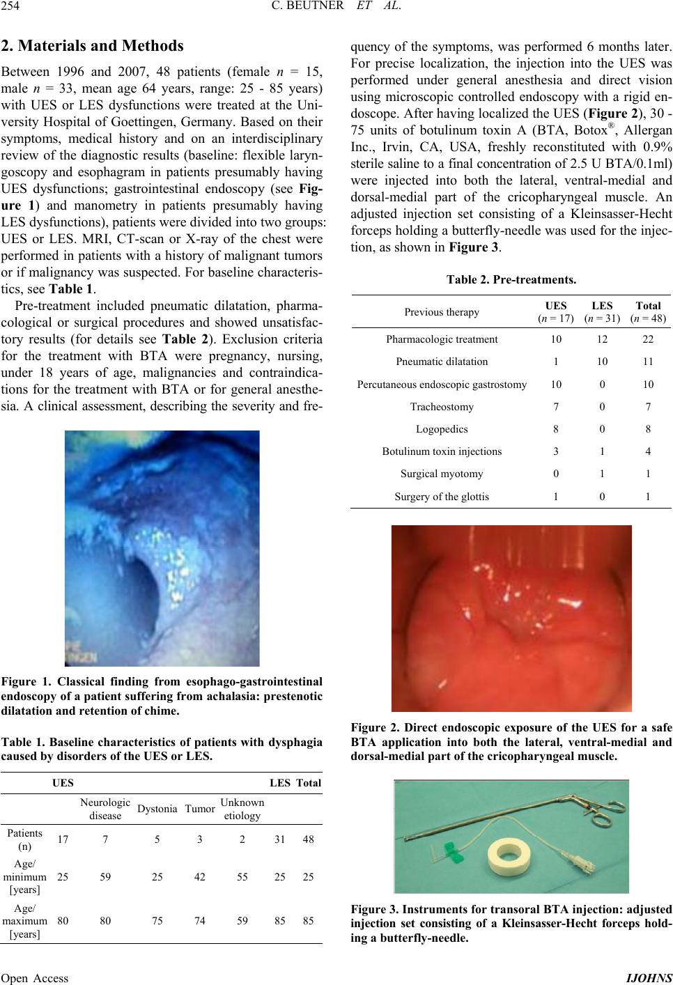 C. BEUTNER ET AL. 254 2. Materials and Methods Between 1996 and 2007, 48 patients (female n = 15, male n = 33, mean age 64 years, range: 25 - 85 years) with UES or LES dysfunctions were treated at the Uni- versity Hospital of Goettingen, Germany. Based on their symptoms, medical history and on an interdisciplinary review of the diagnostic results (baseline: flexible laryn- goscopy and esophagram in patients presumably having UES dysfunctions; gastrointestinal endoscopy (see Fig- ure 1) and manometry in patients presumably having LES dysfunctions), patients were divided into two groups: UES or LES. MRI, CT-scan or X-ray of the chest were performed in patients with a history of malignant tumors or if malignancy was suspected. For baseline characteris- tics, see Table 1. Pre-treatment included pneumatic dilatation, pharma- cological or surgical procedures and showed unsatisfac- tory results (for details see Table 2). Exclusion criteria for the treatment with BTA were pregnancy, nursing, under 18 years of age, malignancies and contraindica- tions for the treatment with BTA or for general anesthe- sia. A clinical assessment, describing the severity and fre- Figure 1. Classical finding from esophago-gastrointestinal endoscopy of a patient suffering from ac halasia: prestenotic dilatation and retention of chime. Table 1. Baseline characteristics of patients with dysphagia caused by disorders of the UES or LES. UES LES Total Neurologic disease DystoniaTumor Unknown etiology Patients (n) 17 7 5 3 2 3148 Age/ minimum [years] 25 59 25 42 55 2525 Age/ maximum [years] 80 80 75 74 59 8585 quency of the symptoms, was performed 6 months later. For precise localization, the injection into the UES was performed under general anesthesia and direct vision using microscopic controlled endoscopy with a rigid en- doscope. After having localized the UES (Figure 2), 30 - 75 units of botulinum toxin A (BTA, Botox®, Allergan Inc., Irvin, CA, USA, freshly reconstituted with 0.9% sterile saline to a final concentration of 2.5 U BTA/0.1ml) were injected into both the lateral, ventral-medial and dorsal-medial part of the cricopharyngeal muscle. An adjusted injection set consisting of a Kleinsasser-Hecht forceps holding a butterfly-needle was used for the inj ec- tion, as shown in Figure 3. Table 2. Pre-treatments. Previous therapy UES (n = 17) LES (n = 31)Total (n = 48) Pharmacologic treatment 10 12 22 Pneumatic dilatation 1 10 11 Percutaneous endo scopic gastrostomy 10 0 10 Tracheostomy 7 0 7 Logopedics 8 0 8 Botulinum toxin inj e c t i o n s 3 1 4 Surgical myotomy 0 1 1 Surgery of the glottis 1 0 1 Figure 2. Direct endoscopic exposure of the UES for a safe BTA application into both the lateral, ventral-medial and dorsal-medial part of the cricopharyngeal muscle. Figure 3. Instruments for transoral BTA injection: adjusted injection set consisting of a Kleinsasser-Hecht forceps hold- ing a butterfly-needle. Open Access IJOHNS 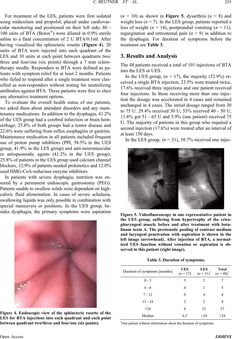 C. BEUTNER ET AL. 255 For treatment of the LES, patients were first sedated using midazolam and propofol, placed under cardiovas- cular monitoring and positioned on their left side; 80 - 100 units of BTA (Botox®) were diluted in 0.9% sterile saline to a final concentration of 2 U BTA/0.1ml. After having visualized the sphincteric rosette (Figure 4), 20 units of BTA were injected into each quadrant of the LES and 10 units at each point between quadrants two/ three and four/one (six points) through a 7 mm sclero- therapy needle. Responders to BTA were defined as pa- tients with symptom relief for at least 2 months. Patients who failed to respond after a single treatment were clas- sified as non-responders without testing for neutralizing antibodies against BTA. These patients were free to elect any alternative treatment options. To evaluate the overall health status of our patients, we asked them about attendant disorders and any main- tenance medications. In addition to the dysphagia, 41.2% of the UES group had a cerebral infarction or brain hem- orrhage; 25.8% of both groups had a tumor disease and 22.6% were suffering from reflux esophagitis or gastritis. Maintenance medication in all patients included frequent use of proton pump inhibitors (PPI, 58.5% in the UES group, 41.9% in the LES group) and anti-neuromuscular or antispasmodic agents (41.2% in the UES group); 25.8% of patients in the LES group used calcium channel blockers, 12.9% of patients needed prokinetics and 12.0 % used HMG-CoA-reductase enzyme inhibitors. In patients with severe dysphagia, nutrition was en- sured by a permanent endoscopic gastrostomy (PEG). Patients unable to swa llo w so lids were dependent on high - caloric fluid alimentation. In cases of severe achalasia, swallowing liquids was only possible in combination wit h special maneuvers or positions. In the UES group, be- sides dysphagia, the primary symptoms were aspiration Figure 4. Endoscopic view of the sphincteric rosette of the LES for BTA injections into each quadrant and each point between quadr ant two/three and four/one (six points). (n = 10) as shown in Figure 5, dysarthria (n = 8) and weight loss (n = 7). In the LES group, patients repo rted a loss of weight (n = 14), postprandial vomiting (n = 11), regurgitation and retrosternal pain (n = 9) in addition to the dysphagia. For duration of symptoms before the treatment see Table 3. 3. Results and Analysis The 48 patients received a total of 101 injections of BTA into the LES or UES. In the UES group, (n = 17), the majority (52.9%) re- ceived a single BTA injection, 23.5% were treated twice, 17.6% received three injections and one patient received four injections. In those receiving more than one injec- tion the dosage was accelerated in 4 cases and remained unchanged in 4 cases. The initial dosage ranged from 30 to 75 U. 29.4% received 30 U, 53% received 40 - 50 U, 11.8% got 51 - 65 U and 5.9% (one patient) received 75 U. The majority of patients in this group who required a second injection (17.6%) were treated after an interval of at least 150 days. In the LES group, (n = 31), 38.7% received one injec- Figure 5. Videofluoroscopy in one representative patient in the UES group, suffering from hypertrophy of the crico- pharyngeal muscle before and after treatment with botu- linum toxin A. The prestenotic pooling of contrast medium and laryngeal penetration with aspiration is shown in the left image (arrowhead). After injection of BTA, a normal- ized UES function without retention or aspiration is ob- served in this patient (right image). Table 3. Duration of symptoms. Duration of symptoms [months]UES (n = 17) LES (n = 31)*Total (n = 48)* 0 - 3 5 2 7 4 - 6 4 1 5 7 - 12 0 4 4 13 - 24 2 2 4 >24 6 21 27 Median 6.5 >24 >24 *One patient without inform a tion about the dura tion of sym ptoms. Open Access IJOHNS 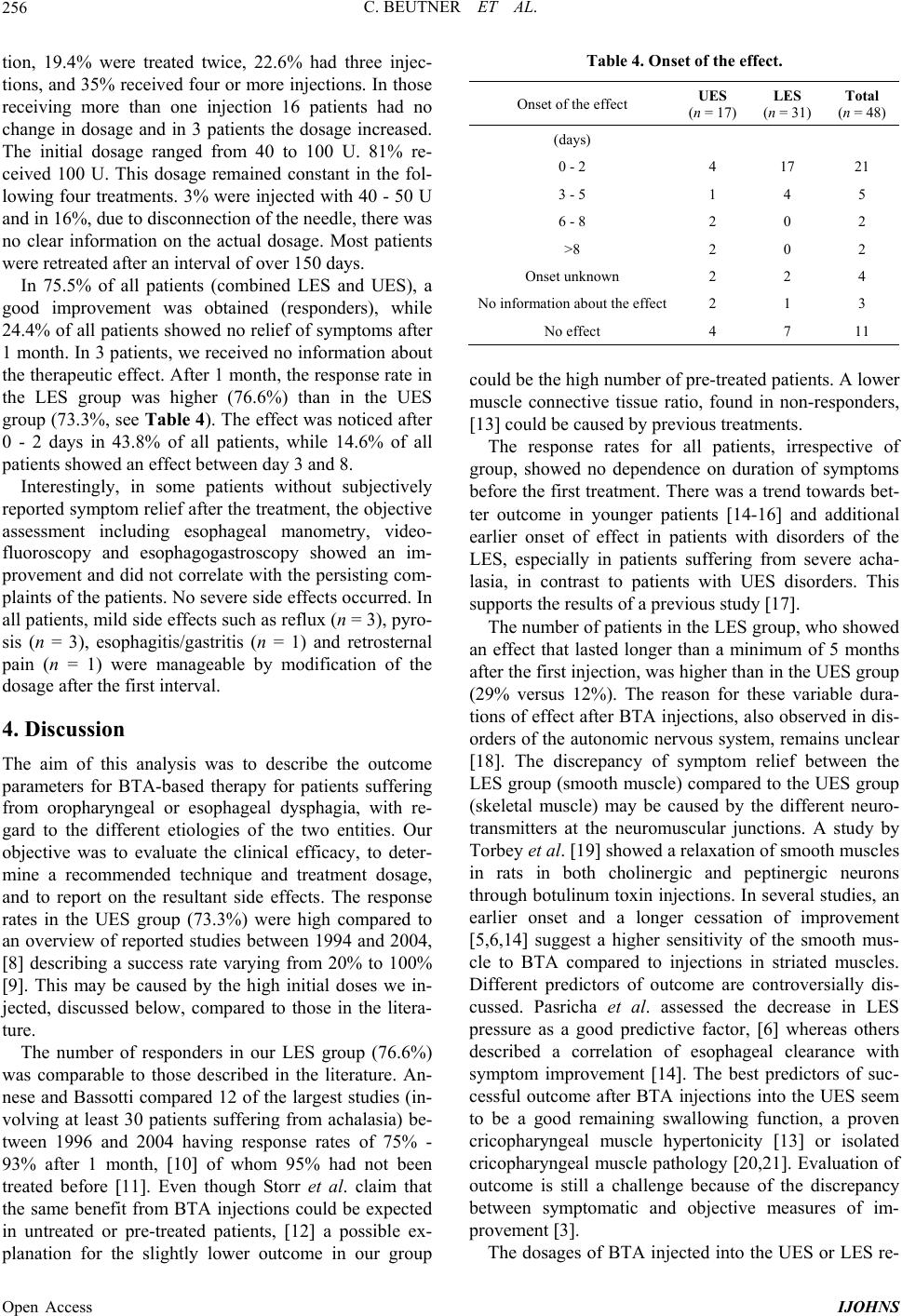 C. BEUTNER ET AL. 256 tion, 19.4% were treated twice, 22.6% had three injec- tions, and 35% received four or more injections. In those receiving more than one injection 16 patients had no change in dosage and in 3 patients the dosage increased. The initial dosage ranged from 40 to 100 U. 81% re- ceived 100 U. This dosage remained constant in the fol- lowing four treatments. 3% were injected with 40 - 50 U and in 16%, due to disconnection of the needle, there was no clear information on the actual dosage. Most patients were retreated after an interval of over 150 days. In 75.5% of all patients (combined LES and UES), a good improvement was obtained (responders), while 24.4% of all patients showed no relief of symptoms after 1 month. In 3 patients, we received no information about the therapeutic effect. After 1 month, the respon se rate in the LES group was higher (76.6%) than in the UES group (73.3%, see Table 4). The effect was noticed after 0 - 2 days in 43.8% of all patients, while 14.6% of all patients showed an effect between day 3 and 8. Interestingly, in some patients without subjectively reported symptom relief after the treatment, the objective assessment including esophageal manometry, video- fluoroscopy and esophagogastroscopy showed an im- provement and did not correlate with the persisting com- plaints of the patients. No severe side effects occurred. In all patients, mild side effects such as reflux (n = 3), pyro- sis (n = 3), esophagitis/gastritis (n = 1) and retrosternal pain (n = 1) were manageable by modification of the dosage after the first interval. 4. Discussion The aim of this analysis was to describe the outcome parameters for BTA-based therapy for patients suffering from oropharyngeal or esophageal dysphagia, with re- gard to the different etiologies of the two entities. Our objective was to evaluate the clinical efficacy, to deter- mine a recommended technique and treatment dosage, and to report on the resultant side effects. The response rates in the UES group (73.3%) were high compared to an overview of reported studies between 1994 and 2004, 8 describing a success rate varying from 20% to 100% 9. This may be caused by the high initial doses we in- jected, discussed below, compared to those in the litera- ture. The number of responders in our LES group (76.6%) was comparable to those described in the literature. An- nese and Bassotti compared 12 of the largest studies (in- volving at least 30 patients suffering from achalasia) be- tween 1996 and 2004 having response rates of 75% - 93% after 1 month, 10 of whom 95% had not been treated before 11. Even though Storr et al. claim that the same benefit from BTA injections could be expected in untreated or pre-treated patients, 12 a possible ex- planation for the slightly lower outcome in our group Table 4. Onset of the effect. Onset of the effect UES (n = 17) LES (n = 31) Total (n = 48) (days) 0 - 2 4 17 21 3 - 5 1 4 5 6 - 8 2 0 2 >8 2 0 2 Onset unknown 2 2 4 No informat i o n a bout the e f f ect2 1 3 No effect 4 7 11 could be the high number of pre-treated patients. A lower muscle connective tissue ratio, found in non-responders, 13 could be caused by previous treatments. The response rates for all patients, irrespective of group, showed no dependence on duration of symptoms before the first treatment. There was a trend towards bet- ter outcome in younger patients 14-16 and additional earlier onset of effect in patients with disorders of the LES, especially in patients suffering from severe acha- lasia, in contrast to patients with UES disorders. This supports the results of a previou s study 17. The number of patients in the LES group, wh o showed an effect that lasted longer than a minimum of 5 months after the fir st injection , was hig her th an in the U ES group (29% versus 12%). The reason for these variable dura- tions of effect after BTA injections, also observed in dis- orders of the autonomic nervous system, remains unclear 18. The discrepancy of symptom relief between the LES group (smooth muscle) compared to the UES group (skeletal muscle) may be caused by the different neuro- transmitters at the neuromuscular junctions. A study by Torbey et al. 19 showed a relaxation of smooth muscles in rats in both cholinergic and peptinergic neurons through botulinum toxin injections. In several studies, an earlier onset and a longer cessation of improvement 5,6,14 suggest a higher sensitivity of the smooth mus- cle to BTA compared to injections in striated muscles. Different predictors of outcome are controversially dis- cussed. Pasricha et al. assessed the decrease in LES pressure as a good predictive factor, 6 whereas others described a correlation of esophageal clearance with symptom improvement 14. The best predictors of suc- cessful outcome after BTA injections into the UES seem to be a good remaining swallowing function, a proven cricopharyngeal muscle hypertonicity 13 or isolated cricopharyngeal muscle pathology 20,21. Evaluation of outcome is still a challenge because of the discrepancy between symptomatic and objective measures of im- provement 3. The dosages of BTA injected into th e UES or LES re- Open Access IJOHNS  C. BEUTNER ET AL. 257 ported in the literature are similar to our initial dosages. Dosages from 10 - 100 units (all dosages transformed to Botox®) with different injections techniques (transoral, percuateneous with or without electromyography, per- cuataneous CT-guided, with rigid or flexible endoscopes or in open techniqu e during vocal cord lateralization) are repor ted concerning the UES 7,9,20-24. We decided to inject dosages of BTA ranging from 30 to 75 U into the UES, with exact localization under gen- eral anesthesia, based upon evaluation of the results from dose-range studies. These studies showed a dosage-re- lated effect and relief of symptoms 25,26 without ob- servation of severe complications 8. Injections of com- parable high dosages are usually administered under general anesthesia and direct endoscopic view to ensure safe injection and correct localization of the needle to minimize the risk of complications. The dosages of BTA injection in patients with disorders of the LES, especially of achalasia, vary from 80 U 6,14 to 200 U 1,11. Since the mean dosage used by most centers was about 100 U BTA, in the majority of our patients, we chose to inject this amount. There was no clear dose/effect rela- tionship described in the first injection (investigation of 118 patients suffering from achalasia) 11. To inhibit the complete nerve regeneration in the smooth muscle, which is responsible for the recurrence of symptoms, a timely re-injection might be useful. Re-injection with 100 U BTA into the LES is recommended after 30 days 11. Nevertheless, we have to consider that short re-in- jection intervals and the amount of toxin might risk a receptor blockage by antibodies. The role of antibodies as a result of autoimmune response possibly leading to variable duration of the effect has to be clarified. The number of complications reported after BTA in- jection into the UES or LES is low. Uncontrolled diffu- sion into the hypopharyngeal musculature 22, transient chest pain (also occurring in the placebo group) 14, unilateral vocal fold paresis 9, abnormal acid reflux 27 and aspiration with death after delayed medical in- tervention 13 have been described. Mediastinitis and ulceration of the esophagus without perforation after BTA injection are rare complications 28. In our patients, no severe side effects or systemic effects were noted. The necessity for BTA re-injection, because of its re- versible effect, seems disadvantageous, but no additional adverse effects have been described after long-term ap- plication 4. Likewise, the indication for revision in surgery must also be considered. The predictive value of outcome of BTA injection into the UES compared to a myotomy is still controversially discussed 7,20. While evaluating the different treatment methods available for a patient’s relief of dysfunctions of the UES or LES causing dysphagia, numerous factors must be taken into account. These include the endoscopist’s or surgeon’s experience 13, the risk of complications in surgery, the patient’s age, coexisting diseases 29, the overall morbidity of the patient, long-term results and economic factors. In conclusion, comparing the two groups of esophag- eal dysfunction with fundamentally different etiologies (UES/LES), intramuscular BTA injections represent a safe and feasible technique with a lack of significant side effects in both groups, especially for older patients with co-morbid illness. Symptom relief, onset and duration of clinical benefit were better in dysfunctions of the LES, explainable by the history of malignant tumor resection in the UES group with scarred tissue and the presumably better effect of BTA in smooth muscles. Further studies are needed providing objective data that correlate with symptom improvement. 5. Summary The pharmacological and surgical treatment of severe oropharyngeal or esophageal dysphagia often gives un- satisfactory results. This paper reports an alternative treatment option, using endoscopic applied intramuscular injection of Botulinum toxin A in the upper or lower esophageal sphincter. 48 treated patients suffering from oropharyngeal or esophageal dysphagia confirmed the clinical efficacy of this method. The recommended dos- age and technique of injection showed no significant side effects. REFERENCES [1] J. E. Leyden, A. C. Moss and P. Mac Mathuna, “Endo- scopic Pneumatic Dilatation versus Botulinum Toxin In- jection in the Management of Primary Achalasia,” Coch- rane Database of Systematic Reviews, Vol. 18, No. 4, 2006, Article ID: CD005046. http://dx.doi.org/10.1002/14651858.CD005046.pub2 [2] Z. H. Wen, E. Gardener and Y. P. Wang, “Nitrates for Achalasia,” Cochrane Database of Systematic Reviews, 2004, No. 1, Article ID: CD002299. http://dx.doi.org/10.1002/14651858.CD002299.pub2 [3] N. Pehlivanov and P. J. Pasricha, “Achalasia: Botox, Dilatation or Laparoscopic Surgery in 2006,” Neurogas- troenterology and Motility, Vol. 18, No. 9, 2006, pp. 799- 804. http://dx.doi.org/10.1111/j.1365-2982.2006.00802.x [4] D. Dressler, “Pharmacological Aspects of Therapeutic Botulinum Toxin Preparations,” Nervenarzt, Vol. 77, No. 8, 2006, pp. 912-921. http://dx.doi.org/10.1007/s00115-006-2090-2 [5] P. J. Pasricha, W. J. Ravich, T. R. Hendrix, S. Sostre, B. Jones and A. N. Kallo, “Intrasphincteric Botulinum Toxin for the Treatment of Achalasia,” New England Journal of Medicine, Vol. 322, No. 12, 1995, pp. 774-778. http://dx.doi.org/10.1056/NEJM199503233321203 [6] P. J. Pasricha, R. Rai, W. J. Ravich, T. R. Hendrix and A. N. Kalloo, “Botulinum Toxin for Achalasia: Long-Term Open Access IJOHNS  C. BEUTNER ET AL. Open Access IJOHNS 258 Outcome and Predictors of Response,” Gastroenterology, Vol. 110, No. 5, 1996, pp. 1410-1415. http://dx.doi.org/10.1053/gast.1996.v110.pm8613045 [7] I. Schneider, W. Thumfart and H. E. Eckel, “Treatment of Dysfunction of the Cricopharyngeal Muscle with Bot u li n u m Toxin. Introduction of a New, Non-Invasive Method,” Annals of Otology, Rhinology, and Laryngology, Vol. 103, 1994, pp. 31-35. [8] M. B. J. Moermann and B. J. Mieke, “Cricopharyngeal Botox Injection: Indication and Technique,” Current Opin- ion in Otolaryngology & Head and Neck Surgery, Vol. 14, No. 6, 2006, pp. 431-436. http://dx.doi.org/10.1097/MOO.0b013e328010b85b [9] S. I. Atkinson and J. Rees, “Botulinum Toxin for Crico- pharyngeal Dysphagia: Case Reports of CT-Guided In- jection,” Journal of Otolaryngology, Vol. 26, No. 4, 1997, pp. 273-276. [10] V. Annese and G. Bassotti, “Non-Surgical Treatment of Esophageal Achalasia,” World Journal of Gastroenterol- ogy, Vol. 12, No. 36, 2006, pp. 5763-5766. [11] V. Annese, G. Bassotti, G. Coccia, M. Dinelli, V. D’Onofrio, G. Gatto, et al. and the GISMAD Achalasia Study Group, “A Multicentre Randomised Study of Intrasphincteric Botulinum Toxin in Patient s wit h Oesophageal A cha l asia,” Gut, Vol. 46, No. 5, 2000, pp. 597-600. http://dx.doi.org/10.1136/gut.46.5.597 [12] M. Storr, P. Born, E. Frimberger, N. Weigert, T. Rösch, A. Meining, et al., “Treatment of Achalasia: The Short- Term Response to Botulinum Toxin Injection Seems to be Independent of Any Kind of Pre-Treatment,” BMC Gastroenterology, Vol. 2, 2002, p. 19. http://dx.doi.org/10.1186/1471-230X-2-19 [13] G. Zaninotto, R. Marchese-Ragona, C. Briani, M. Costan- tini, C. Rizzetto, G. Portale, et al., “The Role of Bo t ul i n um Toxin Injection and Upper Esopha geal Sphincter Myotomy in Treating Oropharyngeal Dysphagia,” Journal of Gas- trointestinal Surgery, Vol. 8, No. 8, 2004, pp. 997-1006. http://dx.doi.org/10.1016/j.gassur.2004.09.037 [14] C. Cuillere, P. Ducrotté, F. Zerbib, E. H. Metman, D. de Looze, F. Guillemot, et al., “Achalasia: Outcome of Pa- tients Treated with Intrasphincteric Injection of Botulinu m Toxin,” Gut, Vol. 41, No. 1, 1997, pp. 87-92. http://dx.doi.org/10.1136/gut.41.1.87 [15] V. M. Fishman, H. P. Parkman, T. D. Schiano, C. Hills, M. A. Dabezies, S. Cohen, et al., “Symptomatic Improve- ment in Achalasia after Botulinum Toxin Injection of the Lower Esophageal Sphincter,” American Journal of Gas- troenterology, Vol. 91, No. 9, 1996, pp. 1724-1730. [16] J. Kolbasnik, E. W. Wat erfall, B. Fachni e, Y. Chen and G. Tougas, “Long-Term Efficacy of Botulinum Toxin in Cl as- sical Achalasia: A Prospective Study,” American Journal of Gastroenterology, Vol. 94, No. 12, 2004, pp. 3434- 3439. http://dx.doi.org/10.1111/j.1572-0241.1999.01605.x [17] V. F. Eckardt, C. Aignherr and G. Bernhard, “Predictors of Outcome in Patients with Achalasia Treated by Pneu- matic Dilation,” Gastroenterology, Vol. 103, 1992, pp. 1732-1738. [18] M. Naumann, W. H. Jost and K. V. Toyka, “Botulinum Toxin in the Treatment of Neurological Disorders of the Autonomic Nervous System,” Archives of Neurology, Vol. 56, 1999, pp. 914-916. http://dx.doi.org/10.1001/archneur.56.8.914 [19] C. F. Torbey, C. Singaram, P. Bass and E. A. Gaumnitz, “Action of Botulinum Toxin on Gut Smooth Muscle In- volves Cholinergic and Non-C holinergic Neural Pathway ,” Gastroenterology, Vol. 112, 1997, p. A840. [20] A. Blitzer and M. F. Brin, “Use of Botulinum Toxin for Diagnosis and Management of Cricopharyngeal Ach a l as i a, ” Otolaryngology—Head and Neck Surgery, Vol. 116, No. 3, 1997, pp. 328-330. http://dx.doi.org/10.1016/S0194-5998(97)70267-5 [21] G. Y. Shaw and J. P. Searl, “Botulinum Toxin Treatment for Cricopharyngeal Dysfunction,” Dysphagia, Vol. 16, No. 3, 2001, pp. 161-167. http://dx.doi.org/10.1007/s00455-001-0074-8 [22] J. Alberty, M. Oelrich, K. Ludwig, S. Hartmann and W. Stoll, “Efficacy of Botulinum Toxin A for the Treatment of Upper Esophageal Sphincter Dysfunction,” Laryngo- scope, Vol. 110, No. 7, 2000, pp. 1151-1156. http://dx.doi.org/10.1097/00005537-200007000-00016 [23] M. Moermann, Y. Callier, C. Dick and H. Vermeersch, “Botulinum Toxin for Dysphagia Due to Cricopharyngeal Dysfunction,” European Archives of Oto-Rhino-Laryn- gology, Vol. 259, No. 1, 2002, pp. 1-3. http://dx.doi.org/10.1007/PL00007520 [24] R. Marchese-Ragona, G. Marioni, D. A. Restivo and A. Staffieri, “Solving Dysphagia Due to Cricopharyngeal Muscle Dysfunction with Botulinum Toxin,” European Archives of Oto-Rhino-Laryngology, Vol. 262, No. 3, 2005, pp. 250-251. http://dx.doi.org/10.1007/s00405-004-0776-2 [25] F. Mancini, G. Sandrini, A. Moglia, G. Nappi and C. Pacchetti, “A Randomized, Double-Blind, Dose-Ranging Study to Evaluate Efficacy and Safety of Three Doses of Botulinum Toxin Type A (Botox) for the Treatment of Spastic Foot,” Neurological Sciences, Vol. 26, No. 1, 2005, pp. 26-31. http://dx.doi.org/10.1007/s10072-005-0378-9 [26] D. Dressler and J. C. Rothwell, “Electromyographic Quantification of the Paralysing Effect of Botulinum Toxin in the Sternomastoid Muscle,” European Neurol- ogy, Vol. 43, No. 1, 2000, pp. 13-16. http://dx.doi.org/10.1159/000008122 [27] W. E. Waterfall, G. Tougas, B. Fachnie and U. Chauhan, “Clinical and Manometric Response to Botulinum Toxin (Botox) Used as Primary Treatment for Achalasia,” Gas- troenterology, Vol. 108, 1995, p. A225. [28] R. Mac Ever, M. Liptay and Y. Johnson, “A Case of Me- diastinitis Following Botulinum Toxin Type A Treatment for Achalasia,” Nature Clinical Practice Gastroenterol- ogy & Hepatology, Vol. 4, No. 10, 2007, pp. 579-582. http://dx.doi.org/10.1038/ncpgasthep0951 [29] L. Dughera, E. Battaglia, D. Maggio, P. Cassolino, P. R. Mioli, A. Morelli, G. Emanuelli, et al., “Botulinum Toxin Treatment of Oesophageal Achalasia in the Old Old and Oldest Old: A 1-Year Follow-Up Study,” Drugs Aging, 2005, Vol. 22, No. 9, pp. 779-783. http://dx.doi.org/10.2165/00002512-200522090-00006 |