

Paper Menu >>
Journal Menu >>
 International Journal of Otolaryngology and Head & Neck Surgery, 2013, 2, 236-239 Published Online November 2013 (http://www.scirp.org/journal/ijohns) http://dx.doi.org/10.4236/ijohns.2013.26049 Open Access IJOHNS Evaluation and Management of Massive Endotracheal Hemorrhage in Pediatric Patients: A Case Report and Review of Literature Cory Vaughn, Mona Shete, Rose Mary Stocks, Jerome Thompson University of Tennessee Health Science Center, Memphis, USA Email: [email protected] Received August 5, 2013; revised September 2, 2013; accepted October 1, 2013 Copyright © 2013 Cory Vaughn et al. This is an open access article distributed under the Creative Commons Attribution License, which permits unrestricted use, distribution, and reproduction in any medium, provided the original work is properly cited. ABSTRACT Massive hemoptysis is a frightening and life-threatening event in children. Prompt, aggressive evaluation and manage- ment are necessary. The most common cause of hemoptysis in a pediatric patient is infectious, but other various etiolo- gies including tracheotomy related problems, cystic fibrosis, bronchiectasis, congenital an omalies of the cardiopulmon- ary vasculature and suction trauma must be considered as well. Presented is a report of a case of acute, massive en- dotracheal hemorrhage with multiple methods of management includ ing balloon tamponade, highly selective emboliza- tion, and mainstem occlusion. This case is an addition to our previously reported case series of nine pediatric patients with massive pulmonary hemorrhage. Various diagnostic and management techniques of hemoptyis in pediatric patients are discussed with an extensive review of the literature. Keywords: Pediatric; Massive; Hemoptysis; Endotracheal Tube; Management 1. Introduction Hemoptysis, a relatively common event in the adult population, is a rare occurrence in the pediatric setting. The most common cause of hemoptysis in the pediatric patients is infectious, but a multitude of etiologies must be considered [1]. In massive pulmonary hemorrhage, the threat of asphyxia from airway obstruction is greater than the threat of dying from exsanguination. Successful air- way management is paramount not only to prevent im- mediate respiratory failure, but also to the swift determi- nation of the location of bleeding through clear visualiza- tion and unobstructed instrumentation. Here a case of massive hemoptysis is discussed along with new tech- niques for its management. This case is an addition to the previously published nine case experiences of large pul- monary hemorrhage in the pediatric population. 2. Case Report A 14-year-old male pedestrian was admitted to LeBon- heur Children’s Medical Center in Memphis, TN with a diagnosis of a closed head injury and associated intracra- nial bleed after being struck by a vehicle. The patient was intubated for a period of 2 weeks, whereupon he devel- oped brisk bleeding from the endotracheal tube (ETT) as a sequela of a coughing spell during central line place- ment. The patient’s past medical history was insignifi- cant. The patient’s family history was significant for an unexplained gastrointestinal hemorrhage resulting in his father’s demise. Subsequently, the patient was noted to require pro- gressively higher ventilator settings to achieve appropri- ate oxygenation. Suction yielded a moderate amount of bright red blood. A flexible fiberoptic bronchoscopy demonstrated a significant amount of bright red blood in tracheobronchial tree bu t no specific site of bleeding was identified. In the following hours, the rate of bleeding intensified and clots began to partially obstruct the en- dotracheal tube. Multiple attempts at clot removal using suction catheters through the endotrach eal tube failed. At that point ventilation became progressively more difficult, then impossible which resulted in rapid desaturation. Direct suction to the endotracheal tube was applied with concurrent extubation yielding a coagulated cast ofthe tracheo-bronchial tree. Subsequently, the patient’s venti- lation dramatically improved and oxygenation was per- formed via face mask. The patient was then re-intubated due to the instability of the airway. This process was re- 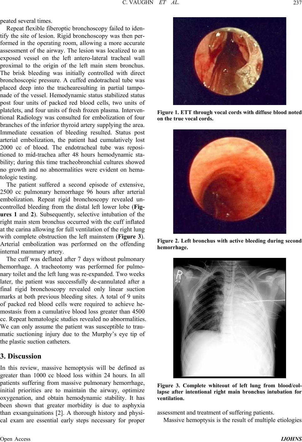 C. VAUGHN ET AL. 237 peated sev eral times. Repeat flexible fiberop tic bronchoscopy failed to iden- tify the site of lesion. Rigid bronchoscopy was then per- formed in the operating room, allowing a more accurate assessment of the airway. The lesion was localized to an exposed vessel on the left antero-lateral tracheal wall proximal to the origin of the left main stem bronchus. The brisk bleeding was initially controlled with direct bronchoscopic pressure. A cuffed endotracheal tube was placed deep into the trachearesulting in partial tampo- nade of the vessel. Hemodynamic status stabilized status post four units of packed red blood cells, two units of platelets, and four units of fresh frozen plasma. Interven- tional Radiology was consulted for embolization of four branch es of the in ferior thyroid artery supplying the area. Immediate cessation of bleeding resulted. Status post arterial embolization, the patient had cumulatively lost 2000 cc of blood. The endotracheal tube was reposi- tioned to mid-trachea after 48 hours hemodynamic sta- bility; during this time tracheobronchial cultures showed no growth and no abnormalities were evident on hema- tologic testing. The patient suffered a second episode of extensive, 2500 cc pulmonary hemorrhage 96 hours after arterial embolization. Repeat rigid bronchoscopy revealed un- controlled bleeding from the distal left lower lobe (Fig- ures 1 and 2). Subsequently, selective intubation of the right main stem bronchus occurred with the cuff inflated at the carina allowing for full ventilation of the right lung with complete obstruction the left mainstem (Figure 3). Arterial embolization was performed on the offending internal mammary artery. The cuff was deflated after 7 days without pulmonary hemorrhage. A tracheotomy was performed for pulmo- nary toilet and the left lung was re-expanded. Two weeks later, the patient was successfully de-cannulated after a final rigid bronchoscopy revealed only linear suction marks at both previous bleeding sites. A total of 9 units of packed red blood cells were required to achieve he- mostasis from a cumulative blood loss greater than 4500 cc. Repeat hematologic studies revealed no abnormalities. We can only assume the patient was susceptible to trau- matic suctioning injury due to the Murphy’s eye tip of the plastic suction catheters. 3. Discussion In this review, massive hemoptysis will be defined as greater than 1000 cc blood loss within 24 hours. In all patients suffering from massive pulmonary hemorrhage, initial priorities are to maintain the airway, optimize oxygenation, and obtain hemodynamic stability. It has been shown that greater morbidity is due to asphyxia than exsanguinations [2]. A thorough history and physi- cal exam are essential early steps necessary for proper Figure 1. ETT through vocal cords with diffuse blood noted on the true vocal cords. Figure 2. Left bronchus with active bleeding during second hemorrhage. Figure 3. Complete whiteout of left lung from blood/col- lapse after intentional right main bronchus intubation for ventilation. assessment and treatment of suffering patients. Massive hemoptysis is the result of multiple etiologies Open Access IJOHNS  C. VAUGHN ET AL. 238 which can be associated with various patient risk factors. Infection is widely known as the most common cause of massive hemoptysis in the pediatric population. While the majority of infectious hemorrhagic episodes are the result of S. aureus, all varieties of infection have been reported [3]. Interestingly, there is no correlation be- tween the severity of infection and rate or volume of bleeding. Further etiologic consid erations should includ e, but not be limited to: bleeding disorders, congenital vas- cular abnormalities, tracheobronchial papillomatosis, foreign body, tracheostomy tube related, diabetes, and cystic fibrosis (a 1% yearly incidence) [1,3,4]. In the previously published series from our institution, we en- countered infection as the most frequent cause of hemoptysis, followed by laryngotracheal papillomatosis, trauma and one case of unknown etiology [1]. In a re- cently published retrospective review of 15 patients, in- fection was also found to be the leading cause followed by tracheotomy related problems [5]. These risk factors and etiologies can differ greatly from those seen in the adult population where the incidence of massive pulmo- nary hemorrhage is much greater. Many reported cases of massive hempotysis are without known etiology or loca- tion of lesion . Endoscopic evaluation frequently allows for both identification of the lesion site and initial therapy simul- taneously, especially in cases of tracheal or bronchial injury. Flexible laryngoscopy and bronchoscopy provides the ability to examine the distal bronchial tree, to per- form tracheobronchial washings, and to obtain small bi- opsies. As in the case presented, flexible endoscopy car- ries an increased risk of missing view of the lesion. In general, caution should be exercised using flexible fi- beroptic bronchoscopy in small children because of the inability to simultaneously ventilate effectively against airway resistance and the difficulty of removing large clots through the small suction lumens or small endotra- cheal tubes as used in infants. Rigid bronchoscopy with a ventilating bronchoscope and telescopic capabilities has been shown to offer the best chance massive hemorrhage localization in a pediatric patient [2]. Once the site of bleeding is localized, method of in- tervention can be determined. In patien ts with a high rate of bleeding or diagnosed tuberculosis, surgical interven- tion has been shown to improve mortality compared to medical intervention [6]. During rigid bronchoscopy, options of endo-bronchial treatment become a considera- tion. Bleeding may be controlled with topical lavage therapy (i.e. epipherine), rigid bronchoscopic pressure, or endobronchialtamponade. Topical therapy is unfortu- nately associated with hemodynamic consequences of concentrated epinephrine. Valipour et al. reported bron- choscopy guided topical hemostatic tamponade with oxi- dized regenerated cellulose mesh is arrests hemoptysis in 98% of 57 patients for the first 48 hours with only 10.5% of patients experiencing a recurrence of hemoptysis and to a lesser degree 6 [7]. In a report of seventy-six con- secutive patients with massive hemoptysis unresponsive to the bronchoscopic wedging technique, cold saline so- lution lavage or instillation of regional vasoconstrictors [7]. Electrical current coagulation still has a role if tech- nically feasible. Laser coagulation therapy also is a consideration in specific circumstances. Use of laser coagulation is a suit- able option for patients with vascular lesions, tracheo- bronchial papillomatosis, or other tumor-like growths. In our previously reported series, a potassium titanyl phos- phate (KTP) laser was used. It, however, cannot be used during active bleeding [1]. In a retrospective review of 14 cases of children with life-threatening pulmonary hemorrhage, both CO2 and Nd-Yag laser use were per- formed on patients suffering from hemoptysis due to vascular lesions or tumor-like growths. In all patients who underwent Nd-Yag laser therapy, disappointing re- sults revealed only temporary control on discrete bleed- ing areas and thus required further management for per- manent hemostasis [8]. If bleeding continues or cannot be localized, emergent arteriography and embolization is indicated. Bronchial artery embolization (BAE) is an established treatment for massive and seriou s hemoptysis with good results and an acceptable rate of complications and morbidity [9-11]. Occlusion of the bleeding bronchial vessels should be considered for both short term and long term definitive therapy as we proved in the presented case report. Remy et al. treated 49 patients with arterial embolization during active hemoptysis, 41 of whom had an initial arrest of bleeding [12]. 6 of those 41 patients suffered a reoccur- rence of hemorrhage between 2 and 7 months of initial therapy, 83.3% of which w ere of a lesser severity. In a 20 year retrospective review of patients with cystic fibrosis, massive pulmonary hemorrhage was more likely to be treated with BAE [4]. While BAE was highly effective (98% success rate) at controlling symptoms temporarily, re-embolization was required in 46% of these patients. In another review, 22 patients underwent BAE with a reoc- currence rate of 50%. Reported absolute contraindica- tions to bronchial arterial embolization include the spinal artery arising from a variant position, relative contrain- dications include sepsis, coagulopathy, and renal-insuf- ficiency [13]. All reported episodes of re-bleeding occur within 3 years of initial embolization. Emergency open surgical interv ention for this problem should be considered when embolization has failed or is not feasible. Various techniques have been utilized to treat persistent, severe hemoptysis. An option for tracheal bleeding is to skeletonize the tracheal wall surgically when embolization has failed. Methods have included Open Access IJOHNS  C. VAUGHN ET AL. Open Access IJOHNS 239 pneumonectomy in an adult, reported by Mehta et al. [14]. Taguchi reported pneumonectomy and embolec- tomy in a 10-year-old child [15]. A staged extra-anat- omic conduit to deliver low-pressure blood to the lung, and interruption of the high-pressure vessels were re- ported in a 14-year-old child by Rankin [16]. Kirkpatrick reported success with a complete anatomic repair of the congenital heart abnormality in the treatment of a hemoptysis in a symptomatic 2-month-old in high-output cardiac failure [17]. 4. Conclusion Hemorrhage into the child’s airway is a frightening and potentially lethal event. The differential diagnosis in children is broad and a systematic approach to diagnosis and management is essential. We add another case report and two new alternative techniques to our series and to the armamentarium for the management of massive hemoptysis in children. REFERENCES [1] J. W. Thompson, C. D. Nguyen, R. H. Lazar, R. M. Stocks, R. A. Schoumacher, F. Hamdan and K. V. Nguyen, “Evaluation and Management of Hemoptysis in Infants and Children,” The Annals of Otology, Rhinology, and Laryngology, Vol. 105, No. 7, 1996, pp. 516-520 [2] L. Tom, R. A. Weisman and S. D. Handler, “Hemoptysis in Children,” The Annals of Otology, Rhinology, and Laryngology, Vol. 89, No. 5, 1980, pp. 419-424. [3] P. A. Flume, J. R. Yankaskis, M. Ebeling, T. Hulsey and L. L. Clark, “Massive Hemoptysis in Cystic Fibrosis,” Chest, Vol. 128, No. 2, 2005, pp. 729-738. http://dx.doi.org/10.1378/chest.128.2.729 [4] J. U. Barben, M. Ditchfield, J. B. Carlin, et al., “Major Hemoptysis in Children with Cystic Fibrosis: A 20-Year Retrospective Study,” Journal of Cystic Fibrosis, Vol. 2, No. 3, 2003, pp. 105-111. http://dx.doi.org/10.1016/S1569-1993(03)00066-3 [5] P. S. Batra and L. D. Holinger, “Etiology and Manage- ment of Pediatrichemoptysis,” Archives of Otolaryngol- ogy—Head and Neck Surgery, Vol. 127, No. 4, 2001, pp. 377-382. http://dx.doi.org/10.1001/archotol.127.4.377 [6] R. Corey and K. M. Hla, “Major and Massive Hemoptysis: Reassessment of Conservative Management,” American Journal of Medicine, Vol. 294, No. 5, 1987, pp. 301-309. http://dx.doi.org/10.1097/00000441-198711000-00003 [7] A. Valipour, A. Kreuzer, H. Koller, W. Koessler and O. C. Burghuber, “Bronchoscopy-Guided Topical Hemostatic Tamponade Therapy for the Management of Life Threat- ening Hemoptysis,” Chest, Vol. 127, No. 6, 2005, pp. 1888-1889. http://dx.doi.org/10.1378/chest.127.6.2113 [8] J. D. Sidman, W. B. Wheeler, A. K. Cabalka, B. Sou me k h , C. A. Brown and J. B. Wright, “Management of Acute Pulmonary Hemorrhage in Children,” Laryngoscope, Vol. 111, No. 1, 2001, pp. 33-35. http://dx.doi.org/10.1097/00005537-200101000-00006 [9] S. Osaki, Y. Nakanishi, H. Wataya, K. Takayama, K. Inoue, Y. Takaki, S. Murayama and N. Hara, “Prognosis of Bronchial Artery Embolisation in the Management of Hemoptysis,” Respiration, Vol. 67, No. 4, 2000, pp. 412- 416. http://dx.doi.org/10.1159/000029540 [10] L. E. Lampmann and T. G. Tjan, “Embolization Therapy in Hemoptysis,” European Journal of Radiology, Vol. 18, 1994, pp. 15-19. http://dx.doi.org/10.1016/0720-048X(94)90356-5 [11] M. E. Hansen and S. Kadir, “Elective and Emergency Embolization Therapy in Children and Adolescents, Effi- cacy and Safet y, ” Radiology, Vol. 30, 1990, pp. 331-336. [12] J. Remy, A. Arnaud, H. Fardou, R. Giraud and C. Voisin, “Treatment of Hemoptysis by Embolization of Bronchial Arteries,” Diagnostic Radiology, Vol. 122, No. 1, 1997, pp. 33-37. [13] J. F. Angle, N. H. Siddiqi, M. J. Wallace, et al., “Quality Improvement Guidelines for Percutaneous Transcatheter Embolization Society of Interventional Radiology Stan- dards of Practice Committee,” Journal of Vascular and Interventional Radiology, Vol. 21, No. 10, 2010, pp. 1479- 1486. http://dx.doi.org/10.1016/j.jvir.2010.06.014 [14] A. C. Mehta, D. R. Livingston, W. Kawalek, J. A. Golish and J. K. O’Donnell, “Pulmonary Artery Agenesis Pre- senting as Massive Hemopty sis,” Angiology, Vol. 38, No. 1, 1987, pp. 67-71. http://dx.doi.org/10.1177/000331978703800110 [15] T. Taguchi, K. Ikeda, K. Kume, et al., “Isolated Unilat- eral Absence of Left Pulmonary Artery with Peribron- chialarteriovenous Malformation Showing Recurrent He- moptysis,” Pediatric Radiology, Vol. 17, 1987, pp. 316- 318. http://dx.doi.org/10.1007/BF02388246 [16] J. S. Rankin, R. J. Sterba, J. H. Karis and H. N. Oldham Jr., “Staged Correction of Pulmonary Atresia and Absent Right Pulmonry Artery,” Archives of Surgery, Vol. 118, 1983, pp. 974-978. http://dx.doi.org/10.1001/archsurg.1983.01390080076019 [17] S. E. Kirkpatrik, D. A. Girod and H. King, “Aortic Origin of the Right Pulmonary Artery. Surgical Repair without a Graft,” Circulation, Vol. 36, No. 5, 1967, pp. 777-782. http://dx.doi.org/10.1161/01.CIR.36.5.777 |