

Paper Menu >>
Journal Menu >>
 International Journal of Otolaryngology and Head & Neck Surgery, 2013, 2, 228-231 Published Online November 2013 (http://www.scirp.org/journal/ijohns) http://dx.doi.org/10.4236/ijohns.2013.26047 Open Access IJOHNS Solitary Fungus Ball of the Sphenoid Sinus Faiz Alouni, Yousof Yousof, Shahram Talebian Khorasani University of Tishreen, Lattakia, Syrian Arab Republic Email: [email protected], [email protected] Received July 18, 2013; revised August 15, 2013; accepted September 10, 2013 Copyright © 2013 Faiz Alouni et al. This is an open access article distributed under the Creative Commons Attribution License, which permits unrestricted use, distribution, and reproduction in any medium, provided the original work is properly cited. ABSTRACT A 50-year-old woman with long standing nonspecific disturbing headaches of the mid-face and rear of the head plus retro-orbital pain for about one year duration was proved to suffer fro m the fungus ball invo lvement of the left sphenoid sinus after operation. The diagnosis was established by histopathologic examination of the specimen removed at the time of operation. Keywords: Nonspecific Headaches; Retro-Orbital Pain; Fungus Ball; Sphenoid Sinus 1. Case Presentation A 50-year-old woman, living in the coastal city of Lat- takia—Syrian Arab Republic, developed chronic head- aches of nonspecific nature for more than a year before referring to our ENT clinic in Al-Asad university hospi- tal. She was complaining of nonspecific headaches and retro-orbital pain that have affected her life and work to a great extent. During the time and before referral to our clinic, she had seen many doctors including ENT spe- cialist but she had been diagnosed as having psychologi- cal headaches and/or as a tension headaches of natural etiology, partly pointing to the stress and hard work she had. She had been given analgesics and painkillers of different natures but indeed with no benefit. She also sought neurologic consultations prior to ENT examina- tion, but they found no clue whatsoever pointing to sphenoid pathology. So she was finally referred to our clinic for furt her worku p and poss i b le diagnosi s. Upon inquiry, she did not give us any prior history of hypertension, diabetes mellitus, any hereditary or occu- pational disorder, as well as any disease or medicines causing immunosuppression. In physical examination and all body systems including neurologic examinations, we found no clue to indicate pathology in the sphenoid sinus at first and also the rest of the physical examination proved quite normally. Nevertheless, ENT examination did not show any significant finding. She underwent complete blood test and chemical analyses to rule out any systemic problems and the results of tests were negative and unrevealed of any predisposing factors or comorbid- ity elements. To reach a diagnosis and take into account the possibility of sinus involvement, CT scanning of the head was done (Figure 1) that showed a radio-opaque mass in the left sphenoid sinus. Taking into account the physical examination, lab an alysis and CT result (Figure 1), our preliminary diagnosis was fungal involvement of the sphenoid sinus. 2. Discussion 2.1. Clinical Presentation SFB produces symptoms as a result of mass effect and paranasal sinus obstructio n. Medical attention is typ ically sought for symptoms consistent with Chronic Rhinosi- nusitis, although the extensive duration and refractoriness to medical therapy of a patient’s facial pain, headache, nasal airway obstruction, chronic cough, or purulent rhi- norrhea may be indicative of a more unusual process. Nasal endoscopy may demonstrate inflammatory polyp disease, which is found in only 10% of patients, but is more likely to reveal normal to mild mucosal inflamma- tion without other revealing characteristics [1-4]. 2.2. Radiology CT scans are more revealing, yet certainly not diagnostic. Single sinus involvement is reported in 59% to 94% of SFB cases, almost always with complete or subtotal opacification of the involved sinus and frequently dem- onstrating radiodensities within such opacifications (41%). Bony sclerosis of the walls of the involved sinus is common, as radiographic evidence of this bony thick- ening is noted in 33% to 62% in different case series. In 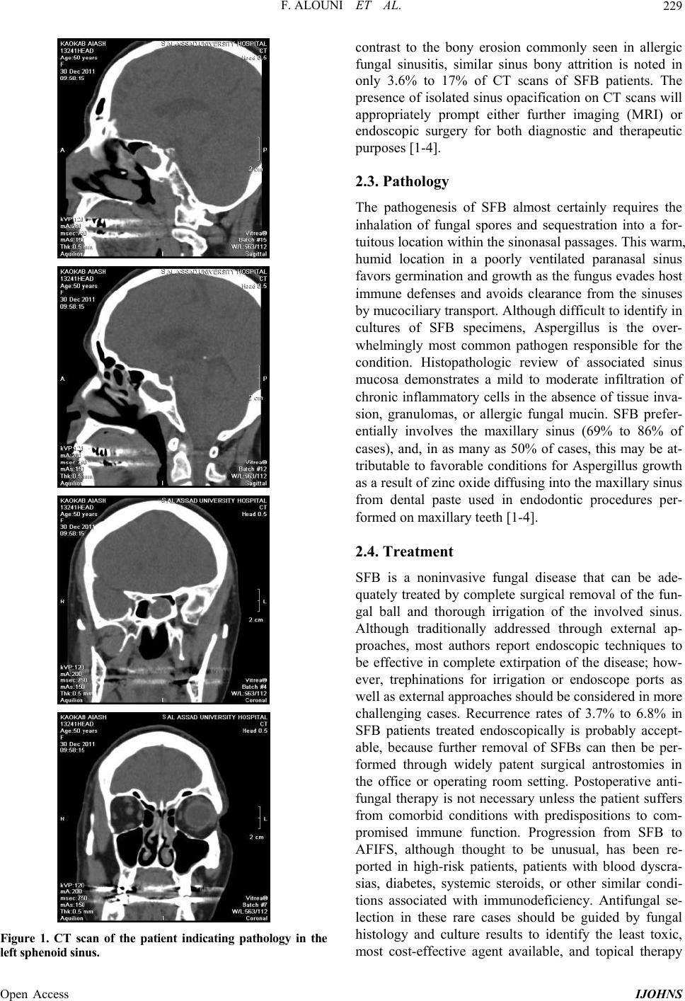 F. ALOUNI ET AL. 229 Figure 1. CT scan of the patient indicating pathology in the left sphenoid sinus. contrast to the bony erosion commonly seen in allergic fungal sinusitis, similar sinus bony attrition is noted in only 3.6% to 17% of CT scans of SFB patients. The presence of isolated sinus opacification on CT scans will appropriately prompt either further imaging (MRI) or endoscopic surgery for both diagnostic and therapeutic purposes [1-4]. 2.3. Pathology The pathogenesis of SFB almost certainly requires the inhalation of fungal spores and sequestration into a for- tuitous location within the sinonasal passages. This warm, humid location in a poorly ventilated paranasal sinus favors germination and growth as the fungus evades ho st immune defenses and avoids clearance from the sinuses by mucociliary transport. Although d ifficult to iden tify in cultures of SFB specimens, Aspergillus is the over- whelmingly most common pathogen responsible for the condition. Histopathologic review of associated sinus mucosa demonstrates a mild to moderate infiltration of chronic inflammatory cells in the absence of tissue inva- sion, granulomas, or allergic fungal mucin. SFB prefer- entially involves the maxillary sinus (69% to 86% of cases), and, in as many as 50% of cases, this may be at- tributable to favorable conditions for Aspergillus growth as a result of zinc oxide diffusing into the maxillary sinus from dental paste used in endodontic procedures per- formed on maxillary teeth [1-4]. 2.4. Treatment SFB is a noninvasive fungal disease that can be ade- quately treated by complete surgical removal of the fun- gal ball and thorough irrigation of the involved sinus. Although traditionally addressed through external ap- proaches, most authors report endoscopic techniques to be effective in complete extirpation of the disease; how- ever, trephinations for irrigation or endoscope ports as well as external approaches should be considered in more challenging cases. Recurrence rates of 3.7% to 6.8% in SFB patients treated endoscopically is probably accept- able, because further removal of SFBs can then be per- formed through widely patent surgical antrostomies in the office or operating room setting. Postoperative anti- fungal therapy is not necessary unless the patient suffers from comorbid conditions with predispositions to com- promised immune function. Progression from SFB to AFIFS, although thought to be unusual, has been re- ported in high-risk patients, patients with blood dyscra- sias, diabetes, systemic steroids, or other similar condi- tions associated with immunodeficiency. Antifungal se- lection in these rare cases should be guided by fungal histology and culture results to identify the least toxic, most cost-effective agent available, and topical therapy Open Access IJOHNS 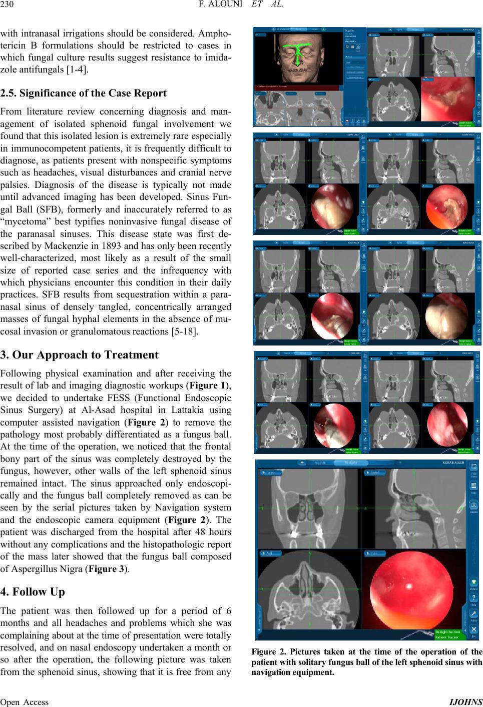 F. ALOUNI ET AL. 230 with intranasal irrigations shou ld be considered. Ampho- tericin B formulations should be restricted to cases in which fungal culture results suggest resistance to imida- zole antifunga ls [1-4]. 2.5. Significance of the Case Report From literature review concerning diagnosis and man- agement of isolated sphenoid fungal involvement we found that this isolated lesion is extremely rare especially in immunocompetent patients, it is frequently difficult to diagnose, as patients present with nonspecific symptoms such as headaches, visual disturbances and cranial nerve palsies. Diagnosis of the disease is typically not made until advanced imaging has been developed. Sinus Fun- gal Ball (SFB), formerly and inaccurately referred to as “mycetoma” best typifies noninvasive fungal disease of the paranasal sinuses. This disease state was first de- scribed by Mackenzie in 1893 and has only been recently well-characterized, most likely as a result of the small size of reported case series and the infrequency with which physicians encounter this condition in their daily practices. SFB results from sequestration within a para- nasal sinus of densely tangled, concentrically arranged masses of fungal hyphal elements in the absence of mu- cosal invasion or granulomatous reactions [5-18]. 3. Our Approach to Treatment Following physical examination and after receiving the result of lab and imaging diagnostic workups (Figure 1), we decided to undertake FESS (Functional Endoscopic Sinus Surgery) at Al-Asad hospital in Lattakia using computer assisted navigation (Figure 2) to remove the pathology most probably differentiated as a fungus ball. At the time of the operation, we noticed that the frontal bony part of the sinus was completely destroyed by the fungus, however, other walls of the left sphenoid sinus remained intact. The sinus approached only endoscopi- cally and the fungus ball completely removed as can be seen by the serial pictures taken by Navigation system and the endoscopic camera equipment (Figure 2). The patient was discharged from the hospital after 48 hours without any complications and the histopathologic report of the mass later showed that the fungus ball composed of Aspergillus Nigra (Figure 3). 4. Follow Up The patient was then followed up for a period of 6 months and all headaches and problems which she was complaining about at the time of presentation were total ly resolved, and on nasal endoscopy undertaken a month or so after the operation, the following picture was taken from the sphenoid sinus, showing that it is free from any Figure 2. Pictures taken at the time of the operation of the patient wit h soli tary fungus ball of the left sphenoi d si nus with navigation equipment. Open Access IJOHNS 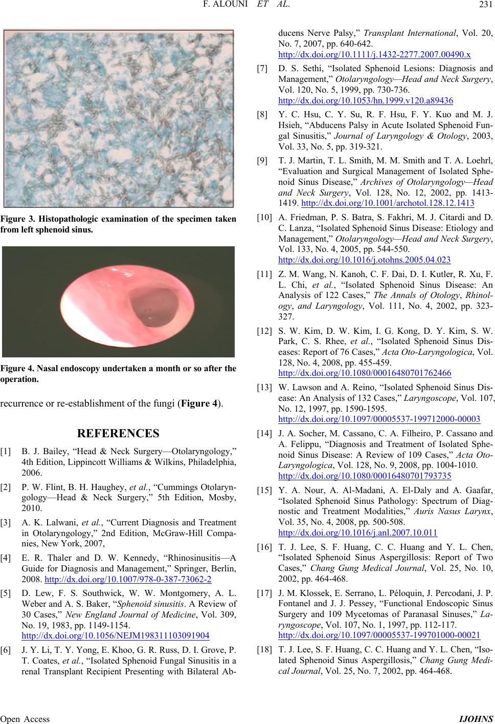 F. ALOUNI ET AL. Open Access IJOHNS 231 Figure 3. Histopathologic examination of the specimen taken from left sphenoid sinus. Figure 4. Nasal endoscopy underta ken a mo nth or so after the operatio n. recurrence or re-establishment of the fungi (Figure 4). REFERENCES [1] B. J. Bailey, “Head & Neck Surgery—Otolaryngology,” 4th Edition, Lippincott Williams & Wilkins, Philadelphia, 2006. [2] P. W. Flint, B. H. Haughey, et al., “Cummings Otolaryn- gology—Head & Neck Surgery,” 5th Edition, Mosby, 2010. [3] A. K. Lalwani, et al., “Current Diagnosis and Treatment in Otolaryngology,” 2nd Edition, McGraw-Hill Compa- nies, New York, 2007, [4] E. R. Thaler and D. W. Kennedy, “Rhinosinusitis—A Guide for Diagnosis and Management,” Springer, Berlin, 2008. http://dx.doi.org/10.1007/978-0-387-73062-2 [5] D. Lew, F. S. Southwick, W. W. Montgomery, A. L. Weber and A. S. Baker, “Sphenoid sinusitis. A Review of 30 Cases,” New England Journal of Medicine, Vol. 309, No. 19, 1983, pp. 1149-1154. http://dx.doi.org/10.1056/NEJM198311103091904 [6] J. Y. Li, T. Y. Yong, E. Khoo, G. R. Russ, D. I. Grove, P. T. Coates, et al., “Isolated Sphenoid Fungal Sinusitis in a renal Transplant Recipient Presenting with Bilateral Ab- ducens Nerve Palsy,” Transplant International, Vol. 20, No. 7, 2007, pp. 640-642. http://dx.doi.org/10.1111/j.1432-2277.2007.00490.x [7] D. S. Sethi, “Isolated Sphenoid Lesions: Diagnosis and Management,” Otolaryngology—Head and Neck Surgery, Vol. 120, No. 5, 1999, pp. 730-736. http://dx.doi.org/10.1053/hn.1999.v120.a89436 [8] Y. C. Hsu, C. Y. Su, R. F. Hsu, F. Y. Kuo and M. J. Hsieh, “Abducens Palsy in Acute Isolated Sphenoid Fun- gal Sinusitis,” Journal of Laryngology & Otology, 2003, Vol. 33, No. 5, pp. 319-321. [9] T. J. Martin, T. L. Smith, M. M. Smith and T. A. Loehrl, “Evaluation and Surgical Management of Isolated Sphe- noid Sinus Disease,” Archives of Otolaryngology—Head and Neck Surgery, Vol. 128, No. 12, 2002, pp. 1413- 1419. http://dx.doi.org/10.1001/archotol.128.12.1413 [10] A. Friedman, P. S. Batra, S. Fakhri, M. J. Citardi and D. C. Lanza, “Isolated Sphenoid Sinus Disease: Etiology and Management,” Otolaryngology—Head and Neck Surgery, Vol. 133, No. 4, 2005, pp. 544-550. http://dx.doi.org/10.1016/j.otohns.2005.04.023 [11] Z. M. Wang, N. Kanoh, C. F. Dai, D. I. Kutler, R. Xu, F. L. Chi, et al., “Isolated Sphenoid Sinus Disease: An Analysis of 122 Cases,” The Annals of Otology, Rhinol- ogy, and Laryngology, Vol. 111, No. 4, 2002, pp. 323- 327. [12] S. W. Kim, D. W. Kim, I. G. Kong, D. Y. Kim, S. W. Park, C. S. Rhee, et al., “Isolated Sphenoid Sinus Dis- eases: Report of 76 Cases,” Acta Oto-Laryngologica, Vol. 128, No. 4, 2008, pp. 455-459. http://dx.doi.org/10.1080/00016480701762466 [13] W. Lawson and A. Reino, “Isolated Sphenoid Sinus Dis- ease: An Analysis of 132 Cases,” Laryngoscope, Vol. 107, No. 12, 1997, pp. 1590-1595. http://dx.doi.org/10.1097/00005537-199712000-00003 [14] J. A. Socher, M. Cassano, C. A. Filheiro, P. Cassano and A. Felippu, “Diagnosis and Treatment of Isolated Sphe- noid Sinus Disease: A Review of 109 Cases,” Acta Oto- Laryngologica, Vol. 128, No. 9, 2008, pp. 1004-1010. http://dx.doi.org/10.1080/00016480701793735 [15] Y. A. Nour, A. Al-Madani, A. El-Daly and A. Gaafar, “Isolated Sphenoid Sinus Pathology: Spectrum of Diag- nostic and Treatment Modalities,” Auris Nasus Larynx, Vol. 35, No. 4, 2008, pp. 500-508. http://dx.doi.org/10.1016/j.anl.2007.10.011 [16] T. J. Lee, S. F. Huang, C. C. Huang and Y. L. Chen, “Isolated Sphenoid Sinus Aspergillosis: Report of Two Cases,” Chang Gung Medical Journal, Vol. 25, No. 10, 2002, pp. 464-468. [17] J. M. Klossek, E. Serrano, L. Péloquin, J. Percodani, J. P. Fontanel and J. J. Pessey, “Functional Endoscopic Sinus Surgery and 109 Mycetomas of Paranasal Sinuses,” La- ryngoscope, Vol. 107, No. 1, 1997, pp. 112-117. http://dx.doi.org/10.1097/00005537-199701000-00021 [18] T. J. Lee, S. F. Huang, C. C. Huang and Y. L. Chen, “Iso- lated Sphenoid Sinus Aspergillosis,” Chang Gung Medi- cal Journal, Vol. 25, No. 7, 2002, pp. 464-468. |