

Paper Menu >>
Journal Menu >>
 Journal of Biosciences and Medicines, 2013, 1, 6-9 JBM http://dx.doi.org/10.4236/jbm.2013.12002 Published Online October 2013 (http://www.scirp.org/journal/jbm/) OPEN ACCESS Towards n ove l regularization approaches to PET i m age reconstruction E. Karali, D. Koutsouris Department of Electrical and Computer Engineering, National Technical University of Athens, Athens, Greece Email: [email protected] Received August 2013 ABSTRACT The purpose of this study is to introduce a novel em- pirical iterative algorithm for medical image recon- struction, under the short name MRP-ISWLS (Me- dian Root Prior Image Space Weighted Least Squares). Further, we assess the performance of the new algo- rithm by comparing it to the simultaneous version of known MRP algorithms. All algorithms are com- pared in terms of cross-correlation and CNRs (Con- trast-to-Noise Ratios). As it turns out, MRP-ISWLS presents higher CNRs than the known algorithms for objects of different size. Also MRP-ISWLS has better noise manipulation. Keywords: Image Reconstruction; Positron Emission Tomography (PET); Small Animal Imaging; Median Root Prior (MRP) 1. INTRODUCTION Iterative techniques are divided into two main categories: algebraic and statistical. Statistical techniques are classi- fied to maximum-likelihood algorithms and least squares methods. The most famous maximum likelihood tech- nique is the expectation maximization maximum likeli- hood (EM-ML) algorithm, which was first presented by Shepp and Vardi. Image Space Reconstruction Algorithm (ISRA) is a least square reconstruction method intro- duced by Daube-Witherspoon and Muehllehner. It shows better noise manipulation than EM-ML. Another least squares algorithm is the Weighted Least Square tech- nique (WLS), due to Anderson et al. WLS assumes that random independent noise variables present different standard deviations. The matrix of these standard devia- tions consists of the expected projection data. WLS ac- celerates the reconstruction process and results in recon- structed images of better spatial resolution [1-3]. For the reduction of the noise many regularization methods have been proposed, which reduce drastically the noise with a small image resolution reduction. These methods take into account a priori information for the radioactivity spatial distribution inside the object under examination [1]. The success of a regularization method depends on the mathematical formula of the prior. Me- dian root prior (MRP) [4] belongs to the most popular priors. It is derived from a Gaussian distribution with mean value the median value of reconstructed image pixels in the vicinity of pixel i. The use of MRP results in noise component reduction while at the same time it preserves the edges. The purpose of this study is on the one hand to intro- duce a new empirical MRP image reconstruction algo- rithm, under the short name MRP-ISWLS (Median Root- Prior-Image Space Weighted Least Squares). MRP- ISWLS will be an MRP version of ISWLS (Image Space Weigthed Least Squares), which was introduced in [5]. ISWLS has ISRA properties in noise manipulation and WLS acceleration of reconstruction process. To assess the performance of the new iterative reconstruction me- thod we have used phantom data produced from simu- lating a prototype small-animal PET system. We com- pared reconstruction data with MRP-EM-ML, MRP- ISRA and MRP-WLS following the OSL (One Step Late) philoshophy [6]. The methods presented here are applied to 2D sinograms. Moreov e r, the simultaneous version of the aforementioned algorithms is implemented. We note that simultaneous versions of reconstruction algorithms, that is, algorithms where all image pixels are simultaneously updated in every iteration, are of great interest because of their ability to be implemented in parallel computing architectures, which decreases dras- tically the total reconstruction time [1]. 2. THEORY In general, every iterative method relies on the hypothe- sis that the projection data y is linearly connected to the image x of radiopharmaceutical spatial distribution, ac- cording to the equation: xy T A = (1) where A is a matrix that characterizes the PET system  E. Karali, D. Koutsouris / Journal of Biosciences and Medicines 1 (2013) 6-9 Copyright © 2013 SciRes. OPEN ACCESS 7 being used for data acquisition. In bibliography this ma- trix is called system or probability matrix and it projects image data to sinogram domain (the term sinogram refers to the projection data matrix) [1]. Every element αij of the system matrix A represents the probability an an- nihilation event emitted in image pixel i to be detected in LORj (Line of Response). The significance of the probability matrix lies on the valuable information re- lated to the data acquisition process, that it can contain (e.g. number of detector rings, number of detector ele- ments in every ring, ring d iameter, diameter of transaxial field of view, detector size, image size, spatial and angu- lar sampling). 2.1. The ISWLS Algorithm Consider an image discretized into N pixels and the measured data y collected by M detector tubes. The updating scheme of the ISWLS algorithm in the kth it- eration is: ( ) 2 1 1 2 1 11 M ij j j kk ii MN k iji ji ji ay xx a ax = − − ′′ ′ = = = ∑ ∑∑ (2) 2.2. The Median Root Prior (MRP) It is derived from a Gaussian distribution with mean value the median value of reconstructed image pixels in the vicinity of pixel i . Suppose that: M M i x b iexf 2 2 )( )( − − ≈ (3) where ),(ixmedMi = , the median value of recon- structed image pixels in the vicinity of pixel i. Then: ),( ),()( )( ixmed ixmedx b x xf xu i ii i i i − = ∂ ∂ = (4) The term b ∈ [0,2] determines the degree of smoo- thing in reconstruction images. If b = 0 no prior is ap- plied. Big values of b cau se over-smoothing, while small values of b result in images with high resolution but with increased noise. 2.3. MRP Algorithms According to the one-step-late philosophy, where the prior is applied to the previous radiopharmaceutical dis- tribution estimation, we can extract an empirical iterative formula for the ISWLS algorithm in combination with MRP prior. The new empirical iterative algorithm has updating scheme: 2 11 11 2 1 '' 11 '1 ( ,) 1( ,) i i M ij j kj ki ikk MN ik iji ji kji ay x x xmed xi ba ax med xi −= −− − −= = =− + ∑ ∑∑ (5) The MRP-EM-ML updating scheme in kth iteration is: 1 11 11 '' 1'1 ( ,) 1( ,) i i kMij j ki ikk N jk iij i ki ay x xxmed xiax bmed xi − −− =− −= =− + ∑∑ (6) The MRP-ISRA updating scheme in kth iteration is: 11 11 1 111 ( ,) 1( ,) ii i M ij j kj ki ikkMN k iiji j kji ay x xxmed xia ax bmed xi ′ −= −− − ′ −′ = = =− + ∑ ∑∑ (7) The MRP-WLS updating s cheme in kth iteration is 2 1 11 2 11 '' 1'1 ( ,) 1( ,) j i i kMij ki ikk N j ik ij i ki ay x xxmed xi bax med xi − −− =− −= =− + ∑ ∑ (8) 3. RESULT S For the evaluation of the iterative reconstruction methods presented in Section 2, projection data of a Derenzo-type phantom have been used. The Derenzo-type phantom consists in sets of rods filled with F18, with diameters 4.8, 4, 3.2, 2.4, 1.6, and 1.2 mm, and the same separation between surfaces in the corresponding sets. The rods were surrounded by plastic (polyethylene). Data were produced using Monte Carlo simulation of a small-ani- mal PET scanner, consisting of two detector heads. Further, 18 × 106 coincidence events were collected. Projection data was binned to a 2D s inogram, 55 pixels × 170 pixels in size, which means that data from 55 TORs (Tube of Response) per rotation angle were collected and 170 totally angular samples were used. Since the two detector heads rotate from 0˚ to 180˚ the angular step size was 1.0647˚. The system matrix was derived from an analytical method and calculated once before reconstruction. Each element ij a was computed as the area of intersection Eij, of TORj (Tube-of-Response) with image pixel i . The calculated system matrix is a sparse matrix. It consists of zero-valued elements in majority that have no contribu- tion during iterative reconstruction process. So, only the non-zero elements were stored, resulting in significant reduction in system matrix size and consequently in re- quired storage. The reconstructed 2D images were 128 pixels × 128 pixels in size, thus the system matrix con- sisted of 55 × 170 × 128 × 128 elements with 4.33% 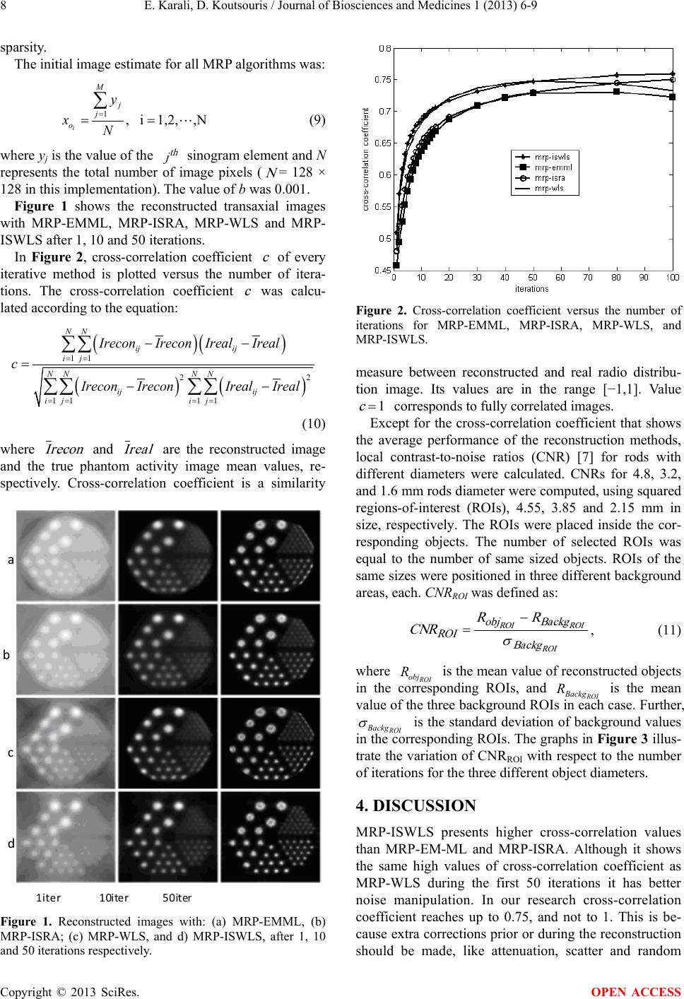 E. Karali, D. Koutsouris / Journal of Bios ci ences and Medicine s 1 (2013) 6-9 Copyright © 2013 SciRes. OPEN ACCESS 8 sparsity. The initial image estimate for all MRP algorithms was: 1 , i1,2,,N i M j j o y xN = = = ∑ (9) where yj is the value of the th j sinogram element and N represents the total number of image pixels ( N = 128 × 128 in this implementation). The value of b was 0.00 1. Figu re 1 shows the reconstructed transaxial images with MRP-EMML, MRP-ISRA, MRP-WLS and MRP- ISWLS after 1, 10 and 50 iterations. In Figure 2, cross-correlation coefficient c of every iterative method is plotted versus the number of itera- tions. The cross-correlation coefficient c was calcu- lated according to the equation: ()( ) () () 11 22 11 11 NN ij ij ij NN NN ij ij ij ij IreconIrecon IrealIreal c Irecon IreconIrealIreal = = = == = −− = −− ∑∑ ∑∑ ∑∑ (10) where reconI and realI are the reconstructed image and the true phantom activity image mean values, re- spectively. Cross-correlation coefficient is a similarity Figure 1. Reconstructed images with: (a) MRP-EMML, (b) MRP-ISRA; (c) MRP-WLS, and d) MRP-ISWLS, after 1, 10 and 50 iterations respectively. Figure 2. Cross-correlation coefficient versus the number of iterations for MRP-EMML, MRP-ISRA, MRP-WLS, and MRP-ISWLS. measure between reconstructed and real radio distribu- tion image. Its values are in the range [−1,1]. Value 1= c corresponds to fully correlated images. Except for the cross-correlation coefficient that shows the average performance of the reconstruction methods, local contrast-to-noise ratios (CNR) [7] for rods with different diameters were calculated. CNRs for 4.8, 3.2, and 1.6 mm rods diameter were computed, using squared regions-of-interest (ROIs), 4.55, 3.85 and 2.15 mm in size, respectively. The ROIs were placed inside the cor- responding objects. The number of selected ROIs was equal to the number of same sized objects. ROIs of the same sizes were positioned in three different background areas, each. CNRROI was defined as: ROI ROIROI Backg Backgobj ROI RR CNR σ − = , (11) where ROI obj R is the mean value of reconstructed objects in the corresponding ROIs, and ROI Backg R is the mean value of the three background ROIs in each case. Further, ROI Backg σ is the standard deviation of background values in the corresponding ROIs. The graphs in Figure 3 illus- trate the variation of CNRROI with respect to the number of iterations for the three different object diameters. 4. DISCUSSIO N MRP-ISWLS presents higher cross-correlation values than MRP-EM-ML and MRP-ISRA. Although it shows the same high values of cross-correlation coefficient as MRP-WLS during the first 50 iterations it has better noise manipulation. In our research cross-correlation coefficient reaches up to 0.75, and not to 1. This is be- cause extra corrections prior or during the reconstruction should be made, like attenuation, scatter and random b a c d 1iter 10ite r50iter 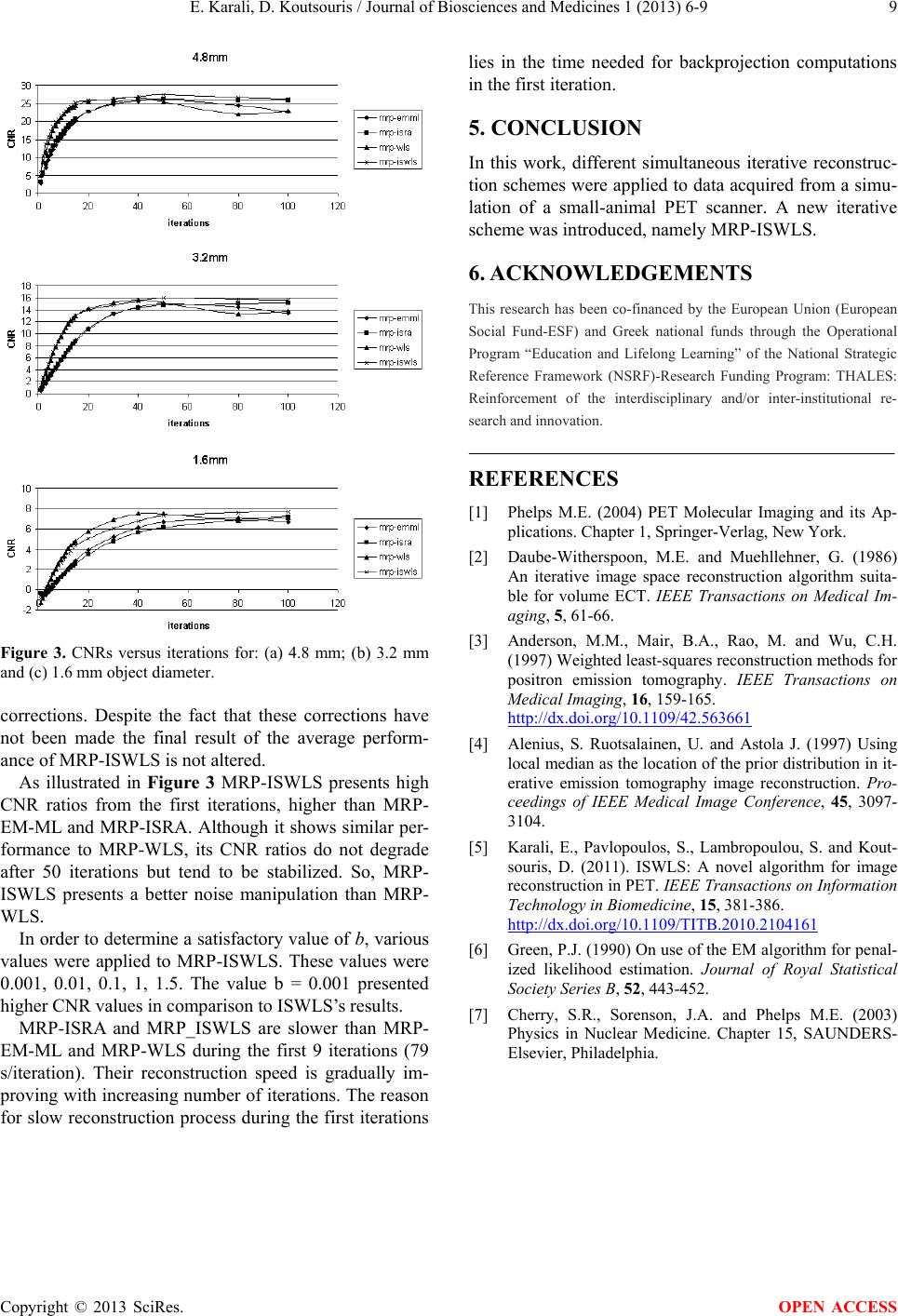 E. Karali, D. Koutsouris / Journal of Biosciences and Medicines 1 (2013) 6-9 Copyright © 2013 SciRes. OPEN ACCESS 9 Figure 3. CNRs versus iterations for: (a) 4.8 mm; (b) 3.2 mm and (c) 1.6 mm object diameter. corrections. Despite the fact that these corrections have not been made the final result of the average perform- ance of MRP-ISWLS is not altered. As illustrated in Figu re 3 MRP-ISWLS presents high CNR ratios from the first iterations, higher than MRP- EM-ML and MRP-ISRA. Although it shows similar per- formance to MRP-WLS, its CNR ratios do not degrade after 50 iterations but tend to be stabilized. So, MRP- ISWLS presents a better noise manipulation than MRP- WLS. In order to determine a satisfactory value of b, various values were applied to MRP-ISWLS. These values were 0.001, 0.01, 0.1, 1, 1.5. The value b = 0.001 presented higher CNR values in comparison to ISWLS’s results. MRP-ISRA and MRP_ISWLS are slower than MRP- EM-ML and MRP-WLS during the first 9 iterations (79 s/iteration). Their reconstruction speed is gradually im- proving with increasing number of iterations. The reason for slow reconstruction process during the first iterations lies in the time needed for backprojection computations in the first iteration. 5. CONCLUSION In this work, different simultaneous iterative reconstruc- tion schemes were applied to data acquired from a simu- lation of a small-animal PET scanner. A new iterative scheme was introduced, namely MRP-ISWLS. 6. ACKNOWLEDGEMENTS This research has been co-financed by the European Union (European Social Fund-ESF) and Greek national funds through the Operational Program “Education and Lifelong Learning” of the National Strategic Reference Framework (NSRF)-Research Funding Program: THALES: Reinforcement of the interdisciplinary and/or inter-institutional re- search and innovation. REFERENCES [1] Phelps M.E. (2004) PET Molecular Imaging and its Ap- plications. Chapter 1, Springer-Verlag, New York. [2] Daube-Witherspoon, M.E. and Muehllehner, G. (1986) An iterative image space reconstruction algorithm suita- ble for volume ECT. IEEE Transactions on Medical Im- aging, 5, 61-66. [3] Anderson, M.M., Mair, B.A., Rao, M. and Wu, C.H. (1997) Weighted least-squares reconstruction methods for positron emission tomography. IEEE Transactions on Medical Imaging, 16, 159-165. http://dx.doi.org/10.1109/42.563661 [4] Alenius, S. Ruotsalainen, U. and Astola J. (1997) Using local median as the location of the prior distribution in it- erative emission tomography image reconstruction. Pro- ceedings of IEEE Medical Image Conference, 45, 3097- 3104. [5] Karali, E., Pavlopoulos, S., Lambropoulou, S. and Kout- souris, D. (2011). ISWLS: A novel algorithm for image reconstruction in PET. IEEE Transactions on Information Technology in Biomedicine, 15, 381-386. http://dx.doi.org/10.1109/TITB.2010.2104161 [6] Green, P. J. (1990) On use of the EM algorithm for penal- ized likelihood estimation. Journal of Royal Statistical Society Series B , 52, 443-452. [7] Cherry, S.R., Sorenson, J.A. and Phelps M.E. (2003) Physics in Nuclear Medicine. Chapter 15, SAUNDE RS- Elsevier, Philadelphia. |