

Paper Menu >>
Journal Menu >>
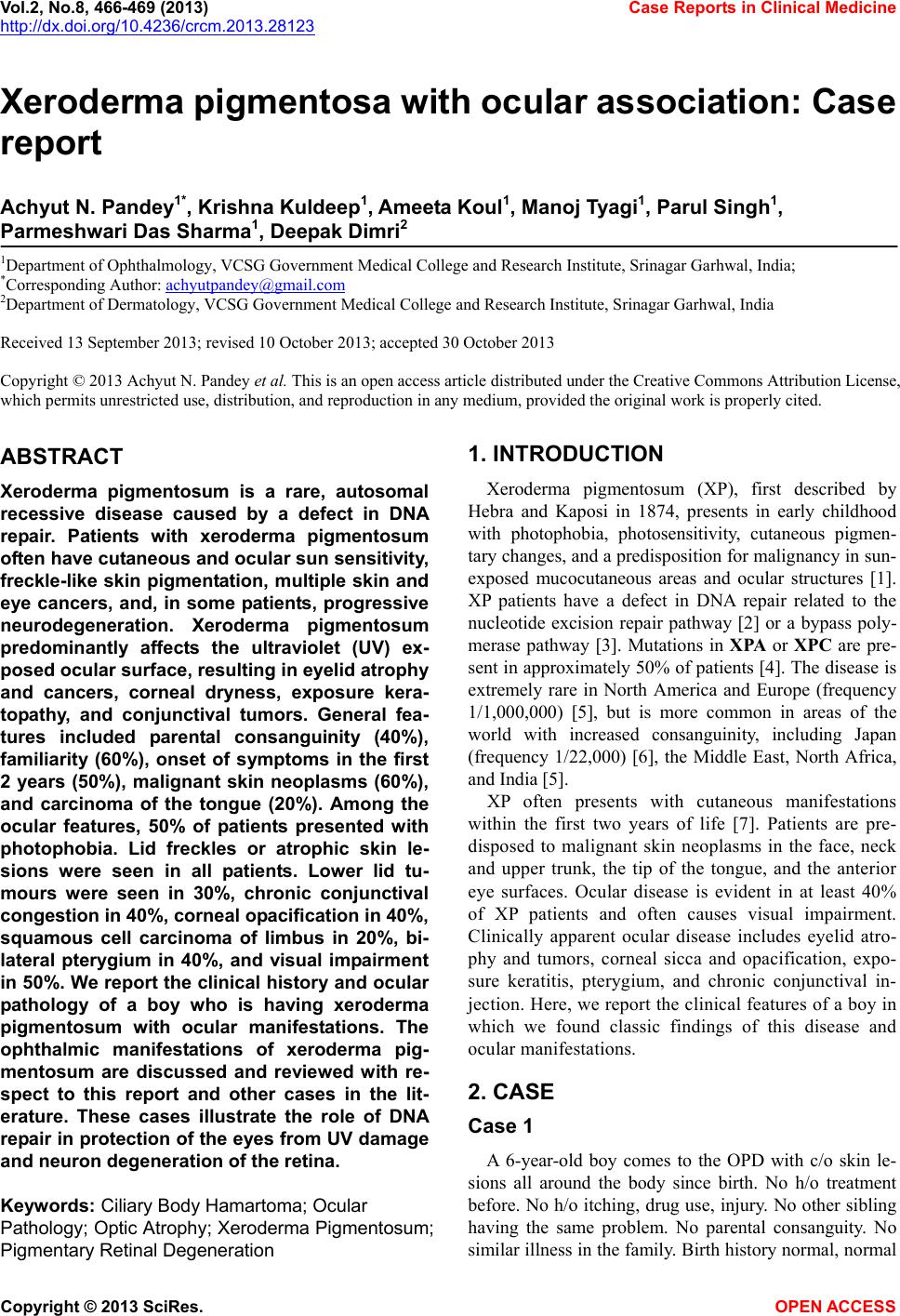 Vol.2, No.8, 466-469 (2013) Case Reports in Clinical Medicine http://dx.doi.org/10.4236/crcm.2013.28123 Xeroderma pigmentosa with ocular association: Case report Achyut N. Pandey1*, Krishna Kuldeep1, Ameet a Koul1, Manoj Tyagi1, Parul Singh1, Parmeshwari Das Sharma1, Deepak Dimri2 1Department of Ophthalmology, VCSG Government Medical College and Research Institute, Srinagar Garhwal, India; *Corresponding Author: [email protected] 2Department of Dermatology, VCSG Government Medical College and Research Institute, Srinagar Garhwal, India Received 13 September 2013; revised 10 October 2013; accepted 30 October 2013 Copy r ight © 2013 Achyut N. Pa ndey et al . This is an open access article distributed under the Creative Commons Attribution License, which permits unrestricted use, distribution, and reproduction in any medium, provided the original work is properly cited. ABSTRACT Xeroderma pigmentosum is a rare, autosomal recessive disease caused by a defect in DNA repair. Patients with xeroderma pigmentosum often have cutaneous and ocular sun sensitivity, freckle-like skin pigmentation, multiple skin and eye cancers, and, in some patients, progressive neurodegeneration. Xeroderma pigmentosum predominantly affects the ultraviolet (UV) ex- posed ocular surface, resulting in eyelid atrophy and cancers, corneal dryness, exposure kera- topathy, and conjunctival tumors. General fea- tures included parental consanguinity (40%), familiarity (60%), onset of symptoms in the first 2 years (50%), malignant skin neoplasms (60%), and carcinoma of the tongue (20%). Among the ocular features, 50% of patients presented with photophobia. Lid freckles or atrophic skin le- sions were seen in all patients. Lower lid tu- mours were seen in 30%, chronic conjunctival congestion in 40%, corneal opacification in 40%, squamous cell carcinoma of limbus in 20%, bi- lateral pterygium in 40%, and visual impairment in 50%. We report the clinical history and ocular pathology of a boy who is having xeroderma pigmentosum with ocular manifestations. The ophthalmic manifestations of xeroderma pig- mentosum are discussed and reviewed with re- spect to this report and other cases in the lit- erature. These cases illustrate the role of DNA repair in protection of the eyes from UV damage and neuron degeneration of the retina. Keywords: Ciliary Body Hamartoma; Ocular Pathology; Optic Atrophy; Xeroderma Pigmentosum; Pigmentary Retinal Degeneration 1. INTRODUCTION Xeroderma pigmentosum (XP), first described by Hebra and Kaposi in 1874, presents in early childhood with photophobia, photosensitivity, cutaneous pigmen- tary changes, and a predisposition for malignancy in sun- exposed mucocutaneous areas and ocular structures [1]. XP patients have a defect in DNA repair related to the nucleotide excision repair pathway [2] or a bypass poly- merase pathway [3]. Mutations in XPA or XPC are pre- sent in approximately 50% of patients [4]. The disease is extremely rare in North America and Europe (frequency 1/1,000,000) [5], but is more common in areas of the world with increased consanguinity, including Japan (frequency 1/22,000) [6], the Middle East, North Africa, and India [5]. XP often presents with cutaneous manifestations within the first two years of life [7]. Patients are pre- disposed to malignant skin neoplasms in the face, neck and upper trunk, the tip of the tongue, and the anterior eye surfaces. Ocular disease is evident in at least 40% of XP patients and often causes visual impairment. Clinically apparent ocular disease includes eyelid atro- phy and tumors, corneal sicca and opacification, expo- sure keratitis, pterygium, and chronic conjunctival in- jection. Here, we report the clinical features of a boy in which we found classic findings of this disease and ocular manifestations. 2. CASE Case 1 A 6-year-old boy comes to the OPD with c/o skin le- sions all around the body since birth. No h/o treatment before. No h/o itching, drug use, injury. No other sibling having the same problem. No parental consanguity. No similar illness in the family. Birth history normal, normal Copyright © 2013 SciRes. OPEN ACCESS 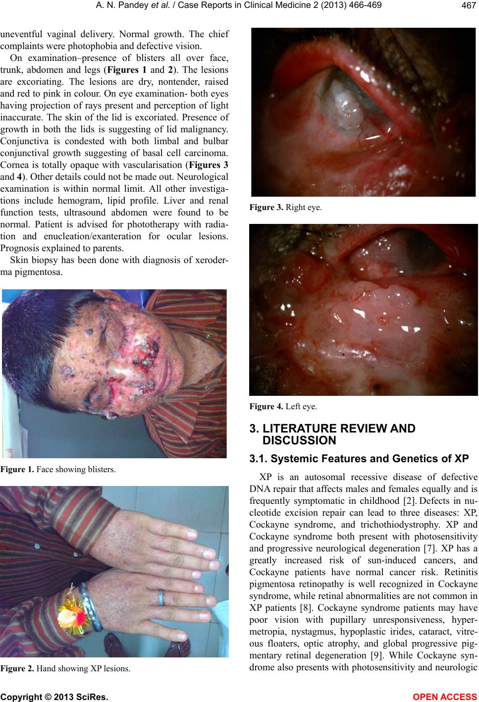 A. N. Pandey et al. / Case Reports in Clinical Medicine 2 (2013) 466-469 467 uneventful vaginal delivery. Normal growth. The chief complaints were photophobia and defective vision. On examination–presence of blisters all over face, trunk, abdomen and legs (Figures 1 and 2). The lesions are excoriating. The lesions are dry, nontender, raised and red to pink in colour. On eye examination- both eyes having projection of rays present and perception of light inaccurate. The skin of the lid is excoriated. Presence of growth in both the lids is suggesting of lid malignancy. Conjunctiva is condested with both limbal and bulbar conjunctival growth suggesting of basal cell carcinoma. Cornea is totally opaque with vascularisation (Figures 3 and 4). Other details could not be made out. Neurological examination is within normal limit. All other investiga- tions include hemogram, lipid profile. Liver and renal function tests, ultrasound abdomen were found to be normal. Patient is advised for phototherapy with radia- tion and enucleation/exanteration for ocular lesions. Prognosis explained to parents. Skin biopsy has been done with diagnosis of xeroder- ma pigmentosa. Figure 1. Face showing blisters. Figure 2. Hand showing XP lesions. Figure 3. Right eye. Figure 4. Left eye. 3. LITERATURE REVIEW AND DISCUSSION 3.1. Systemic Features and Genetics of XP XP is an autosomal recessive disease of defective DNA repair that affects males and females equally and is frequently symptomatic in childhood [2]. Defects in nu- cleotide excision repair can lead to three diseases: XP, Cockayne syndrome, and trichothiodystrophy. XP and Cockayne syndrome both present with photosensitivity and progressive neurological degeneration [7]. XP has a greatly increased risk of sun-induced cancers, and Cockayne patients have normal cancer risk. Retinitis pigmentosa retinopathy is well recognized in Cockayne syndrome, while retinal abnormalities are not common in XP patients [8]. Cockayne syndrome patients may have poor vision with pupillary unresponsiveness, hyper- metropia, nystagmus, hypoplastic irides, cataract, vitre- ous floaters, optic atrophy, and global progressive pig- mentary retinal degeneration [9]. While Cockayne syn- drome also presents with photosensitivity and neurologic Copyright © 2013 SciRes. OPEN ACCESS 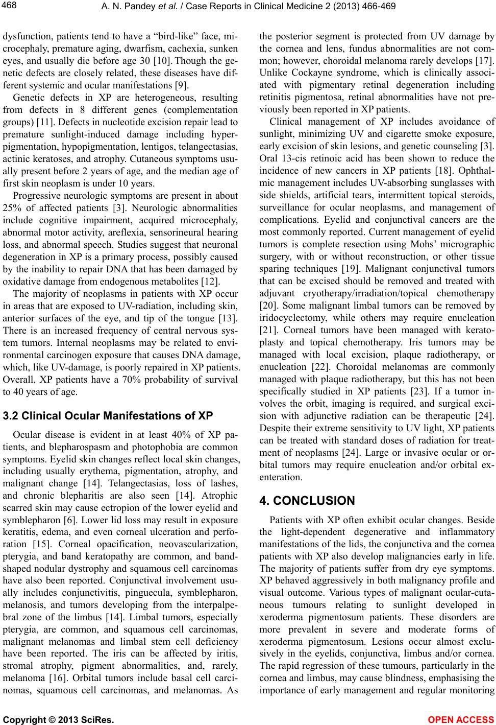 A. N. Pandey et al. / Case Reports in Clinical Medicine 2 (2013) 466-469 468 dysfunction, patients tend to have a “bird-like” face, mi- crocephaly, premature aging, dwarfism, cachexia, sunken eyes, and usually die before age 30 [10]. Though the ge- netic defects are closely related, these diseases have dif- ferent systemic and ocular manifestations [9]. Genetic defects in XP are heterogeneous, resulting from defects in 8 different genes (complementation groups) [11]. Defects in nucleotide excision repair lead to premature sunlight-induced damage including hyper- pigmentation, hypopigmentation, lentigos, telangectasias, actinic keratoses, and atrophy. Cutaneous symptoms usu- ally present before 2 years of age, and the median age of first skin neoplasm is under 10 years. Progressive neurologic symptoms are present in about 25% of affected patients [3]. Neurologic abnormalities include cognitive impairment, acquired microcephaly, abnormal motor activity, areflexia, sensorineural hearing loss, and abnormal speech. Studies suggest that neuronal degeneration in XP is a primary process, possibly caused by the inability to rep air DNA that has been damaged by oxidative damage from endogenous metabolites [12]. The majority of neoplasms in patients with XP occur in areas that are exposed to UV-radiation, including skin, anterior surfaces of the eye, and tip of the tongue [13]. There is an increased frequency of central nervous sys- tem tumors. Internal neoplasms may be related to envi- ronmental carcinogen exposure that causes DNA damage, which, like UV-damage, is poorly repaired in XP patients. Overall, XP patients have a 70% probability of survival to 40 years of age. 3.2 Clinical Ocular Manifestations of XP Ocular disease is evident in at least 40% of XP pa- tients, and blepharospasm and photophobia are common symptoms. Eyelid skin changes reflect local skin chang es, including usually erythema, pigmentation, atrophy, and malignant change [14]. Telangectasias, loss of lashes, and chronic blepharitis are also seen [14]. Atrophic scarred skin may cause ectropion of the lower eyelid and symblepharon [6]. Lower lid loss may result in exposure keratitis, edema, and even corneal ulceration and perfo- ration [15]. Corneal opacification, neovascularization, pterygia, and band keratopathy are common, and band- shaped nodular dystrophy and squamous cell carcinomas have also been reported. Conjunctival involvement usu- ally includes conjunctivitis, pinguecula, symblepharon, melanosis, and tumors developing from the interpalpe- bral zone of the limbus [14]. Limbal tumors, especially pterygia, are common, and squamous cell carcinomas, malignant melanomas and limbal stem cell deficiency have been reported. The iris can be affected by iritis, stromal atrophy, pigment abnormalities, and, rarely, melanoma [16]. Orbital tumors include basal cell carci- nomas, squamous cell carcinomas, and melanomas. As the posterior segment is protected from UV damage by the cornea and lens, fundus abnormalities are not com- mon; however, choroidal melanoma rarely develops [17]. Unlike Cockayne syndrome, which is clinically associ- ated with pigmentary retinal degeneration including retinitis pigmentosa, retinal abnormalities have not pre- viously been reported in XP patients. Clinical management of XP includes avoidance of sunlight, minimizing UV and cigarette smoke exposure, early excision of skin lesions, and genetic counseling [3]. Oral 13-cis retinoic acid has been shown to reduce the incidence of new cancers in XP patients [18]. Ophthal- mic management includes UV-absorbing sunglasses with side shields, artificial tears, intermittent topical steroids, surveillance for ocular neoplasms, and management of complications. Eyelid and conjunctival cancers are the most commonly reported. Current management of eyelid tumors is complete resection using Mohs’ micrographic surgery, with or without reconstruction, or other tissue sparing techniques [19]. Malignant conjunctival tumors that can be excised should be removed and treated with adjuvant cryotherapy/irradiation/topical chemotherapy [20]. Some malignant limbal tumors can be removed by iridocyclectomy, while others may require enucleation [21]. Corneal tumors have been managed with kerato- plasty and topical chemotherapy. Iris tumors may be managed with local excision, plaque radiotherapy, or enucleation [22]. Choroidal melanomas are commonly managed with plaque radiotherapy, but this has not been specifically studied in XP patients [23]. If a tumor in- volves the orbit, imaging is required, and surgical exci- sion with adjunctive radiation can be therapeutic [24]. Despite their extreme sensitivity to UV light, XP patients can be treated with standard doses of radiation for treat- ment of neoplasms [24]. Large or invasive ocular or or- bital tumors may require enucleation and/or orbital ex- enteration. 4. CONCLUSION Patients with XP often exhibit ocular changes. Beside the light-dependent degenerative and inflammatory manifestations of the lids, the conjunctiva and the cornea patients with XP also develop malignancies early in life. The majority of patients suffer from dry eye symptoms. XP behaved aggressively in both malignancy profile and visual outcome. Various types of malignant ocular-cuta- neous tumours relating to sunlight developed in xeroderma pigmentosum patients. These disorders are more prevalent in severe and moderate forms of xeroderma pigmentosum. Lesions occur almost exclu- sively in the eyelids, conjunctiva, limbus and/or cornea. The rapid regression of these tumours, particularly in the cornea and limbus, may cause blindness, emphasising the importance of early management and regular monitoring Copyright © 2013 SciRes. OPEN ACCESS 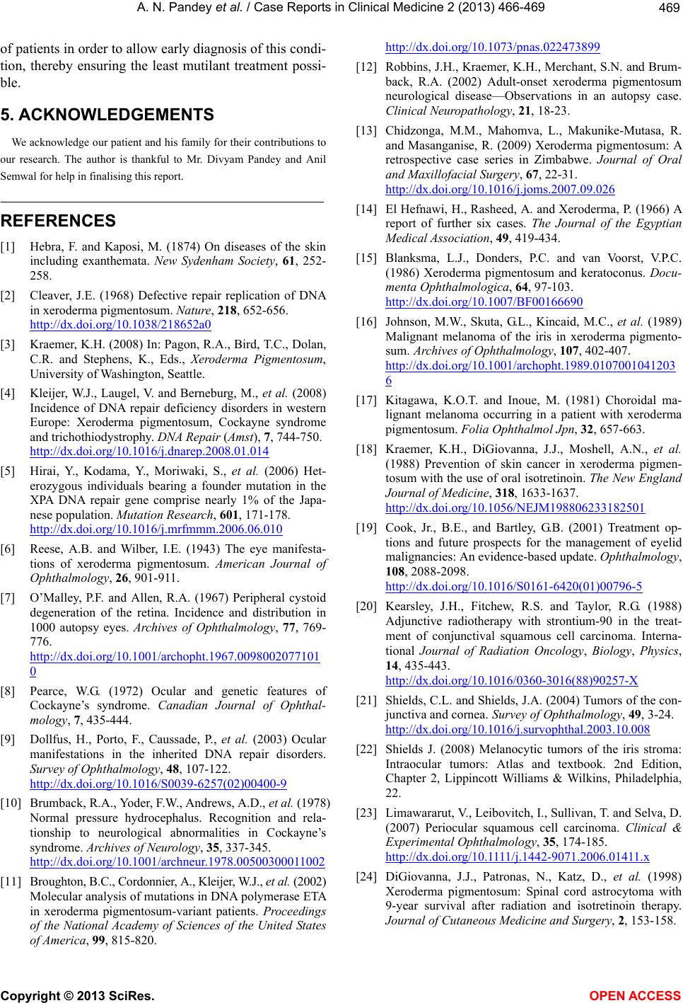 A. N. Pandey et al. / Case Reports in Clinical Medicine 2 (2013) 466-469 Copyright © 2013 SciRes. 469 of patients in order to allow early diagnosis of this cond i- tion, thereby ensuring the least mutilant treatment possi- ble. OPEN A CCESS 5. ACKNOWLEDGEMENTS We acknowledge our patient and his family for their contributions to our research. The author is thankful to Mr. Divyam Pandey and Anil Semwal for help in finalising this report. REFERENCES [1] Hebra, F. and Kaposi, M. (1874) On diseases of the skin including exanthemata. New Sydenham Society, 61, 252- 258. [2] Cleaver, J.E. (1968) Defective repair replication of DNA in xeroderma pigmentosum. Nature, 218, 652-656. http://dx.doi.org/10.1038/218652a0 [3] Kraemer, K.H. (2008) In: Pagon, R.A., Bird, T.C., Dolan, C.R. and Stephens, K., Eds., Xeroderma Pigmentosum, University of Washington, Seattle. [4] Kleijer, W.J., Laugel, V. and Berneburg, M., et al. (2008) Incidence of DNA repair deficiency disorders in western Europe: Xeroderma pigmentosum, Cockayne syndrome and trichothiodystrophy. DNA Repair (Amst), 7, 744-750. http://dx.doi.org/10.1016/j.dnarep.2008.01.014 [5] Hirai, Y., Kodama, Y., Moriwaki, S., et al. (2006) Het- erozygous individuals bearing a founder mutation in the XPA DNA repair gene comprise nearly 1% of the Japa- nese population. Mutation Research, 601, 171-178. http://dx.doi.org/10.1016/j.mrfmmm.2006.06.010 [6] Reese, A.B. and Wilber, I.E. (1943) The eye manifesta- tions of xeroderma pigmentosum. American Journal of Ophthalmology, 26, 901-911. [7] O’Malley, P.F. and Allen, R.A. (1967) Peripheral cystoid degeneration of the retina. Incidence and distribution in 1000 autopsy eyes. Archives of Ophthalmology, 77, 769- 776. http://dx.doi.org/10.1001/archopht.1967.0098002077101 0 [8] Pearce, W.G. (1972) Ocular and genetic features of Cockayne’s syndrome. Canadian Journal of Ophthal- mology, 7, 435-444. [9] Dollfus, H., Porto, F., Caussade, P., et al. (2003) Ocular manifestations in the inherited DNA repair disorders. Survey of Ophthalmology, 48, 107-122. http://dx.doi.org/10.1016/S0039-6257(02)00400-9 [10] Brumback, R.A., Yoder, F.W., Andrews, A.D., et al. (1978) Normal pressure hydrocephalus. Recognition and rela- tionship to neurological abnormalities in Cockayne’s syndrome. Archives of Neurology, 35, 337-345. http://dx.doi.org/10.1001/archneur.1978.00500300011002 [11] Broughton, B.C., Cordonnier , A ., Kleijer, W.J., et al. (2002) Molecular analysis of mutations in DNA polymerase ETA in xeroderma pigmentosum-variant patients. Proceedings of the National Academy of Sciences of the United States of America, 99, 815-820. http://dx.doi.org/10.1073/pnas.022473899 [12] Robbins, J.H., Kraemer, K.H., Merchant, S.N. and Brum- back, R.A. (2002) Adult-onset xeroderma pigmentosum neurological disease—Observations in an autopsy case. Clinical Neuropathology, 21, 18-23. [13] Chidzonga, M.M., Mahomva, L., Makunike-Mutasa, R. and Masanganise, R. (2009) Xeroderma pigmentosum: A retrospective case series in Zimbabwe. Journal of Oral and Maxillofacial Surgery, 67, 22-31. http://dx.doi.org/10.1016/j.joms.2007.09.026 [14] El Hefnawi, H., Rasheed, A. and Xeroderma, P. (1966) A report of further six cases. The Journal of the Egyptian Medical Association, 49, 419-434. [15] Blanksma, L.J., Donders, P.C. and van Voorst, V.P.C. (1986) Xeroderma pigmentosum and keratoconus. Docu- menta Ophthalmologica, 64, 97-103. http://dx.doi.org/10.1007/BF00166690 [16] Johnson, M.W., Skuta, G.L., Kincaid, M.C., et al. (1989) Malignant melanoma of the iris in xeroderma pigmento- sum. Archives of Ophthalmology, 107, 402-407. http://dx.doi.org/10.1001/archopht.1989.0107001041203 6 [17] Kitagawa, K.O.T. and Inoue, M. (1981) Choroidal ma- lignant melanoma occurring in a patient with xeroderma pigmentosum. Folia Ophthalmol Jpn, 32, 657-663. [18] Kraemer, K.H., DiGiovanna, J.J., Moshell, A.N., et al. (1988) Prevention of skin cancer in xeroderma pigmen- tosum with the use of oral isotretinoin. The New England Journal of Medicine, 318, 1633-1637. http://dx.doi.org/10.1056/NEJM198806233182501 [19] Cook, Jr., B.E., and Bartley, G.B. (2001) Treatment op- tions and future prospects for the management of eyelid malignancies: An evidence-based update. Ophthalmology, 108, 2088-2098. http://dx.doi.org/10.1016/S0161-6420(01)00796-5 [20] Kearsley, J.H., Fitchew, R.S. and Taylor, R.G. (1988) Adjunctive radiotherapy with strontium-90 in the treat- ment of conjunctival squamous cell carcinoma. Interna- tional Journal of Radiation Oncology, Biology, Physics, 14, 435-443. http://dx.doi.org/10.1016/0360-3016(88)90257-X [21] Shields, C.L. and Shields, J.A. (2004) Tumors of the con- junctiva and cor n ea. Survey of Ophthalmology, 49, 3-24. http://dx.doi.org/10.1016/j.survophthal.2003.10.008 [22] Shields J. (2008) Melanocytic tumors of the iris stroma: Intraocular tumors: Atlas and textbook. 2nd Edition, Chapter 2, Lippincott Williams & Wilkins, Philadelphia, 22. [23] Limawararut, V., Leibovitch, I., Sullivan, T. and Selva, D. (2007) Periocular squamous cell carcinoma. Clinical & Experimental Ophthalmology, 35, 174-185. h ttp: //dx .do i .org/1 0 .1111/ j.1442-9071.2006.01411.x [24] DiGiovanna, J.J., Patronas, N., Katz, D., et al. (1998) Xeroderma pigmentosum: Spinal cord astrocytoma with 9-year survival after radiation and isotretinoin therapy. Journal of Cutaneous Medicine and Surgery, 2, 153-158. |