

Paper Menu >>
Journal Menu >>
 Journal of Behavioral and Brain Science, 2011, 1, 6-11 doi:10.4236/jbbs.2011.11002 Published Online February 2011 (http://www.SciRP.org/journal/jbbs) Copyright © 2011 SciRes. JBBS Depressive Cognitions May Affect Cingulate Neurochemistry in ADHD Patients Evgeniy Perlov1, Alexandra Philipsen1, Simon Maier1, Martin Buechert2, Bernd Hesslinger1, Dieter Ebert1, Ludger Tebartz van Elst1 1University Hospital Freiburg, Department of Psychia try and Psychotherapy, Hauptstr , Freiburg, Germany 2University Hospital Freiburg, Department of Diag nostic Radiology, Hugstetterstr, Freiburg, Germany E-mail: [email protected] Received December 29, 2010; revised January 28, 2011; accepted Febr uary 14, 2011 Abstract Objectives: The anterior cingulate is thought to be essentially involved in impulsivity and affect regulation and in the pathogenesis of depression as well as of attention deficit/hyperactivity disorder. At the same time alterations in glutamatergic neurotransmission in the frontal forebrain have been found in imaging studies in Attention Deficit Hyperactivity Disorder (ADHD) and in depressive patients. Therefore we hypothesized that glutamate/glutamine (Glx) signals in the anterior cingulate cortex (ACC) of depressed ADHD patients might differ from that in non-depressed patients. Methods: Fourteen male adult patients with ADHD were included into the study. Chemical Shift Imaging of ACC was performed. Subgroups were defined based on scores on the Beck Depression Inventory (BDI) and ratios of metabolites were compared between groups. Additionally correlation analyses of BDI scores with metabolite ratios were calculated. Results: Significantly lower Glx signals and N-acetyl-aspartate (NAA) signals were found in the left anterior cingulate cortex of depressed ADHD patients. The Glx/Cr and NAAX/Cr ratios in the left ACC correlated significantly with BDI-scores. Conclusions: To our knowledge this is the first report about a relationship between depressive symptoms and metabolite disturbances in ACC of adult patients with ADHD. Our preliminary data produce first evi- dence for a putative link between neurochemical alterations in the ACC and depressive symptoms. They should be controlled for in further studies. Keywords: ADHD, MR-Spectroscopy, Depression, Anterior Cingulum, Glutamate 1. Introduction Most psychiatric disorders are known to be overrepre- sented in ADHD patients throughout the life from child- hood into adulthood. Psychiatric comorbidities include alcohol and other substance abuse as well as mood and anxiety disorders and borderline and antisocial personal- ity disorders [1-4] . Co morbidities with differential types of depression – major depressive disorder (MDD), dyst- hymia and brief recurrent depression – are particularly common [5]. This might be due to underachievement in school, further education and professional life as well as to frequent interperso nal proble ms in the family and rela- tionships. Depression in ADHD patients is often atypical. Anhedonia and physiological concomitants of depression are often absent whereas restlessness and inattention seem to be dominant [1,6]. Dopaminergic disturbance is thought to be a crucial pathogenetic mechanism in ADHD [7,8]. Additionally, glutamatergic alterations in the prefrontal cortex have been thought to be involved in the pathogenesis of this disorder in children [9,10] as well as in adults [11]. At the same time glutamatergic disturbances in prefrontal cortex and in particular in the anterior cingulated cortex (ACC) have been reported in MDD [12,13] and in some disorders in association with depressive symptoms [14,15]. Proton-Magnet-Resonanz-Spectroscopy (1H-MRS) is a non-invasive method which allows to detecting signals from neurometabolites in vivo. Amongst others N-ace- tylaspartate (NAA), N-acetylaspartate-glutamine (NAAG) – taken together – NAAx, creatine and phosphocreatine (Cre), choline compounds (Cho), glutamate and gluta- mine (Glx) and myo-inositol (Ins) can be measured with 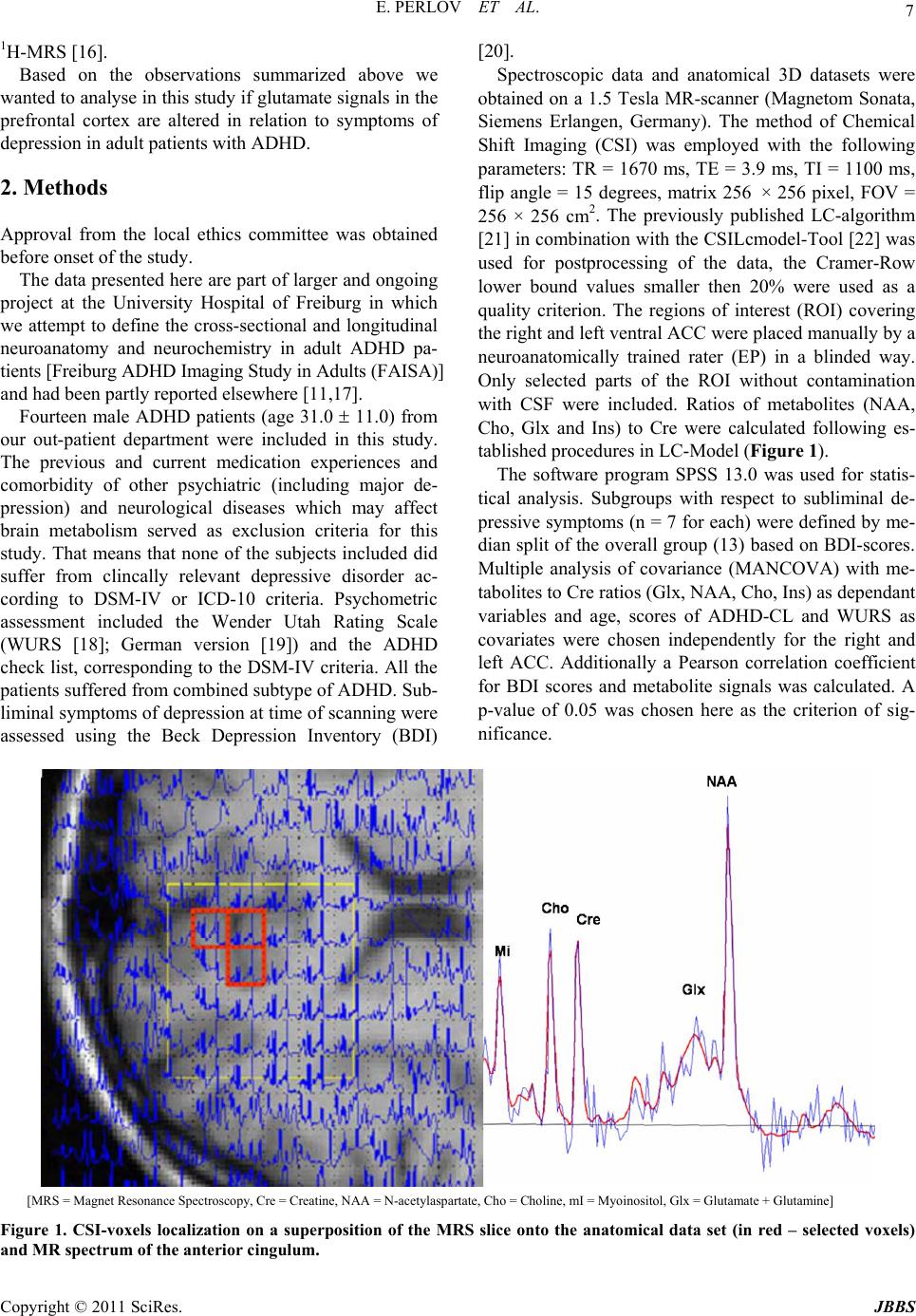 E. PERLOV ET AL. 7 1H-MRS [16]. Based on the observations summarized above we wanted to analyse in this study if glutamate sign als in the prefrontal cortex are altered in relation to symptoms of depression in adult patients with ADHD. 2. Methods Approval from the local ethics committee was obtained before onset of the study. The data presented here are part of larger and ongoing project at the University Hospital of Freiburg in which we attempt to define the cross-sectional and longitudinal neuroanatomy and neurochemistry in adult ADHD pa- tients [Freiburg ADHD Imaging Study in Adults (FAISA)] and had be en partly reported elsewhere [11,17]. Fourteen male ADHD patients (age 31.0 11.0) from our out-patient department were included in this study. The previous and current medication experiences and comorbidity of other psychiatric (including major de- pression) and neurological diseases which may affect brain metabolism served as exclusion criteria for this study. That means that none of the subjects included did suffer from clincally relevant depressive disorder ac- cording to DSM-IV or ICD-10 criteria. Psychometric assessment included the Wender Utah Rating Scale (WURS [18]; German version [19]) and the ADHD check list, corresponding to the DSM-IV criteria. All the patients suffered from combined subtype of ADHD. Sub- liminal symptoms of depression at time of scanning were assessed using the Beck Depression Inventory (BDI) [20]. Spectroscopic data and anatomical 3D datasets were obtained on a 1.5 Tesla MR-scanner (Magnetom Sonata, Siemens Erlangen, Germany). The method of Chemical Shift Imaging (CSI) was employed with the following parameters: TR = 1670 ms, TE = 3.9 ms, TI = 1100 ms, flip angle = 15 degrees, matrix 256 × 256 pixel, FOV = 256 × 256 cm2. The previously published LC-algorithm [21] in combination with the CSILcmodel-Tool [22] was used for postprocessing of the data, the Cramer-Row lower bound values smaller then 20% were used as a quality criterion. The regions of interest (ROI) covering the right and left ventral ACC were placed manually by a neuroanatomically trained rater (EP) in a blinded way. Only selected parts of the ROI without contamination with CSF were included. Ratios of metabolites (NAA, Cho, Glx and Ins) to Cre were calculated following es- tablished procedures in LC-Mo del (Figure 1). The software program SPSS 13.0 was used for statis- tical analysis. Subgroups with respect to subliminal de- pressive symptoms (n = 7 for each) were defined by me- dian split of the overall group (13) based on BDI-scores. Multiple analysis of covariance (MANCOVA) with me- tabolites to Cre ratios (Glx, NAA, Cho, Ins) as dependant variables and age, scores of ADHD-CL and WURS as covariates were chosen independently for the right and left ACC. Additionally a Pearson correlation coefficient for BDI scores and metabolite signals was calculated. A p-value of 0.05 was chosen here as the criterion of sig- nificance. [MRS = Magnet Resonance Spectroscopy, Cre = Creatine, NAA = N-acetylaspartate, Cho = Choline, mI = Myoinositol, Glx = Glutamate + Glutamine] Figure 1. CSI-voxels localization on a superposition of the MRS slice onto the anatomical data set (in red – selected voxels) and MR spectrum of the anterior cingulum. Copyright © 2011 SciRes. JBBS 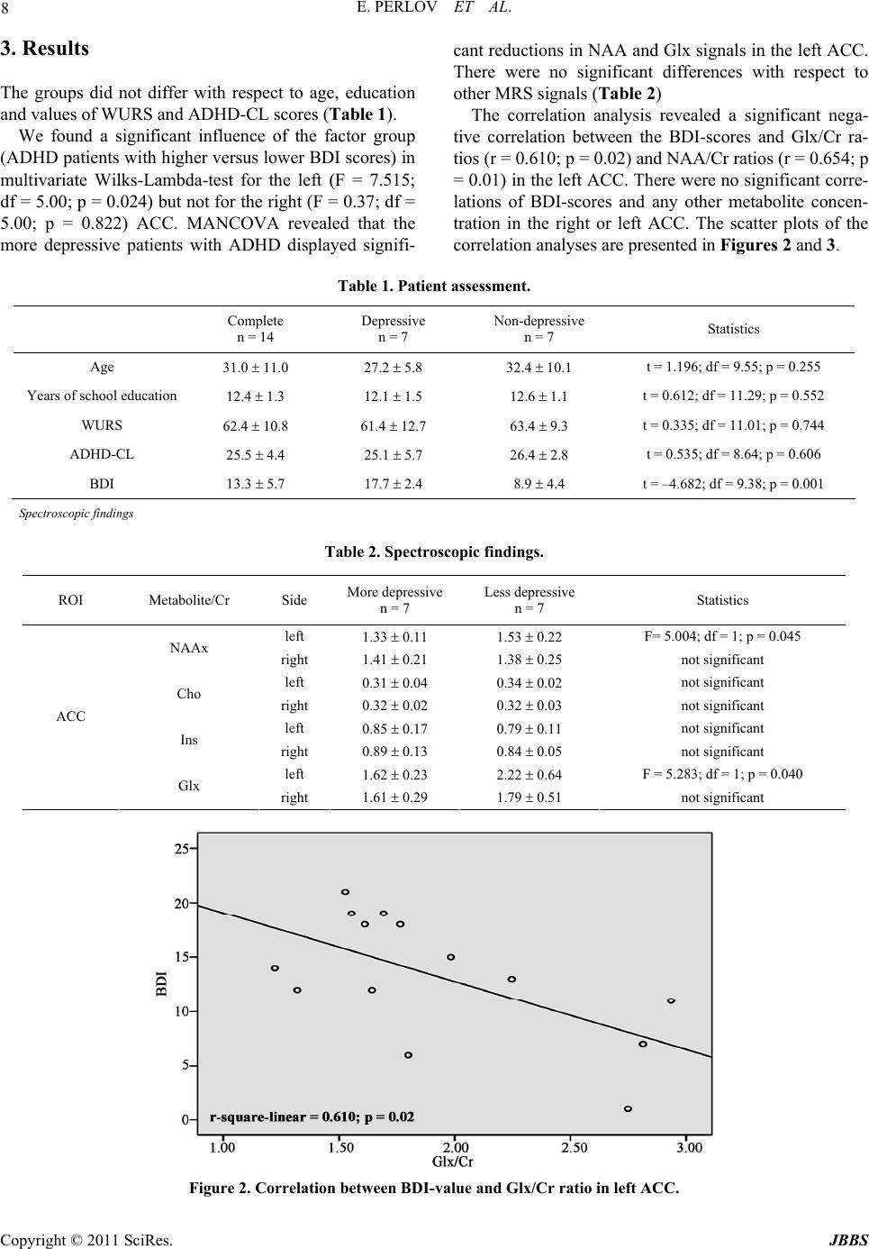 E. PERLOV ET AL. Copyright © 2011 SciRes. JBBS 8 3. Results The groups did not differ with respect to age, education and values of WURS and ADHD-CL scores (Table 1). We found a significant influence of the factor group (ADHD patients with higher versus lower BDI scores) in multivariate Wilks-Lambda-test for the left (F = 7.515; df = 5.00; p = 0.024) but no t for the right (F = 0.37; df = 5.00; p = 0.822) ACC. MANCOVA revealed that the more depressive patients with ADHD displayed signifi- cant reductions in NAA and Glx signals in the left ACC. There were no significant differences with respect to other MRS signals (Table 2) The correlation analysis revealed a significant nega- tive correlation between the BDI-scores and Glx/Cr ra- tios (r = 0.610; p = 0.02) and NAA/Cr ratios (r = 0.654; p = 0.0 1) in t he left ACC. Th ere were no significant corre- lations of BDI-scores and any other metabolite concen- tration in the right or left ACC. The scatter plots of the correlation analyses are presented in Figures 2 and 3. Table 1. Patient assessment. Complete n = 14 Depressive n = 7 Non-depressive n = 7 Statistics Age 31.0 11.0 27.2 5.8 32.4 10.1 t = 1.196; df = 9.5 5; p = 0.255 Years of school education 12.4 1.3 12.1 1.5 12.6 1.1 t = 0.612; df = 11.29; p = 0.552 WURS 62.4 10.8 61.4 12.7 63.4 9.3 t = 0.335; df = 11 .01; p = 0.744 ADHD-CL 25.5 4.4 25.1 5.7 26.4 2.8 t = 0.535; df = 8.6 4; p = 0.606 BDI 13.3 5.7 17.7 2.4 8.9 4.4 t = –4.682; df = 9.38; p = 0.001 Spectroscopic findings Table 2. Spectroscopic findings. Figure 2. Correlation between BDI-value and Glx/Cr ratio in left ACC. ROI Metabolite/Cr Side More depressive n = 7 Less depressive n = 7 Statistics left 1.33 0.11 1.53 0.22 F= 5.004; df = 1; p = 0 .045 NAAx right 1.41 0.21 1.38 0.25 not significant left 0.31 0.04 0.34 0.02 not significant Cho right 0.32 0.02 0.32 0.03 not significant left 0.85 0.17 0.79 0.11 not significant Ins right 0.89 0.13 0.84 0.05 not significant left 1.62 0.23 2.22 0.64 F = 5.283; df = 1; p = 0.040 ACC Glx right 1.61 0.29 1.79 0.51 not significant 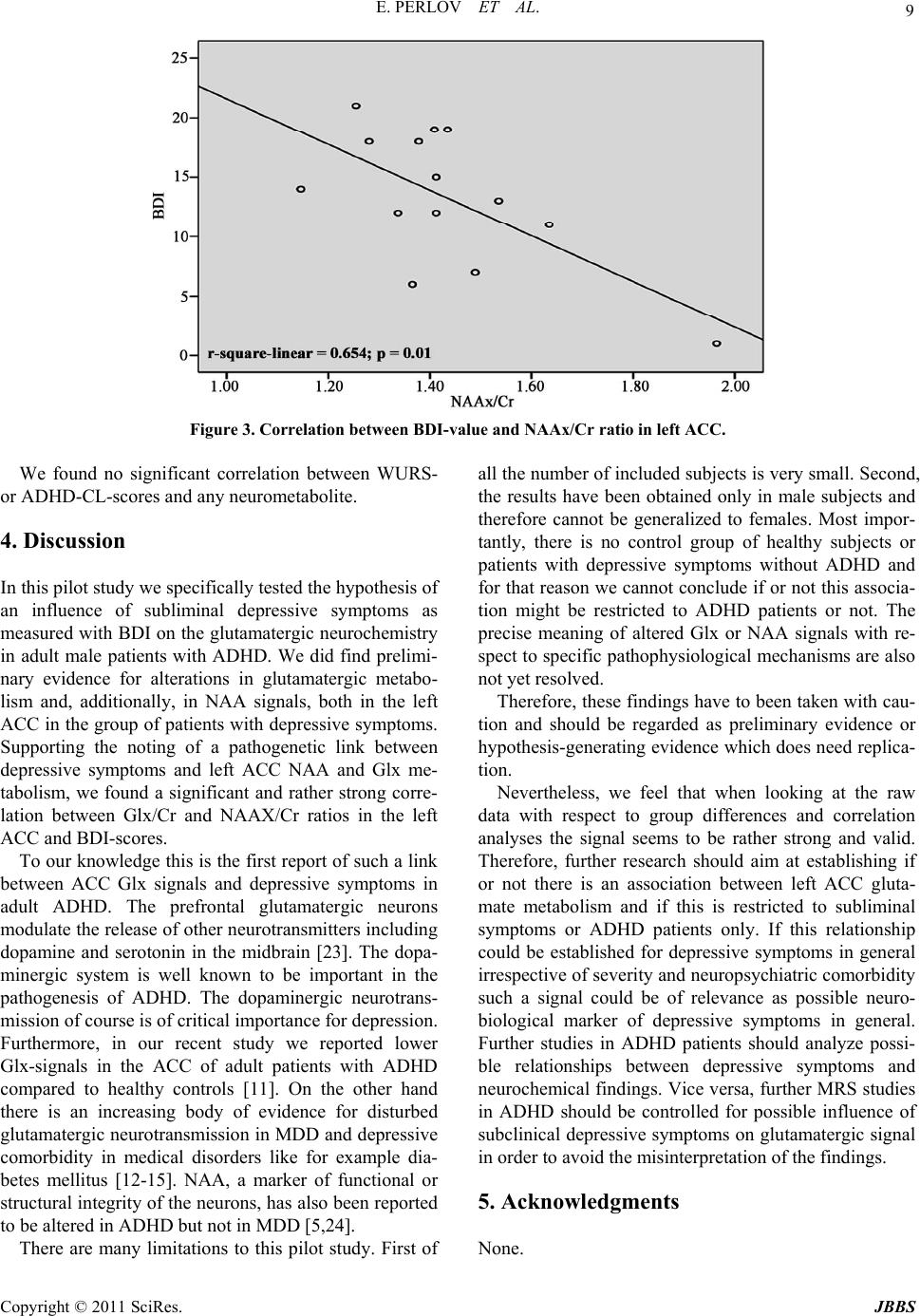 E. PERLOV ET AL. 9 Figure 3. Correlation between BDI-value and NAAx/Cr ratio in left ACC. We found no significant correlation between WURS- or ADHD-CL-scor es and any neurometabolite. 4. Discussion In this pilot study we specifically tested th e hypo th esis of an influence of subliminal depressive symptoms as measured with BDI on the glutamatergic neurochemistry in adult male patients with ADHD. We did find prelimi- nary evidence for alterations in glutamatergic metabo- lism and, additionally, in NAA signals, both in the left ACC in the group of patients with depressive symptoms. Supporting the noting of a pathogenetic link between depressive symptoms and left ACC NAA and Glx me- tabolism, we found a significant and rather strong corre- lation between Glx/Cr and NAAX/Cr ratios in the left ACC and BDI-scores. To our knowledge this is th e first report of such a link between ACC Glx signals and depressive symptoms in adult ADHD. The prefrontal glutamatergic neurons modulate the release of other neurotransmitters including dopamine and serotonin in the midbrain [23]. The dopa- minergic system is well known to be important in the pathogenesis of ADHD. The dopaminergic neurotrans- mission of course is of critical importance for depression. Furthermore, in our recent study we reported lower Glx-signals in the ACC of adult patients with ADHD compared to healthy controls [11]. On the other hand there is an increasing body of evidence for disturbed glutamatergic neurotransmission in MDD and depressive comorbidity in medical disorders like for example dia- betes mellitus [12-15]. NAA, a marker of functional or structural integrity of the neurons, has also been reported to be altered in ADHD but not in MDD [5,24]. There are many limitations to this pilot study. First of all the number of included subjects is very small. Second, the results have been obtained only in male subjects and therefore cannot be generalized to females. Most impor- tantly, there is no control group of healthy subjects or patients with depressive symptoms without ADHD and for that reason we cannot conclude if or not this associa- tion might be restricted to ADHD patients or not. The precise meaning of altered Glx or NAA signals with re- spect to specific pathophysiological mechanisms are also not yet resolved. Therefore, these findings have to been taken with cau- tion and should be regarded as preliminary evidence or hypothesis-generating evidence which does need replica- tion. Nevertheless, we feel that when looking at the raw data with respect to group differences and correlation analyses the signal seems to be rather strong and valid. Therefore, further research should aim at establishing if or not there is an association between left ACC gluta- mate metabolism and if this is restricted to subliminal symptoms or ADHD patients only. If this relationship could be established for depressive symptoms in general irrespective of severity and neuropsychiatric comorbidity such a signal could be of relevance as possible neuro- biological marker of depressive symptoms in general. Further studies in ADHD patients should analyze possi- ble relationships between depressive symptoms and neurochemical findings. Vice versa, further MRS studies in ADHD should be controlled for possible influence of subclinical depressive symptoms on glutamatergic signal in order to avoid the misinterpretation of the findings. 5. Acknowledgments None. Copyright © 2011 SciRes. JBBS  10 E. PERLOV ET AL. 6. References [1] J. Biederman, S. V. Faraone, T. Spencer, T. Wilens, E. Mick and K. A. Lapey, “Gender Differences in a Sample of Adults with Attention Deficit Hyperactivity Disorder,” Psychiatry Research, Vol. 53, No. 1, 1994, pp. 13-29. doi:10.1016/0165-1781(94)90092-2 [2] J. E. Alpert, A. Maddocks, A. A. Nierenberg, R. O’Sulli- van, J. A. Pava, J. J. Worthington, J. Biederman, J. F. Rosenbaum and M. Fava, “Attention Deficit Hyperacti- vity Disorder in Childhood among Adults with Major Depression,” Psychiatry Research, Vol. 62, No. 3, 1996, pp. 213-219. doi:10.1016/0165-1781(96)02912-5 [3] R. A. Barkley, M. Fischer, L. Smallish and K. Fletcher, “The Persistence of Attention-Deficit/Hyperactivity Dis- order into Young Adulthood as a Function of Reporting Source and Definition of Disorder,” Journal of Abnormal Psychology, Vol. 111, No. 2, 2002, pp. 279-289. doi:10.1037/0021-843X.111.2.279 [4] T. E. Wilens, “Impact of ADHD and Its Treatment on Substance Abuse in Adults,” Journal of Clinical Psy- chiatry, Vol. 65, No. 3, 2004, pp. 38-45. [5] B. Hesslinger, T. Thiel, L. T. van Elst, J. Hennig and D. Ebert, “Attention-Deficit Disorder in Adults with and without Hyperactivity - Where is the Difference? A Study Using Short Echo 1H-magnetic-resonance Spectro- scopy,” Neuroscience Letters, Vol. 304, No. 1, 2001, pp. 117-119. doi:10.1016/S0304-3940(01)01730-X [6] B. Hesslinger, v. E. Tebartz, F. Mochan and D. Ebert, “A Psychopathological Study into the Relationship between Attention Deficit Hyperactivity Disorder in Adult Pa- tients and Recurrent Brief Depression,” Acta Psychiatrica Scandinavica, Vol. 107, No. 5, 2003, pp. 385-389. doi:10.1034/j.1600-0447.2003.00112.x [7] D. D. Dougherty, A. A. Bonab, T. J. Spencer, S. L. Rauch, B. K. Madras and A. J. Fischman, “Dopamine Trans- porter Density in Patients with Attention Deficit Hyper- activity Disorder,” Lancet, Vol. 354, No. 9196, 1999, pp. 2132-2133. doi:10.1016/S0140-6736(99)04030-1 [8] S. V. Faraone, J. Biederman, T. Spencer, T. Wilens, L. J. Seidman, E. Mick and A. E. Doyle, “Attention-Deficit/ Hyperactivity Disorder in Adults: An Overview,” Bio- logical Psychiatry, Vol. 48, No. 1, 2000, pp. 9-20. doi:10.1016/S0006-3223(00)00889-1 [9] F. P. MacMaster, N. Carrey, S. Sparkes and V. Kusu- makar, “Proton Spectroscopy in Medication-free Pedia- tric Attention-Deficit/Hyperactivity Disorder,” Biological Psychiatry, Vol. 53, No. 2, 2003, pp. 184-187. doi:10.1016/S0006-3223(02)01401-4 [10] C. M. Moore, J. Biederman, J. Wozniak, E. Mick, M. Aleardi, M. Wardrop, M. Dougherty, T. Harpold, P. Hammerness, E. Randall and P. F. Renshaw, “Differ- ences in Brain Chemistry in Children and Adolescents with Attention Deficit Hyperactivity Disorder with and without Comorbid Bipolar Disorder: A Proton Magnetic Resonance Spectroscopy Study,” American Journal of Psychiatry, Vol. 163, No. 2, 2006, pp. 316-318. doi:10.1176/appi.ajp.163.2.316 [11] E. Perlov, A. Philipsen, B. Hesslinger, M. Buechert, J. Ahrendts, B. Feige, E. Bubl, J. Hennig, D. Ebert and v. E. Tebartz, “Reduced Cingulate Glutamate/Glutamine-to- Creatine Ratios in Adult Patients with Attention Defi- cit/hyperactivity Disorder - A Magnet Resonance Spec- troscopy Study,” Journal of Psychiatric Research, Vol. 41, No. 11, 2007, pp. 934-941. doi:10.1016/j.jpsychires.2006.12.007 [12] M. Walter, A. Henning, S. Grimm, R. F. Schulte, J. Be ck, U. Dydak, B. Schnepf, H. Boeker, P. Boesiger and G. Northoff, “The Relationship between Aberrant Neuronal Activation in the Pregenual Anterior Cingulate, Altered Glutamatergic Metabolism, and Anhedonia in Major De- pression,” Archives of General Psychiatry, Vol. 66, No. 5, 2009, pp. 478-486. doi:10.1001/archgenpsychiatry.2009.39 [13] D. P. Auer, “Reduced Glutamate in the Anterior Cingu- late Cortex in Depression: An in vivo Proton Magnetic Resonance Spectroscopy Study,” Biological Psychiatry, Vol. 47, No. 4, 2000, pp. 305-313. doi:10.1016/S0006-3223(99)00159-6 [14] I. K. Lyoo, S. J. Yoon, G. Musen, D. C. Simonson, K. Weinger, N. Bolo, C. M. Ryan, J. E. Kim, P. F. Renshaw and A. M. Jacobson, “Altered Prefrontal Glutamate- Glutamine-Gamma-Aminobutyric Acid Levels and Rela- tion to Low Cognitive Performance and Depressive Symptoms in Type 1 Diabetes Mellitus,” Archives of General Psychiatry, Vol. 66, No. 8, 2009, pp. 878-887. doi:10.1001/archgenpsychiatry.2009.86 [15] P. Ohrmann, A. Kersting, T. Suslow, J. Lalee -Mentzel, U. S. Donges, M. Fiebich, V. Arolt, W. Heindel and B. Pfleiderer, “Proton Magnetic Resonance Spectroscopy in Anorexia Nervosa: Correlations with Cognition,” Neuro- Report, Vol. 15, No. 3, 2004, pp. 549-553. doi:10.1097/00001756-200403010-00033 [16] B. Ross and S. Bluml, “Magnetic Resonance Spectro- scopy of the Human Brain,” Anatomical Record, Vol. 265, No. 2, 2001, pp. 54-84. doi:10.1002/ar.1058 [17] E. Perlov, v. E. Tebarzt, M. Buechert, S. Maier, S. Mat- thies, D. Ebert, B. Hesslinger and A. Philipsen, “H(1)- MR-spectroscopy of Cerebellum in Adult Attention Defi- cit/Hyperactivity Disorder,” Journal of Psychiatric Re- search, Vol. 44, No. 1414, 2010, pp. 938-943. [18] M. F. Ward, P. H. Wender and F. W. Reimherr, “The Wender Utah Rating Scale: An Aid in the Retrospective Diagnosis of Childhood Attention Deficit Hyperactivity Disorder,” American Journal of Psychiatry, Vol. 150, No. 6, 1993, pp. 885-890. [19] K. H. Krause, J. Krause and G. E. Trott, “Hyperkinetic Syndrome (Attention Deficit/Hyperactivity Disorder) in Adulthood,” Nervenarzt, Vol. 69, No. 7, 1998, pp. 543-556. doi:10.1007/s001150050311 [20] A. T. Beck, R. A. Steer and M. G. Carbin, “Psychometric Properties of the Beck Depression Inventory: Twenty-five Years of Evaluation,” Clinical Psychology Review, Vol. 8, No. 1, 1988, pp. 77-100. doi:10.1016/0272-7358(88)90050-5 [21] S. W. Provencher, “Estimation of Metabolite Concentra- Copyright © 2011 SciRes. JBBS  E. PERLOV ET AL. Copyright © 2011 SciRes. JBBS 11 tions from Localized in vivo Proton NMR Spectra,” Magnetic Resonance in Medicine, Vol. 30, No. 6, 1993, pp. 672-679. doi:10.1002/mrm.1910300604 [22] C. W. Ko, B. Kreher and M. Buchert, “GUI for Auto- matic Post Processing and Display of 2D-SI Data Sets with LC Model,” ISMRM, 11th Annual Meeting, 2003. [23] T. A. Slotkin, E. C. McCook, J. C. Ritchie, B. J. Carroll and F. J. Seidler, “Serotonin Transporter Expression in Rat Brain Regions and Blood Platelets: Aging and Glu- cocorticoid Effects,” Biological Psychiatry, Vol. 41, No. 2, 1997, pp. 172-183. doi:10.1016/S0006-3223(96)00215-6 [24] A. Yildiz-Yesiloglu and D. P. Ankerst, “Review of 1H Magnetic Resonanc e Spec trosc opy Findings in Major De- pressive Disorder: A Meta-analysis,” Psychiatry Re- search, Vol. 147, No. 1, 2006, pp. 1-25. doi:10.1016/j.pscychresns.2005.12.004 |