

Paper Menu >>
Journal Menu >>
 Journal of Cosmetics, Dermatological Sciences and Applications, 2013, 3, 16-25 Published Online November 2013 (http://www.scirp.org/journal/jcdsa) http://dx.doi.org/10.4236/jcdsa.2013.33A2005 Open Access JCDSA PUVA versus NB-UVB in Management of Vitiligo, Clinico-Immuno-Pathological Study Sameh K. Attia, Sherif S. Awad, Siham Yacoub Department of Dermatology and Venereology, Minia University, Minya, Egypt. Email: [email protected] Received September 3rd, 2013; revised October 1st, 2013; accepted October 9th, 2013 Copyright © 2013 Sameh K. Attia et al. This is an open access article distributed under the Creative Commons Attribution License, which permits unrestricted use, distribution, and reproduction in any medium, provided the original work is properly cited. ABSTRACT Background: Phototherapy is the most common ly used modality in the treatment of vitiligo. Oral PUVA is the classi- cal treatment and the NB-UVB is a recently introduced form excluding the shorter erythemogenic wavelengths. Aims: This study was designed to compare the effects of PUVA and NB-UVB clinically and imm uno-pat hologi cally i n managing non segmental vitiligo. Patients/Methods: Thirty vitiligo patients were divided randomly into two groups and treated either by oral PUVA or by narrow band UVB for 4 months, and evaluation was done clinically and immuno-pathologi- cally. Results: One patient in PUVA group (3.3%) failed to respond to therapy while 29 patients (93.3%) improved including all NB-UVB cases. Excellent repigmentation was achieved in 6.7% in PUVA group and 66.6% in NB-UVB group; good repigmentation was achieved in 60% in PUVA and 20% in NB-UVB while 26.7% in PUVA and 13.3% in NB-UVB showed mild repigmentation. The color matching was excellen t in all NB-UVB patients. Recurrence and ac- tivation of vitiligo were demonstrated in some NB-UVB cases and were less in PUVA treated cases. Microscopic ex- amination revealed persistence of dermal lympho-histiocytic infiltrate and the interface changes in biopsies from vitiliginous lesions treated with NB-UVB more than with PUVA. Conclusions: NB-UVB provides significant better results than oral PUVA in managing non-segmental vitiligo. Although NB-UVB therapy gives a rapid effect, yet the observation of recurrences and d evelopment of new lesions of vitiligo were less sign ificant with PUVA. It was also ob- served that PUVA has better immuno-modulatory effect on vitiligo than NB-UVB and may give better response on a longer period of time. Keywords: PUVA; Vitiligo; NB-UVB 1. Introduction Vitiligo is an acqu ired pigmentary disorder characterized by loss of melanocytes from the skin and subsequent development of depigmented patches of variable sizes, which may enlarge and coalesce to form extensive areas of leukoderma [1]. Treatment modalities aim to stimulate melanocyte pro- liferation or interfere with inflammatory factors affecting melanocyte structure or function; however, no single treatment method has been found to be consistently ef- fective, with relatively few side effects [2]. Ultraviolet irradiation is among the many possible approaches and is divided into ultraviolet A (UVA) (320 - 400 nm), ultra- violet B (UVB) (290 - 320 nm) and ultraviolet C (UVC) (200 - 290 nm) [3]. PUVA (psoralen followed by irradia- tion with UVA) has be en a well established management of non-segmental vitiligo since the last century [4]. Nar- row-band UV-B therapy was introduced recently as new light therapy which emits a concentrated UVB source of 311 nm and has been shown to have a profound thera- peutic efficacy for the treatment of skin conditions in- cluding vitiligo [5]. The aim of this work is to study and to compare the effects of PUVA versus narrow band UVB (NB-UVB) treatment on vitiligo patients clinically and immuno- pathologically. 2. Patients and Methods This study was conducted on 30 vitiligo patients attend- ing the out-patient clinic of Dermatology & Venereology Department, Minia University Hospital after obtaining their consen t and approv al of the local ethical committee. These patients were divided randomly into two groups: 15 patients were treated by PUVA and 15 patients were  PUVA versus NB-UVB in Management of Vitiligo, Clinico-Immuno-Pathological Study 17 treated by NB-UVB. Vitiligo patients included in the study had non seg- mental vitiligo in generalized or localized forms. Exclu- sion criteria enclosed: patients receiving treatment in the last 2 months, patients with light sensitive or aggravated dermatoses, patients with or giving past history of skin tumors, patients with aphakia or cataract. PUVA treat- ment group also excluded children, pregnant or lactating female patients and patients with abnormal liver or renal functions. All patients were subjected to full history taking in- cluding, general clinical examination including: abdomi- nal, chest, cardiac, ophthalmological and ENT examina- tion, dermatological examination including the type and extent of vitiligo and complete routine laboratory inves- tigations, including complete blood picture and liver and kidney function tests. Repeated laboratory tests were performed every two months after starting treatment in patients w ho were treated by PU VA. In PUVA group, the patient ingested 8-MOP at a dose of 0.4 mg/kg of body weight/session on a full stomach 1 and half hour before UVA exposure. The UVA cabinet used was UV compact irradiation cabinet COSMEDICO Medizintechnik, GP-42, Germany. Patients received 2 PUVA sessions/week. The starting dose was 2 J/cm2, which is a common starting dose for patients in similar skin type [6] and 25% dose increment was provided every next session [7 ] guided by the p atients response till asymptomatic erythema was observed. Erythema reading for PUVA treated patients was performed at 72 hours after UVA irradiation [6]. The last previous reading was utilized as the therapeutic dose throughout the 4 months of therapy. Patients were instructed to wear protective goggles during UVA exposure and sunglasses for 12 hours after the session. The genital area was shielded in all cases. In NB-UVB group, patients received 2 NB-UVB ses- sions/week. The starting dose was 0.574 J/cm2, which is the starting dose of skin type IV, according to manufac- ture instructions and 25% dose increment was done every next session guided by th e patients’ response, till asymp- tomatic erythema was observed. Erythema reading for NB-UVB treated patients was performed at 24 hours after UVB irradiation [8], the previous dose was utilized as the therapeutic dose throughout the 4 months of ther- apy. NB-UVB phototherapy cabinet used was COS- MEDICO Medizintechnik, GH-8 ST, Germany, contain- ing fluorescent TL-01 (100 W) tubes as the source of irradiation. Every patient was photographed before the start of treatment then repeatedly every two months throughout PUVA and NB-UVB treatment. Skin biopsies were harvested from each patient before PUVA and NB-UVB therapy (1 biopsy from the center of the vitiliginou s area) and after 4 months of PUVA and NB-UVB therapy (2 biopsies; one from the non-responding areas and the other biopsy from the repigmented areas). Three and half millimeter punch skin biopsies were used to obtain all samples after Mepivacaine HCL 3% local anesthesia. Biopsies were fixed in 10% formalin and embedded in paraffin blocks and sectioned by microtome into 5 µm thick sections. The resulting sections were mounted on glass slides and subjected to histopathologi- cal examination using Hematoxyline-Eosin, Fontana Masson and immunohistochemical staining using mela- noma antigen recognized by T-lymphocyte (MART-1), CD3, CD20 and CD68. MART-1 antibody (1:100, Neomarker, Fremont, CA 94539, USA) recognizes an 18 KD molecular weight MART-1 antigen which is present in endoplasmic re- ticulum of melanocytes. It has been found to localize to melanosomes suggesting a role in melanosomes biogene- sis and it is useful for biological studies on melanocytes and melanoma cells as well as for the development and monitoring of immunotherapy for the patients with melanoma [9]. CD3 marker (1:200, Dako, code No./M 7254) is a Pan-T lymphocyte marker. It recognizes CD3 antigen which is a pan-T cell antigen, composed of 5 invariable polypeptide chains. CD20 marker (1:400, Da- ko, code No./M 0755) is a pan-B lymphocyte marker. It recognizes CD20 antigen which is present in the B lym- phocyte. CD68 marker (1:300, Neomarkers Fremont, CA, Lot: 397P601A) recognizes CD68 antigen which is pre- sent in the cytoplasm of histiocytes. This marker is im- portant for identifying histiocytes in tissue sections. After 2 and 4 months of therapy, we evaluated the pa- tients subjectively into improvement or failure. The pa- tients who showed improvement were evaluated objec- tively by using an instrument called planimeter. The im- proved patients were divided into three groups; excellent group of patients when improvement ranged between 70% - 100%, good repigmentation group of patients when improvement ranged between 40% & 70%, and mild repigmentation group of patients when improve- ment is less than 40%. Planimeter is a measuring instru- ment used to measure the area of an irregular plane fig- ure on a map or photograph . A pointer on the planimeter is used to trace around the boundary of the shape. This induces a movement in another part of the instrument and a reading of this is used to establish the area of the figure. The resulting data were analyzed with SPSS statistical package version 12.0 (SPSS Inc., Chicago) for windows (Microsoft Corporation, Redmond). Data were expressed as mean ± standard deviation. Comparison between groups of data was done by independent samples (un- paired) t-test or ANOVA (between 2 or more groups). Comparison between baseline data and after treatment was done by paired t-test. The difference between compared groups was expressed as probability of value (p value). Open Access JCDSA  PUVA versus NB-UVB in Management of Vitiligo, Clinico-Immuno-Pathological Study 18 The difference was considered significant if p < 0.05. 3. Results The present study was conducted on 27 female patients (90%) and 3 male patients (10%). The age of these pa- tients at the time of examination ranged from 7 to 52 years old. The duration of the disease prior to photother- apy ranged from 2 months to 8 years. A positive family history of vitiligo was present in 5 (16.6%). Regarding skin Fitzpatrick types; 2 patients (6.6%) were skin type III, 20 patients (66.7%) were skin type IV, 8 patients (26.7%) were skin type V. In PUVA treated patients, 13 female patients (86.7%) and 2 male patients (13.3%) were included. The age of these patients at the time of examination ranged from 21 to 52 years, with a mean of 29.8 (±1 2.09) years. The du- ration of the disease prior to PUVA ranged from 2 months to 8 years, with a mean of 37.73 months ± 29 SD. A positive family history of vitiligo was present in 4 pa- tients (26.7%). The erythemogenic dose ranged from 4.883 to 7.629 J/cm2 and the therapeutic dose of PUVA ranged from 3.906 to 6.104 J/ cm2 (Table 1). As regard skin type, 1 patient (6.7%) was skin type III, 11 patients (73.3%) were skin type IV and 3 patients (20%) were skin type V (Table 1). In NB-UVB treated patients, 14 female patients (93.3%) and 1 male patient (6.7%) were in this study. The age of these patients at the time of examination ranged from 7 to 45 years with a mean of 16.2 years ± 11.02 SD. The duration of the disease prior to NB-UVB ranged from 2 months to 8 years with a mean of 30.53 months ± 26.27 SD. A positive family history of vitiligo was present in 1 patient (6.7%). The erythemogenic dose ranged from 1.752 to 3.422 J/cm2 and the therapeutic dose of UVB ranged from 1.402 to 2.737 J/cm2 (Table 1). Regarding skin typ e, 1 patient (6.7%) was skin type III, 9 patients (60%) were skin type IV, and 5 patients (33.3%) were skin type V (Table 1). At the end of the second month of PUVA therapy im- provement of the lesions occurred in 14 patients while 1 patient did not improve at all. Improvement ranged from 6 % to 37% with a mean of 17.1 3 ± 10.08 SD. At the end of the fourth month of therapy 1 patient (3.3%) failed to respond to therapy and the 14 patients (93.3%) showed improvement which ranged from 10% to 70% with a mean of 39.2 7 ± 18 .4 1 SD (Table 2). At the end of the second month of NB-UVB therapy all 15 patients showed improvement ranging from 12% to 90% with a mean of 45.4 ± 21.57 SD. This difference in the range of improvement between the two study groups (PUVA and NB-UVB) after 2 months therapy was statistically significant; p value = 0.0005. At the end of the fourth month of therapy all 15 pa- tients showed improvement which ranged from 23% to Table 1. Data of the patients included in each group. Items PUVA NB-UVB Age of patients at the start of treatment21 to 52 yrs Mean of 29.8 ± 12.09 7 to 45 yrs Mean of 16.2 ± 11.02 Sex 13 females (86.7%) and 2 males (13.3%) 14 females (93.3%) and 1 male (6.7%) Family history 4 patients (26.7%) 1 patient (6.7%) Skin type Type III: 1 (6.7%) Type IV: 11 (73.3%) Type V: 3 (20%) Type III: 1 (6.7%) Type IV: 9 (60%) Type V: 5 (33.3%) Duration of vitiligo prior to treatment2 ms to 8 yrs Mean of 37.73 ± 29 2 ms to 8 yrs Mean of 30.53 ± 26.27 The therapeutic dose3.906 to 6.104 J/cm2 1.402 to 2.737 J/cm2 Table 2. Percentage of improvement after 2 and 4 months. Study groups Percentage of improvementNB-UVB PUVA p-value* Range 12 - 90 6 - 37 Mean ± SD45.4 ± 21.57 17.13 ± 10.080.0005 Percentage of improvement after 2 monthsp-value - - - Range 23 - 98 10 - 70 Mean ± SD73.27 ± 24.94 39.27 ± 18.410.0002 Percentage of improvement after 4 monthsp-value** 0. 0001 0.0005 - *Comparison between NB-UVB and PUVA groups; **Comparison between % of improvement after 2 months and after 4 months. 98% with a mean of 73.2 7 ± 24 .94 SD . This d if f erence in the range of improvement between the two study groups (PUVA and NB-UVB) after 4 months therapy was statis- tically significant; p value = 0.0 002 (Table 2). Improved patients were divided into three groups (Ta- ble 3): Excellent repigmentation (70% - 100%): in 1 patient (6.7%) in PUVA group and in 10 patients (66.7%) in NB-UVB gr oup. Good repigmentation (40% - 70%): in 9 patients (60%) in PUVA group and in 3 patients (20%) in NB-UVB group. Mild repigmentation (less than 40%): in 4 patients (26.7%) in PUVA group and in 2 patients (13.3%) in NB-UVB gr oup. The perifollicular repigmentation was the predominant type of repigmentation observed in both PUVA and NB- UVB treated groups. It occurred in all improved patients (100%) who were treated either by PUVA or NB-UVB. Only 1 patient (6.7%), who was treated with NB-UVB, showed combination of both perifollicular repigmenta- tion and peripheral repigmentation. Diffuse repigmenta- tion was not observed in any patien t. The assessment of color match in comparison with the Open Access JCDSA  PUVA versus NB-UVB in Management of Vitiligo, Clinico-Immuno-Pathological Study 19 Table 3. Degree of repigmentation after treatment. Degree of repigmentation PUVA group N = 14 NB-UVB group N = 15 Excellent repigmentation 1 patient (6.7%) 10 patien t s (66.7%) Good repigme n t a t i o n 9 patients (60%) 3 patients (20%) Mild repigmentation 4 patients (26.7%) 2 patients (13.3%) patient’s unaffected sk in was excellent for all 15 patients (100%) in the NB-UVB group and for only 8 patients (57.2%) out of the 14 improved patients in the PUVA group while the remaining 6 patients (42.8%) showed dark hyperpigmentation of the treated vitiliginous area than the patient’s unaffected skin. The side effects were minimal and did not warrant dis- continuation of the therapy in any of the 30 patients. All patients in each group developed erythema, showing that both regimens were equally erythemogenic. Pruritus oc- curred in 4 patients (26.7%) in the PUVA group com- pared with 3 patients (20%) in the NB-UVB group. Pho- todermatitis with scaliness and eczematous reaction was observed only in 1 patient (6.7%) who was treated with NB-UVB. Throughout the study, recurrence of the vitiliginous patches at the same site of treatment occurred in 1 patient (6.7%) in the PUVA group compared with 3 patients (20%) in the NB-UVB group. New lesions developed in other sites in 1 patient (6.7%) in the PUVA group com- pared with 4 patients (26.7%) in the NB-UVB group. Seven patients (46.7%) who were treated with PUVA, developed nausea during the course of therapy and mi- nimization of the dose of 8-MOP in those patients was performed to decrease the sensation of nausea. One fe- male patient (6.7%) who was treated with PUVA th erapy developed elevation in the liver enzymes after 4 months of treatment and stopped the therapy (Table 4). Histopathological Findings Before treatment (Table 5, Figure 1), microscopic ex- amination of involved skin in vitiligo showed no basal melanization in 22 (73.3%) of the 30 biopsies while 8 (26.7%) showed basal melanization. With Fontana- Masson stain, similar ratio of basal pigmentation was demonstrated. Hyperkeratosis was present in 12 biopsies (40%). Interface changes and epidermal vacuolization was found in 4 biopsies (13.3%), all were included in PUVA group of patients. Dermal infiltrates composed mainly of mononuclear cells (Lympho-histiocytic); seven biopsies (46.7%) in PUVA group and 7 biopsies (46.7%) in NB-UVB group showed sparse infiltrate, 7 biopsies (46.7%) in PUVA group and 5 biopsies (33.3%) in NB-UVB group showed moderate infiltrate, and 1 biopsy (6.7%) in PUVA group Table 4. Side effects reported in the study groups. NB-UVB group N = 15 PUVA group N = 15 Side effects 3 patients (20%)4 patients (26.7%) Pruritus 1 patient (6.7%)0 (0%) Photo dermatitis 0 (0%) 7 patients (46.7 %) Nausea 0 (0%) 1 patient (6.7%) Elevation in the liver enzymes 3 patients (20%)1 patient (6.7%) Recurrence at the same site of treatment 4 patients (26.7%)1 patient (6.7%) New lesions in other sites Table 5. Histopathological findings in the center of the vitiliginous lesions before treatment. Vitiligo center Histopathological chan gesPUVA (n = 15) NB-UVB (n = 15) Total (n = 30) Hyperkeratosis 7 biopsies (46.7%) 5 biopsies (33.3%) 12 biopsies (40%) Basal melanization 5 biopsies (33.3%) 3 biopsies (20%) 8 biopsies (26.7%) Interface changes 4 biopsies (26.7%) 0 (0%) 4 biopsies (13.3%) Melanophages 7 biopsies (46.7%) 4 biopsies (26.7%) 11 biopsies (36.7%) Eosinophils 1 biopsy (6.7%) 0 (0%) 1 biopsies (3.3%) Sparse 7 biopsies (46.7%) 7 biopsies (46.7%) 14 biopsies (46.7) Moderate 7 biopsies (46.7%) 5 biopsies (33.3%) 12 biopsies (40%) Lymphohistiocytic infiltrate Dense 1 biopsy (6.7%) 3 biopsies (20%) 4 biopsies (13.3%) Figure 1. The center of vitiliginous area before treatment; H&E staining showing almost no basal melanization, hy- perkeratosis and sparse superficial dermal lympho-histio- cytic infiltrate (upper left, ×10). CD20 stained section showing negative lymphocytic staining (upper right, ×20). CD3 stained section showing mild positive lymphocytic staining (lower left, ×20). CD68 stained section showing strong positive lymphocytic staining (lower right, ×20). Open Access JCDSA 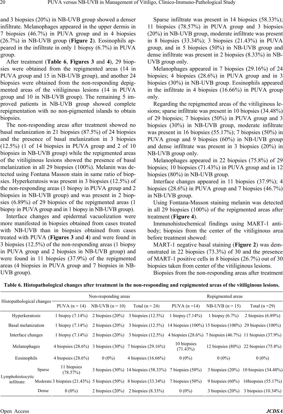 PUVA versus NB-UVB in Management of Vitiligo, Clinico-Immuno-Pathological Study Open Access JCDSA 20 and 3 biopsies (20%) in NB-UVB group showed a denser infiltrate. Melanophages appeared in the upper dermis in 7 biopsies (46.7%) in PUVA group and in 4 biopsies (26.7%) in NB-UVB group (Figure 2). Eosinophils ap- peared in the infiltrate in only 1 biopsy (6.7%) in PUVA group. After treatment (Table 6, Figures 3 and 4), 29 biop- sies were obtained from the repigmented areas (14 in PUVA group and 15 in NB-UVB group), and anothe r 24 biopsies were obtained from the non-responding depig- mented areas of the vitiliginous lesions (14 in PUVA group and 10 in NB-UVB group). The remaining 5 im- proved patients in NB-UVB group showed complete repigmentation with no non-pigmented islands to obtain biopsies. The non-responding areas after treatment showed no basal melanization in 21 biopsies (87.5%) of 24 biopsies and the presence of basal melanization in 3 biopsies (12.5%) (1 of 14 biopsies in PUVA group and 2 of 10 biopsies in NB-UVB group) while the repigmented areas of the vitiliginous lesions showed the presence of basal melanization in all 29 biopsies (100%). Melanin was de- tected using Fontana Masson stain in same ratio of biop- sies. Hyperkeratosis was present in 3 biopsies (12.5%) of the non-responding areas (1 biopsy in PUVA gro up and 2 biopsies in NB-UVB group) and was present in 2 biop- sies (6.89%) of 29 biopsies of the repigmented areas (1 biopsy in PUVA group and in 1 biopsy in NB-UVB group). Interface changes and epidermal vacuolization were more manifested in biopsies obtained from cases treated with NB-UVB than in biopsies obtained from cases treated with PUVA (Figures 3 and 4) an d were found in 3 biopsies (12.5%) of the non-responding areas (1 biopsy in PUVA group and 2 biopsies in NB-UVB group) and were found in 11 biopsies (37.9%) of the repigmented areas (4 biopsies in PUVA group and 7 biopsies in NB- UVB group). Sparse infiltrate was present in 14 biopsies (58.33%); 11 biopsies (78.57%) in PUVA group and 3 biopsies (20%) in NB-UVB group, moderate infiltrate was present in 8 biopsies (33.34%); 3 biopsies (21.43%) in PUVA group, and in 5 biopsies (50%) in NB-UVB group and dense infiltrate was present in 2 biopsies (8.33%) in NB- UVB group only. Melanophages appeared in 7 biopsies (29.16%) of 24 biopsies; 4 biopsies (28.6%) in PUVA group and in 3 biopsies (30%) in N B-UVB group. Eosinophils app eared in the infiltrate in 4 biopsies (16.66%) in PUVA group only. Regarding the repigmented areas of the vitilig inous le- sions; sparse infiltrate was present in 10 biopsies (3 4.48%) of 29 biopsies; 7 biopsies (50%) in PUVA group and 3 biopsies (30%) in NB-UVB group, moderate infiltrate was present in 16 biopsies (55.17%); 7 biopsies (50%) in PUVA group and 9 biopsies (60%) in NB-UVB group and dense infiltrate was present in 3 biopsies (20%) in NB-UVB group only. Melanophages appeared in 22 biopsies (75.8%) of 29 biopsies; 10 biopsies (71.43%) in PUVA group and in 12 biopsies (80%) in NB-UVB group. Interface changes appeared in 11 biopsies (37.9%); 4 biopsies (28.6%) in PUVA group and 7 b iopsies (46.7%) in NB-UVB group. Using Fontana-Masson staining melanin was detected in all 29 biopsies (100%) of the repigmented areas after treatment (Figure 4). Immunohistochemical findings using MART-1 anti- body; biopsies from the center of the vitiliginous area before treatment showed: MART-1 negative basal staining (Figure 2) was dem- onstrated in 22 biopsies (73.3%) of 30 and the presence of MART-1 positive cells in 8 biopsies (26.7%) out of 30 biopsies taken from center of the vitiliginous lesions. Biopsies from the non- responding ar eas after treatment Table 6. Histopathological changes after treatment in the non-responding and repigmented areas of the vitiliginous lesions. Non-responding areas Repigmented areas Histopathological chan ges PUVA (n = 14) NB-UVB (n = 10)Total (n = 24) PUVA (n =14) NB-UVB (n = 15) Total (n =29) Hyperkeratosis 1 biopsy (7.14%) 2 biopsies (20%)3 biopsies (12.5%)1 biopsy (7.14%)1 biopsy (6.7%) 2 biopsies (6.89%) Basal melanization 1 biopsy (7.14%) 2 biopsies (20%)3 biopsies (12.5%)14 biopsies (100%)15 biopsies (100%) 29 biopsies (100%) Interface changes 1 biopsy (7.14%) 2 biopsies (20%)3 biopsies (12.5%)4 biopsies (28.6%)7 biopsies (46.7%) 11 biopsies (37.9%) Melanophages 4 biopsies (28.6%) 3 biopsies (30% )7 biopsies (29.16%)10 biopsies (71.43%) 12 biopsies (80%) 22 biopsies (75.8%) Eosinophils 4 biopsies (28.6%) 0 ( 0%) 4 biopsies (16.66%)0 (0%) 0 (0%) 0 (0%) Sparse 11 biopsies (78.57%) 3 biopsies (30%)14 biopsies (58.33%)7 biopsies (50%)3 biopsies (2 0 % ) 10 biopsies (34.48%) Moderate 3 biopsies (21.43%) 5 biopsies (50%)8 biopsies (33.34%)7 biopsies (50%)9 biopsies (60%) 16biopsies (55.17%) Lymphohistiocytic infiltrate: Dense 0 (0%) 2 biopsies (20%)2 biopsies (8.33%)0 (0%) 3 biopsies (2 0 % ) 3 biopsies (10.34%) 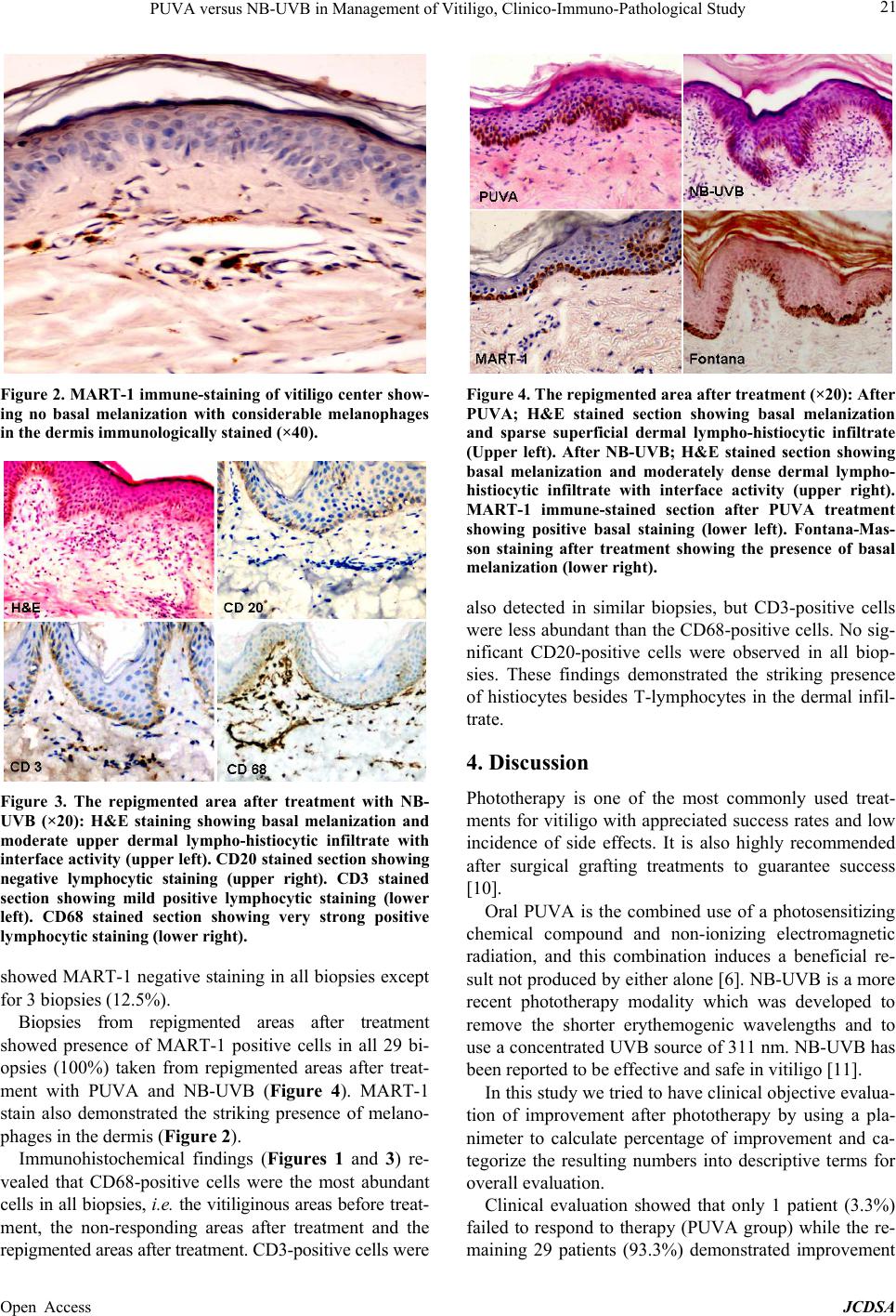 PUVA versus NB-UVB in Management of Vitiligo, Clinico-Immuno-Pathological Study 21 Figure 2. MART-1 immune-staining of vitiligo center show- ing no basal melanization with considerable melanophages in the dermis immunologically stained (×40). Figure 3. The repigmented area after treatment with NB- UVB (×20): H&E staining showing basal melanization and moderate upper dermal lympho-histiocytic infiltrate with interface activity (upper left). CD20 stained section showing negative lymphocytic staining (upper right). CD3 stained section showing mild positive lymphocytic staining (lower left). CD68 stained section showing very strong positive lymphocytic staining (lower right). showed MART-1 negative staining in all biopsies except for 3 biopsies (12.5%). Biopsies from repigmented areas after treatment showed presence of MART-1 positive cells in all 29 bi- opsies (100%) taken from repigmented areas after treat- ment with PUVA and NB-UVB (Figure 4). MART-1 stain also demonstrated the striking presence of melano- phages in the dermis (Figure 2). Immunohistochemical findings (Figures 1 and 3) re- vealed that CD68-positive cells were the most abundant cells in all b iopsies, i.e. the vitiliginous areas before trea t- ment, the non-responding areas after treatment and the repigmented areas after treatment. CD3-positive cells were Figure 4. The repigmented area after treatment (×20): After PUVA; H&E stained section showing basal melanization and sparse superficial dermal lympho-histiocytic infiltrate (Upper left). After NB-UVB; H&E stained section showing basal melanization and moderately dense dermal lympho- histiocytic infiltrate with interface activity (upper right). MART-1 immune-stained section after PUVA treatment showing positive basal staining (lower left). Fontana-Mas- son staining after treatment showing the presence of basal melanization (lower right). also detected in similar biopsies, but CD3-positive cells were less abundant than the CD68-positive cells. No sig- nificant CD20-positive cells were observed in all biop- sies. These findings demonstrated the striking presence of histiocytes besides T-lymphocytes in the dermal infil- trate. 4. Discussion Phototherapy is one of the most commonly used treat- ments for vitiligo with appreciated success rates and low incidence of side effects. It is also highly recommended after surgical grafting treatments to guarantee success [10]. Oral PUVA is the combined use of a photosensitizing chemical compound and non-ionizing electromagnetic radiation, and this combination induces a beneficial re- sult not produced by either alone [6]. NB-UVB is a more recent phototherapy modality which was developed to remove the shorter erythemogenic wavelengths and to use a concentrated UVB source of 311 nm. NB-UVB has been reported to be effective and safe in vitiligo [11]. In this study we tried to have clinical objectiv e evalua- tion of improvement after phototherapy by using a pla- nimeter to calculate percentage of improvement and ca- tegorize the resulting numbers into descriptive terms for overall evaluation. Clinical evaluation showed that only 1 patient (3.3%) failed to respond to therapy (PUVA group) while the re- maining 29 patients (93.3%) demonstrated improvement Open Access JCDSA 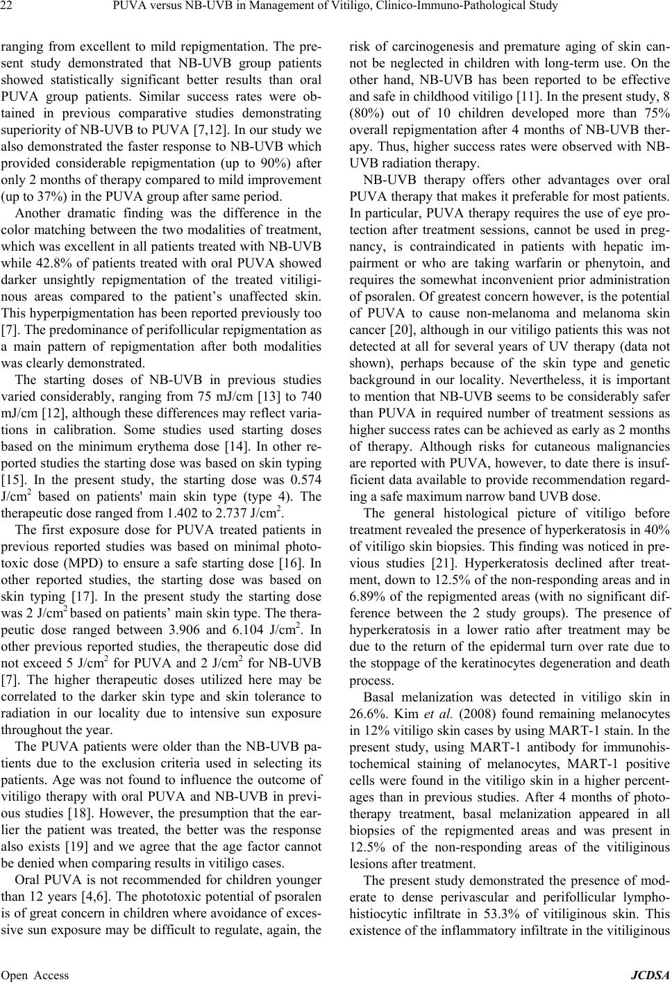 PUVA versus NB-UVB in Management of Vitiligo, Clinico-Immuno-Pathological Study 22 ranging from excellent to mild repigmentation. The pre- sent study demonstrated that NB-UVB group patients showed statistically significant better results than oral PUVA group patients. Similar success rates were ob- tained in previous comparative studies demonstrating superiority of NB-UVB to PUVA [7,12]. In our study we also demonstrated the faster response to NB-UVB which provided considerable repigmentation (up to 90%) after only 2 months of therapy compared to mild improvement (up to 37%) in the PUVA group after same period. Another dramatic finding was the difference in the color matching between the two modalities of treatment, which was excellent in all patients treated with NB-UVB while 42.8% of patients treated with oral PUVA showed darker unsightly repigmentation of the treated vitiligi- nous areas compared to the patient’s unaffected skin. This hyperpigmentation has been reported previously too [7]. The predominan ce of perifollicular repigmentation as a main pattern of repigmentation after both modalities was clearly demonstrated. The starting doses of NB-UVB in previous studies varied considerably, ranging from 75 mJ/cm [13] to 740 mJ/cm [12], although these differences may reflect varia- tions in calibration. Some studies used starting doses based on the minimum erythema dose [14]. In other re- ported studies the starting dose was based on skin typing [15]. In the present study, the starting dose was 0.574 J/cm2 based on patients' main skin type (type 4). The therapeutic dose range d from 1.40 2 t o 2.737 J/cm2. The first exposure dose for PUVA treated patients in previous reported studies was based on minimal photo- toxic dose (MPD) to ensure a safe starting dose [16]. In other reported studies, the starting dose was based on skin typing [17]. In the present study the starting dose was 2 J/cm2 based on patients’ main skin type. The thera- peutic dose ranged between 3.906 and 6.104 J/cm2. In other previous reported studies, the therapeutic dose did not exceed 5 J/cm2 for PUVA and 2 J/cm2 for NB-UVB [7]. The higher therapeutic doses utilized here may be correlated to the darker skin type and skin tolerance to radiation in our locality due to intensive sun exposure throughout th e year. The PUVA patients were older than the NB-UVB pa- tients due to the exclusion criteria used in selecting its patients. Age was not found to influence the outcome of vitiligo therapy with oral PUVA and NB-UVB in previ- ous studies [18]. However, the presumption that the ear- lier the patient was treated, the better was the response also exists [19] and we agree that the age factor cannot be denied when co mparing results in vitiligo cases. Oral PUVA is not recommended for children younger than 12 years [4,6]. The phototoxic potential of psoralen is of great concern in children where avoidance of exces- sive sun exposure may be difficult to regulate, again, the risk of carcinogenesis and premature aging of skin can- not be neglected in children with long-term use. On the other hand, NB-UVB has been reported to be effective and safe in childhood vitiligo [11]. In the presen t study, 8 (80%) out of 10 children developed more than 75% overall repigmentation after 4 months of NB-UVB ther- apy. Thus, higher success rates were observed with NB- UVB radiation therapy. NB-UVB therapy offers other advantages over oral PUVA therapy that makes it preferable for most patients. In particular, PUVA therapy requires the use of eye pro- tection after treatment sessions, cannot be used in preg- nancy, is contraindicated in patients with hepatic im- pairment or who are taking warfarin or phenytoin, and requires the somewhat inconvenient prior administration of psoralen. Of greatest concern however, is the potential of PUVA to cause non-melanoma and melanoma skin cancer [20], although in our vitiligo patien ts this was not detected at all for several years of UV therapy (data not shown), perhaps because of the skin type and genetic background in our locality. Nevertheless, it is important to mention that NB-UVB seems to be considerably safer than PUVA in required number of treatment sessions as higher success rates can be achieved as early as 2 months of therapy. Although risks for cutaneous malignancies are reported with PUVA, however, to date there is insuf- ficient data available to provide recommendation regard- ing a safe maximum narrow band UVB dose. The general histological picture of vitiligo before treatment revealed the presence of hyperkeratosis in 40% of vitiligo skin biopsies. Th is finding was noticed in pre- vious studies [21]. Hyperkeratosis declined after treat- ment, down to 12.5% of the non-responding areas and in 6.89% of the repigmented areas (with no significant dif- ference between the 2 study groups). The presence of hyperkeratosis in a lower ratio after treatment may be due to the return of the epidermal turn over rate due to the stoppage of the keratinocytes degeneration and death process. Basal melanization was detected in vitiligo skin in 26.6%. Kim et al. (2008) found remaining melanocytes in 12% vitiligo sk in cases by using MART-1 stain. In the present study, using MART-1 antibody for immunohis- tochemical staining of melanocytes, MART-1 positive cells were found in the vitiligo skin in a higher percent- ages than in previous studies. After 4 months of photo- therapy treatment, basal melanization appeared in all biopsies of the repigmented areas and was present in 12.5% of the non-responding areas of the vitiliginous lesions after treatment. The present study demonstrated the presence of mod- erate to dense perivascular and perifollicular lympho- histiocytic infiltrate in 53.3% of vitiliginous skin. This existence of the inflammatory infiltrate in the vitilig inous Open Access JCDSA 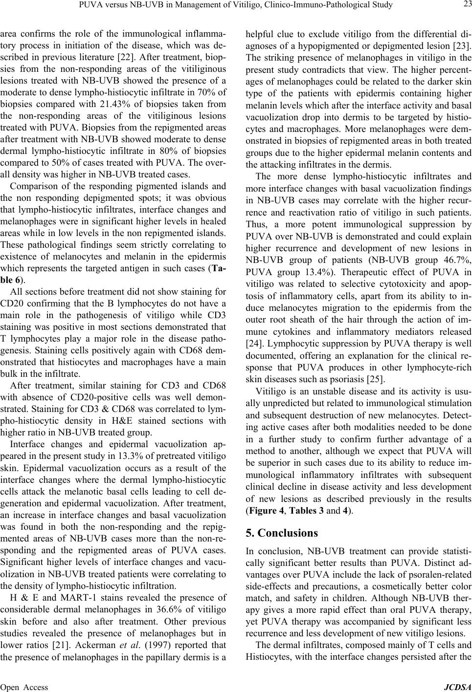 PUVA versus NB-UVB in Management of Vitiligo, Clinico-Immuno-Pathological Study 23 area confirms the role of the immunological inflamma- tory process in initiation of the disease, which was de- scribed in previous literature [22]. After treatment, biop- sies from the non-responding areas of the vitiliginous lesions treated with NB-UVB showed the presence of a moderate to dense lympho-histiocytic infiltrate in 70% of biopsies compared with 21.43% of biopsies taken from the non-responding areas of the vitiliginous lesions treated with PUVA. Biopsies from the repigmented areas after treatment with NB-UVB showed moderate to dense dermal lympho-histiocytic infiltrate in 80% of biopsies compared to 50% of cases treated with PUVA. The over- all density was higher in NB-UVB treated cases. Comparison of the responding pigmented islands and the non responding depigmented spots; it was obvious that lympho-histiocytic infiltrates, interface changes and melanophages were in significant higher levels in healed areas while in low levels in the non repigmented islands. These pathological findings seem strictly correlating to existence of melanocytes and melanin in the epidermis which represents the targeted antigen in such cases (Ta- ble 6). All sections before treatment did not show staining for CD20 confirming that the B lymphocytes do not have a main role in the pathogenesis of vitiligo while CD3 staining was positive in most sections demonstrated that T lymphocytes play a major role in the disease patho- genesis. Staining cells positively again with CD68 dem- onstrated that histiocytes and macrophages have a main bulk in the infiltrate. After treatment, similar staining for CD3 and CD68 with absence of CD20-positive cells was well demon- strated. Staining for CD3 & CD68 was correlated to lym- pho-histiocytic density in H&E stained sections with higher ratio in NB-UVB treated group. Interface changes and epidermal vacuolization ap- peared in the present study in 13.3% of pretreated vitiligo skin. Epidermal vacuolization occurs as a result of the interface changes where the dermal lympho-histiocytic cells attack the melanotic basal cells leading to cell de- generation and epidermal vacuolization. After treatment, an increase in interface changes and basal vacuolization was found in both the non-responding and the repig- mented areas of NB-UVB cases more than the non-re- sponding and the repigmented areas of PUVA cases. Significant higher levels of interface changes and vacu- olization in NB-UVB treated patients were correlating to the density of lympho-histiocytic infiltration. H & E and MART-1 stains revealed the presence of considerable dermal melanophages in 36.6% of vitiligo skin before and also after treatment. Other previous studies revealed the presence of melanophages but in lower ratios [21]. Ackerman et al. (1997) reported that the presence of melanophages in th e papillary dermis is a helpful clue to exclude vitiligo from the differential di- agnoses of a hypopigmented or depigmented lesion [23]. The striking presence of melanophages in vitiligo in the present study contradicts that view. The higher percent- ages of melanophages could be related to the darker skin type of the patients with epidermis containing higher melanin levels which after the in terface activity an d basal vacuolization drop into dermis to be targeted by histio- cytes and macrophages. More melanophages were dem- onstrated in biopsies of repigmented areas in both treated groups due to the higher epidermal melanin contents and the attacking infiltrates in the dermis. The more dense lympho-histiocytic infiltrates and more interface changes with basal vacuolization findings in NB-UVB cases may correlate with the higher recur- rence and reactivation ratio of vitiligo in such patients. Thus, a more potent immunological suppression by PUVA over NB-UVB is demonstrated and could explain higher recurrence and development of new lesions in NB-UVB group of patients (NB-UVB group 46.7%, PUVA group 13.4%). Therapeutic effect of PUVA in vitiligo was related to selective cytotoxicity and apop- tosis of inflammatory cells, apart from its ability to in- duce melanocytes migration to the epidermis from the outer root sheath of the hair through the action of im- mune cytokines and inflammatory mediators released [24]. Lymphocytic suppression by PUVA therapy is well documented, offering an explanation for the clinical re- sponse that PUVA produces in other lymphocyte-rich skin diseases such as psoriasis [25]. Vitiligo is an unstable disease and its activity is usu- ally unpredicted but related to immunological stimulation and subsequent destruction of new melanocytes. Detect- ing active cases after both modalities needed to be done in a further study to confirm further advantage of a method to another, although we expect that PUVA will be superior in such cases due to its ability to reduce im- munological inflammatory infiltrates with subsequent clinical decline in disease activity and less development of new lesions as described previously in the results (Figure 4, Tables 3 and 4). 5. Conclusions In conclusion, NB-UVB treatment can provide statisti- cally significant better results than PUVA. Distinct ad- vantages over PUVA include the lack of psoralen-related side-effects and precautions, a cosmetically better color match, and safety in children. Although NB-UVB ther- apy gives a more rapid effect than oral PUVA therapy, yet PUVA therapy was accompanied by significant less recurrence and less development of new vitiligo lesions. The dermal infiltrates, composed mainly of T cells and Histiocytes, with the interface changes persisted after th e Open Access JCDSA  PUVA versus NB-UVB in Management of Vitiligo, Clinico-Immuno-Pathological Study 24 NB-UVB repigmentation more than in the PUVA treated areas. Thus, PUVA may provide better immuno-modu- latory effect on vitiligo than NB-UVB and may give bet- ter response regarding the disease reactivation and recur- rence. A combination of UVA and NB-UVB bulbs in same treating cabinets maybe recommended obtaining better treatment results. REFERENCES [1] S. O. Kovacs, “Vitiligo,” Journal of the American Acad- emy of Dermatology, Vol. 38, No. 5, 1998, pp. 647-666. [2] M. D. Njoo, W. Westerhof, J. D. Bos and P. M. Bossuyt, “The Development of Guidelines for the Treatment of Vitiligo,” Archiv es of Dermatology, Vol. 135, No. 12, 1999, pp. 1514-1521. http://dx.doi.org/10.1001/archderm.135.12.1514 [3] T. M. Runger, “Ultraviolet Light,” In: J. L. Bolognia, J. L. Jorizzo, R. P. Rapini, T. D. Horn, A. J. Maoncini, J. M. Mascaro, S. J. Salasche, J. H. Saurat and G. Stingl, Eds., Dermatology, Mosby, Edinburgh, London, New York, Oxford, Philadelphia, St. Louis, Sydney, Toronto, Vol. 1, 2003, pp. 1353-1364. [4] M. El-Mofty and H. El-Sawaalhy, “Clinical Study of a New Preparation of 8-Methoxypsoralen in Photochemo- therapy,” International Journal of Dermatology, Vol. 33, No. 8, 1994, pp. 688-703. http://dx.doi.org/10.1111/j.1365-4362.1994.tb02904.x [5] D. Parsad, A. J. Kanwar and B. Kumar, “Psoralen-Ultra- violet A vs. Narrowband Ultraviolet B Phototherapy for the Treatment of Vitiligo,” Journal of the European Aca- demy of Dermatology and Venereology, Vol. 20, No. 2, 2006, pp. 175-177. http://dx.doi.org/10.1111/j.1468-3083.2006.01413.x [6] A. K .Gupta and T. F. Anderson, “Psoralen Photochemo- therapy,” Journal of the American Academy of Derma- tology, Vol. 17, No. 5, 1987, pp. 703-734. http://dx.doi.org/10.1016/S0190-9622(87)70255-2 [7] S. S. Yones, D. Der, A. Roy, et al., “Efficacy of Psora le n- UV-A Therapy vs Narrowband-UV-B Therapy,” Archives of Dermatology, Vol. 143, No. 5, 2007, pp. 578-584. http://dx.doi.org/10.1001/archderm.143.5.578 [8] R. S. Dawe, J. Ferguson and S. H. Ibbotson, “A Study of the Characteristics of Erythema Induced by Bath and Oral Methoxsalen Photochemotherapy and Narrowband Ultra- violet,” Journal of Photochemistry and Photobiology, Vol. 78, No. 1, 2003, pp. 55-60. http://dx.doi.org/10.1562/0031-8655(2003)078<0055:AIS OTC>2.0.CO;2 [9] Y. Kawakami, K. Jane, K. Takeshi, et al., “Production of Recombinant MART -1 Protie ns and Specific AntiMART - 1 Polyclonal and Monoclonal Antibodies: Use in the Cha- racterization of the Human Melanoma Antigen MART- 1,” Journal of Immunological Methods, Vol. 202, No. 1, 1997, pp. 13-25. http://dx.doi.org/10.1016/S0022-1759(96)00211-6 [10] S. S. Awad, H. Abdel-Raof, W. Hosam El-Din and M. El-Domyati, “Epithelial Grafting for Vitiligo Requires Ultraviolet A Phototherapy to Increase Success Rate,” Journal of Cosmetic Dermatology, Vol. 6, No. 2, 2007, pp. 119-124. http://dx.doi.org/10.1111/j.1473-2165.2007.00307.x [11] M. D. Njoo, J. D. Bos and W. Westerhof, “Treatment of Generalized Vitiligo in Children with Narrow-Band (TL- 01) UVB Radiation Therapy,” Journal of the American Academy of Dermatology, Vol. 42, No. 2, 2000, pp. 245- 253. http://dx.doi.org/10.1016/S0190-9622(00)90133-6 [12] M. El Mofty, W. Mostafa, S. Esmat, et al., “Narrow Band Ultraviolet B 311 nm in the Treatment of Vitiligo: Two Right-Left Comparison Studie s,” Photodermato logy, Photo- immunology & Photomedicine, Vol. 22, No. 1, 2006, pp. 6-11. http://dx.doi.org/10.1111/j.1600-0781.2006.00189.x [13] W. Westerhof and L. Nieuweboer-Krobotova, “Treatment of Vitiligo with UV-B Radiation vs Topical Psoralen Plus UV-A,” Archives of Dermatology, Vol. 133, No. 12, 1997, pp. 1525-1528. http://dx.doi.org/10.1001/archderm.1997.03890480045006 [14] I. Hamzavi, H. Jain, D. McLean, et al., “Parametric Mod- eling of Narrowband UV-B Phototherapy for Vitiligo Using a Novel Quantitative Tool,” Archives of Derma- tology, Vol. 140, No. 6, 2004, pp. 677-683. http://dx.doi.org/10.1001/archderm.140.6.677 [15] A. Simin, S. Sedef, B. Gonca and K. Fikret, “No Addi- tional Effect of Topical Calcipotriol on Narrow Band UVB Phototherapy in Patients with Generalized Vitiligo,” Photodermatology, Photoimmunology & Photomedicine, Vol. 21, No. 2, 2005, pp. 79-83. http://dx.doi.org/10.1111/j.1600-0781.2005.00139.x [16] S. M. Halpern, A. Anstey and R. S. Dawe, “Guidelines for Topical PUVA: A Report of a Workshop of the Brit- ish Photodermatology Group,” British Journal of Der- matology, Vol. 142, No. 1, 2000, pp. 22-31. http://dx.doi.org/10.1046/j.1365-2133.2000.03237.x [17] S. Valkova, M. Trashlieva and P. Christova, “Treatment of Vitiligo with Local Khellin and UVA: Comparison with Systemic PUVA,” Clinical and Experimental Der- matology, Vol. 29, No. 2, 2004, pp. 180-184. http://dx.doi.org/10.1111/j.1365-2230.2004.01462.x [18] S. Sahin, U. Hindioglu and A. Karaduman, “PUVA Treatment of Vitiligo: A Retrospective Study of Turikish Patients,” International Journal of Dermatology, Vol. 38, No. 7, 1999, pp. 542-545. http://dx.doi.org/10.1046/j.1365-4362.1999.00654.x [19] T. S. Anbar, W. Westerhof, A. T. Abdel-Rahman and M. A. El-Khayyat, “Evaluation of the Effects of NB-UVB in Both Segmental and Non-Segmental Vitiligo Affecting Different Body Sites,” Photodermatology, Photoimmu- nology & Photomedicine, Vol. 22, No. 3, 2006, pp. 157- 163. http://dx.doi.org/10.1111/j.1600-0781.2006.00222.x [20] R. S. Stern, “The Risk of Melanoma in Association with Long-Term Exposure to PUVA,” Journal of the Ameri- can Academy of Dermatology, Vol. 44, No. 5, 2001, pp. 755-761. http://dx.doi.org/10.1067/mjd.2001.114576 [21] Y. C. Kim, H. Y. Kang, S. Seonghyang and E. S. Lee, “Histopathologic Features in Vitiligo,” The American Journal of Dermatopathology, Vol. 30, No. 2, 2008, pp. 112-115. Open Access JCDSA  PUVA versus NB-UVB in Management of Vitiligo, Clinico-Immuno-Pathological Study Open Access JCDSA 25 http://dx.doi.org/10.1097/DAD.0b013e3181651511 [22] L. F. Montes, J. Abulafia, W. H. Wilborn, et al., “Value of Histopathology in Vitiligo,” International Journal of Dermatology, Vol. 42, No. 1, 2003, pp. 57-61. http://dx.doi.org/10.1046/j.1365-4362.2003.01743.x [23] A. B. Ackerman, N. Chongchitant, J. Sanchez, Y. Guo, B. Bennin, M. Reichel and A. B. Randall, “Embryologic, Histologic and Anatomic Ascpects,” In: A. B. Ackerman, N. Chongchitant, J. Sanchez, Y. Guo, B. Bennin, M. Rei- chel and A. B. Randall, Eds., Histologic Diagnosis of In- flammatory Skin Disease s, 2nd Edition, Williams and Wil - kins, Baltimore, Phildelphia, London, Paris, Banckok, Hong Kong, Sydney, Tokyo, 1997, p. 2. [24] T. B . F i tzp at ric k, “Me c ha ni sm s of P ho to th er apy o f Vitiligo ,” Archives of Dermatology, Vol. 133, No. 12, 1997, pp. 1591-1592. http://dx.doi.org/10.1001/archderm.1997.03890480113020 [25] R. M. Halder and C. M. Young, “New and Emerging Therapies for Vitiligo,” Dermatologic Clinics, Vol. 18, No. 1, 2000, pp. 79-83. http://dx.doi.org/10.1016/S0733-8635(05)70149-2 |